
Contents:
Pseudotumor cerebri is a condition associated with increased pressure inside the skull despite the absence of a brain tumor. Today, this condition is generally referred to as idiopathic intracranial hypertension. Headache, temporary visual blackouts, double vision, and hearing a pulse-synchronous sound in the ear are common complaints. Increased intracranial pressure may cause swelling of the optic nerve head known as papilledema. Since the course of the disease varies from person to person, diagnosis and treatment should be planned through evaluation by the relevant specialties, particularly ophthalmology and neurology.
What Is Pseudotumor Cerebri?
Pseudotumor cerebri is a condition in which intracranial pressure is elevated, but no brain tumor, hydrocephalus, or other structural cause explaining the pressure increase is detected. The term "idiopathic" indicates that the cause of the increased pressure cannot be determined. If a known medication, vascular disease, or systemic condition explains the increase in intracranial pressure, the condition is not directly classified as idiopathic. Ruling out secondary causes that may produce similar findings is therefore an important part of the diagnosis.
Increased intracranial pressure may cause swelling of the part of the optic nerve visible at the back of the eye. Papilledema is an important finding that can be detected during an eye examination and requires close monitoring of visual function. If the pressure remains elevated for a long time, the visual field may be affected, and some patients may develop permanent vision loss.
Who Develops Pseudotumor Cerebri?
The condition may occur at any age and in any sex. However, it is known to be more common among women of childbearing age and people with obesity. Significant recent weight gain may also be considered during the assessment. Being female or overweight alone is not sufficient to establish a diagnosis. Secondary causes may need to be investigated in greater detail in men, children, older people, or patients without obesity.
Certain medications and systemic diseases are among the conditions that may be associated with increased intracranial pressure. Tetracycline antibiotics, vitamin A derivatives, and medications such as growth hormone may be investigated during the assessment. Cerebral venous thrombosis, anemia, and certain hormonal or metabolic diseases may also cause a similar condition. However, not every reported association can be said to cause the disease definitively. Regularly used medications should not be discontinued or their doses changed without medical evaluation.
What Are the Symptoms of Pseudotumor Cerebri?
Symptoms may vary from person to person, and the diagnosis cannot be established based on headache alone. Visual complaints may be more prominent in some patients, while prolonged headaches may be more noticeable in others. The characteristics of the headache may resemble migraine or change over time.
Common symptoms include:
- Recurrent or prolonged headache.
- Temporary visual blackouts lasting several seconds.
- Blurred vision or narrowing of the visual field.
- Newly developed double vision.
- A pulsing or ringing sound in the ear synchronized with the heartbeat.
- Nausea and, in some cases, vomiting.
- A feeling of pressure in the neck, shoulders, or around the eyes.
In some people, headaches may increase in the morning, when bending forward, coughing, or straining. However, these characteristics do not occur in every patient and are not specific to pseudotumor cerebri. Hearing a pulse-synchronous sound in the ear may also be a notable complaint. Since similar symptoms may occur with different neurological and eye diseases, the assessment should not be delayed.
Why Are Visual Problems Important?
One of the most important risks of increased intracranial pressure is its effect on the optic nerve. Patients may describe brief visual blackouts, blurred vision, double vision, or reduced peripheral vision. When visual field loss develops gradually, the person may not initially notice the change. Peripheral visual field impairment may be present even when central visual acuity is normal.
Swelling and functional changes in the optic nerve can be monitored through fundus examination, visual field testing, and optical coherence tomography. Optical coherence tomography helps measure changes in the retinal nerve fiber layer. Rather than relying on a single test result, changes in visual acuity, color vision, visual field, and optic nerve findings over time are evaluated together.
How Is Pseudotumor Cerebri Diagnosed?
The diagnosis is not based solely on a headache or a single lumbar puncture result. Symptoms, eye and neurological examinations, brain imaging, and evaluation of the cerebrospinal fluid are considered together. The aim is to demonstrate increased intracranial pressure and rule out other diseases that could explain the pressure increase.
The diagnostic process generally consists of 4 components:
- A detailed eye examination including visual acuity, fundus examination, and visual field testing.
- A neurological examination and evaluation of accompanying symptoms.
- Investigation of structural causes using brain MRI or CT when appropriate.
- Measurement of opening pressure and examination of cerebrospinal fluid through lumbar puncture following appropriate imaging.
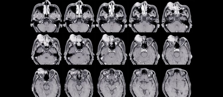
MR venography or CT venography may be required to rule out thrombosis in the cerebral veins. Imaging may reveal findings associated with increased intracranial pressure, such as an empty sella, enlargement around the optic nerve, or narrowing of the venous sinuses. These findings alone are not sufficient to establish a diagnosis of pseudotumor cerebri. The pressure measured during lumbar puncture must also be interpreted together with the conditions under which the procedure was performed and the other clinical findings.
How Is Pseudotumor Cerebri Treated?
Treatment has 3 main objectives: managing modifiable factors associated with the disease, protecting visual function, and reducing the effect of headaches on daily life. The treatment plan is developed by considering the degree of papilledema, changes in the visual field, characteristics of the headache, and the person's general health.
The Role of Weight Management in Treatment
For suitable patients who are overweight or have obesity, weight management is one of the main approaches that may affect the course of the disease. The target should be determined safely and sustainably by considering the person's general health. Instead of rapid and uncontrolled weight-loss methods, a plan may be developed through nutrition, physical activity, and support from the relevant specialties when necessary. The same approach is not expected to be applied to people with a normal body weight.
What Can Be Expected from Medication?
Acetazolamide is one of the medications considered for suitable patients to help reduce cerebrospinal fluid production. The need for treatment and the dosage are determined by considering the optic nerve findings in particular. Side effects such as tingling in the hands or feet, changes in taste, fatigue, and gastrointestinal complaints may occur. Kidney disease, plans for pregnancy, and other medications being used should be evaluated before treatment.
Different medication options may be considered for some patients. Headache treatment is planned separately according to whether the pain has migraine-like characteristics and whether medication overuse is present. A medication intended to reduce intracranial pressure is not expected to eliminate every headache. Medications should not be started, discontinued, or have their dosage changed without medical guidance.
Is Lumbar Puncture Used for Treatment?
Lumbar puncture allows the opening pressure to be measured and the cerebrospinal fluid to be examined during diagnosis. Some patients may experience temporary relief from headaches after the procedure. However, since cerebrospinal fluid is produced again, this effect may not be permanent. Repeated lumbar punctures are not considered a routine long-term treatment for the disease.
Lower back pain or a headache that becomes more pronounced when standing may develop after the procedure. If complaints are severe or prolonged, the healthcare institution that performed the procedure should be contacted.
When Is Surgical Intervention Required?
Surgical options may be considered in patients whose visual function is deteriorating rapidly or whose risk of vision loss continues despite medication. Shunt operations that redirect cerebrospinal fluid to another body cavity and optic nerve sheath fenestration are among these options. The preferred method may vary according to the visual findings, headache, anatomical features, and the center's experience.
Venous sinus stenting may be considered in selected patients with pronounced narrowing of the cerebral veins and suitable pressure characteristics. This method is not performed in every patient. Patient selection, long-term outcomes, and the possibility of recurrent narrowing should be evaluated together. The benefits and risks of the procedure should be considered individually.
How Should Patient Reviews of Pseudotumor Cerebri Be Evaluated?
Patient reviews of pseudotumor cerebri may help people understand the uncertainty experienced during diagnosis, the effect of headaches on daily life, and the importance of regular eye examinations. Some people may state that they attributed their headaches to migraine, stress, or fatigue before diagnosis. Other patients may report that they initially did not consider temporary visual blackouts important. These experiences may demonstrate why headaches and visual changes should be evaluated together.
Medication side effects, temporary relief following lumbar puncture, weight management, and surgical experiences may be described differently in patient reviews. However, a medication, dosage, or procedure that benefits one person may not be suitable for another patient. Disease severity, the degree of papilledema, visual field findings, and accompanying health problems vary from person to person. Online reviews should therefore not be used as criteria for diagnosis, medication selection, or surgical decisions.
How Does the Disease Affect Daily Life?
Prolonged headaches, anxiety about vision loss, and repeated examinations may affect work, school, and social life. Screen use does not directly cause the disease, but it may reduce working comfort when headaches or visual complaints are present. A person's activity level, working routine, and need for rest may vary according to the symptoms. Headaches should be evaluated separately because they may continue in some patients even after intracranial pressure has been brought under control.
Recording symptoms, medications being used, and visual changes may be helpful during follow-up consultations. If anxiety caused by the diagnostic process or loss of daily functioning is significant, psychological support may also be considered part of the overall treatment plan.
Which Tests Are Performed During Follow-Up?
The frequency of follow-up is determined according to the degree of papilledema and changes in visual function. Visual acuity, color vision, fundus examination, visual field testing, and optic nerve imaging may be repeated at specific intervals. Headache frequency, medications being used, side effects, and weight changes are also reviewed during follow-up. If the visual field is deteriorating, reassessment may be required without waiting for the scheduled follow-up appointment.
Which Symptoms Require Urgent Evaluation?
Sudden reduction in vision, rapidly increasing blurred vision, newly developed double vision, or significant narrowing of the visual field should be evaluated without delay. A severe headache accompanied by repeated vomiting, changes in consciousness, weakness, speech impairment, or other neurological symptoms also requires urgent investigation. Increasingly frequent episodes of visual blackout may indicate a greater effect on the optic nerve.
If these symptoms occur, it is important not to wait for the scheduled follow-up appointment. The information provided here is intended for general informational purposes and does not replace an individual examination, diagnosis, or treatment plan.










































