
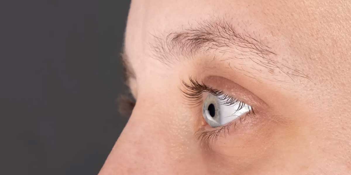
Corneal transplantation is a surgical procedure performed when the transparent corneal layer at the front of the eye has been damaged by disease, infection, trauma or a structural disorder. Corneal transplantation involves replacing all of the damaged tissue or only the affected layer with corneal tissue obtained from a suitable donor. Also known as keratoplasty, this surgery may be planned to improve visual quality, protect the ocular surface or restore the structural integrity of the cornea. Keratoconus, Fuchs endothelial dystrophy, corneal oedema and permanent corneal scarring are among the conditions that may require transplantation. Surgery is not necessary for every corneal disease, and the treatment decision is based on the depth of the damage. The level of vision after transplantation also depends on factors other than the cornea, including the retina, optic nerve and intraocular pressure.
When Is Corneal Transplantation Performed?
Corneal transplantation may be considered when glasses, contact lenses, medication or other treatments do not provide adequate results. Advanced keratoconus can cause pronounced thinning and irregularity of the cornea, seriously affecting vision. In Fuchs endothelial dystrophy, a reduction in the cells on the inner surface of the cornea can lead to tissue oedema and clouding. Bullous keratopathy that develops after cataract surgery may also create a need for transplantation in some patients. Dense scars remaining after infection, trauma or chemical injury may prevent light from entering the eye. Congenital corneal opacities, corneal melt and loss of function in previously transplanted tissue are also among the possible causes. In some patients, the primary aim is to improve vision, while in other cases preserving the integrity of the eye or controlling an infection that is resistant to treatment takes priority. The need for surgery is determined by considering the person's daily life together with the current findings in the eye.
What Are the Types of Corneal Transplantation?
Corneal transplantation methods vary according to which layer of the cornea is damaged. In penetrating keratoplasty, a full-thickness tissue replacement involving all layers of the cornea is performed. Deep anterior lamellar keratoplasty aims to preserve the healthy inner layer when the front and middle layers of the cornea are diseased. In endothelial keratoplasty methods, the damaged layer in the inner part of the cornea is replaced. DMEK and DSAEK are two different partial transplantation methods used for endothelial diseases. Replacing only the diseased layer may affect the recovery process and the likelihood of certain complications in suitable patients. However, no single method is suitable for every corneal disease. The surgical method is selected according to tissue transparency, the condition of the endothelial cells and any previous eye surgery.
What Is a Full-Thickness Corneal Transplant?
Penetrating keratoplasty involves removing the damaged tissue in the centre of the cornea through all its layers and replacing it with healthy donor tissue. This method may be considered in patients whose anterior and posterior corneal layers are both affected or who have deep scarring. The donor tissue is prepared to the appropriate dimensions under an operating microscope and secured to the ocular surface with fine sutures. It may be necessary in cases of advanced corneal scarring, pronounced thinning or loss of function in a previous transplant. Recovery may take longer in some patients than with partial-layer transplants. Since suture-related astigmatism may develop after surgery, the level of vision can change over time. The timing of suture removal is determined individually according to wound healing and the shape of the cornea. A full-thickness transplant should be considered only when the damage cannot be treated with more limited methods.
How Is DALK Corneal Transplantation Performed?
DALK corneal transplantation is a surgical method in which the anterior and middle layers of the cornea are replaced while the person's healthy endothelial layer is preserved. It may be considered particularly in keratoconus and certain corneal scars where the endothelial cells remain healthy. After the damaged outer layers have been separated, appropriately prepared donor tissue is placed in position. Because the person's own inner layer is preserved, the risk of endothelial rejection differs from methods in which this layer is transplanted. However, rejection in the anterior layers, suture problems or irregular astigmatism may still occur. If an unintended opening forms in the deep layers of the cornea during surgery, the surgical plan may change. Visual recovery may take time depending on tissue healing and the condition of the sutures. DALK is not a suitable option for patients with a damaged endothelial layer.
What Is DMEK Corneal Transplantation?
DMEK corneal transplantation involves replacing Descemet's membrane and the endothelial cells on the inner surface of the cornea with suitable donor tissue. This method is most often considered for Fuchs endothelial dystrophy and corneal oedema caused by endothelial failure. Only the inner layer that has lost its function is replaced, while the outer and middle layers of the cornea are preserved. The prepared thin donor tissue is inserted into the eye through a small incision. An air or suitable gas bubble is then introduced into the eye to help the tissue attach to the posterior surface of the cornea. In suitable patients, visual recovery may occur more quickly than after a full-thickness transplant. However, risks such as tissue detachment, the need for another air injection, increased intraocular pressure and tissue rejection are not eliminated entirely. Suitability for the method is determined by taking into account the anatomical structure of the eye and any other accompanying eye diseases.
Differences Between DSAEK and DMEK
DSAEK and DMEK are partial transplantation methods used to treat endothelial failure in the inner layer of the cornea. In DMEK, the donor tissue consists primarily of Descemet's membrane and endothelial cells. In DSAEK, a thin layer of supporting corneal tissue is transplanted in addition to these layers. The thickness of the transplanted tissue and the way it is placed inside the eye are among the main differences between the two techniques. DMEK may provide faster visual recovery in some suitable patients, but unfolding and attaching the thin tissue creates different technical requirements. DSAEK may be considered in eyes with certain anatomical features or in more complex surgical situations. Risks such as tissue detachment, changes in intraocular pressure and transplant rejection should be monitored with both methods. The choice of method is determined according to the type of corneal damage and the overall structure of the eye.
Is Corneal Transplantation Necessary for Keratoconus?
Keratoconus is an eye disease in which the cornea thins and bulges forwards. In the early or moderate stages of the disease, glasses, special contact lenses and corneal cross-linking in suitable patients may be considered. Corneal transplantation is not the first treatment used for every patient with keratoconus. Transplantation may be considered if adequate vision cannot be achieved with contact lenses, pronounced corneal scarring has developed or advanced thinning is present. In patients with a healthy endothelial layer, anterior-layer transplants such as DALK may be considered depending on suitability. A full-thickness transplant may be required if the damage affects all layers of the cornea. The need for glasses or special contact lenses may not disappear completely after surgery. The treatment decision is made by considering the stage of disease progression together with the structural characteristics of the cornea.
When Is Transplantation Necessary for Fuchs Dystrophy?
Fuchs endothelial dystrophy is a disease associated with a gradual reduction in the endothelial cells on the inner surface of the cornea. Because these cells maintain the fluid balance of the cornea, loss of their function can cause the tissue to swell and vision to become blurred. Blurred vision may be more pronounced in the morning for some people. In mild cases, symptoms may be monitored with eye drops, ointments and other supportive methods. Endothelial transplantation may be considered when corneal oedema becomes permanent or vision loss begins to affect daily life. DMEK and DSAEK are options intended to replace the damaged inner layer in suitable patients. If cataracts or other eye problems are also present, the surgical plan may be adjusted accordingly. A diagnosis of Fuchs dystrophy does not mean that every patient will necessarily undergo corneal transplantation.
How Is Donor Corneal Tissue Obtained?
The donor tissue used in corneal transplantation is obtained from deceased donors who have been assessed as suitable. The donated corneas undergo specified medical and laboratory assessments at authorised eye banks. The donor's medical history, risks of infectious disease and the structural characteristics of the cornea are examined. The condition of the endothelial cells is one of the criteria that must be assessed, particularly for inner-layer transplants. Tissue found to be suitable is stored under specified conditions until transplantation. Tissue procurement and allocation are carried out in accordance with the relevant legislation and the procedures followed by authorised centres. Advanced donor age alone does not mean that the tissue cannot be used, and assessment is based on the characteristics of the tissue. Corneal transplantation replaces only the required corneal tissue, not the entire eye.
Is Blood Type Important in Corneal Transplantation?
Under healthy conditions, the cornea is a tissue without prominent blood vessels. For this reason, routine blood type matching is generally not required as it is in kidney or liver transplantation. When selecting donor tissue, priority is given to corneal transparency, adequate endothelial cell quality and infection screening. However, the risk of tissue rejection may increase if there is extensive corneal vascularisation, a history of previous transplants or significant inflammation. In high-risk patients, the transplant plan and preventive treatment are arranged accordingly. The fact that blood type compatibility is not routinely required does not mean that tissue rejection can never occur. The immune response, the type of surgery and the condition of the ocular surface can affect the long-term course of the transplant. The suitability of donor tissue is assessed according to established medical criteria, not personal preference or age.
Who Is Not Suitable for Corneal Transplantation?
Suitability for corneal transplantation is assessed according to the condition of the other structures of the eye as well as the corneal disease. If there is advanced optic nerve damage or serious retinal disease, the expected visual benefit may remain limited even if the cornea becomes clear. Uncontrolled glaucoma, severe ocular surface disease and significant eyelid problems can negatively affect the success of transplantation. In people with an active eye infection, planned transplantation to improve vision is generally postponed until the infection is under control. However, in severe corneal infections that do not respond to medication, a therapeutic transplant may also be considered to preserve the integrity of the eye. Diabetes, autoimmune diseases or immune system disorders do not always constitute an absolute barrier on their own. The level of control of these conditions, wound healing and the risk of infection should be assessed individually. The principal criterion in the transplantation decision is whether the expected benefit is favourable in relation to the potential risks.
How Should You Prepare for Corneal Transplantation?
Before surgery, a detailed eye examination is used to determine which layers of the cornea are affected. Corneal topography, corneal thickness measurement and endothelial cell examination may be used in patients for whom they are considered necessary. Intraocular pressure, the retina and the optic nerve are assessed to investigate the visual benefit that may be expected after surgery. Previous infections, a history of herpes, earlier eye surgeries and regularly used medications are taken into account. Blood thinners or other systemic medications should not be stopped on the person's own initiative. If medication adjustments are required, the decision is made according to the surgical plan and general health status. If eyelid inflammation, dry eye or an active infection is present, treatment of these problems may be planned first. The possible benefits, limitations and follow-up requirements of the surgery should be assessed in detail before the procedure.
How Is Corneal Transplant Surgery Performed?
Corneal transplantation may be performed under local or general anaesthesia, depending on the method to be used and the patient's condition. First, the damaged corneal layer or full-thickness tissue is carefully removed under an operating microscope. In a full-thickness transplant, the donor cornea prepared to the appropriate size is secured in place with fine surgical sutures. During DALK, the person's healthy inner layer is preserved and donor tissue is placed in the anterior layers. In DMEK and DSAEK, donor tissue is introduced into the eye through a small incision. In endothelial transplantation, an air or gas bubble supports the attachment of the new tissue to the posterior surface of the cornea. The duration of the procedure varies according to the selected technique and whether another eye operation is required at the same time. After surgery, the cornea, sutures and intraocular pressure are checked at specified intervals.
Recovery After Corneal Transplantation
Recovery after corneal transplantation varies according to whether a full-thickness or partial-layer transplant has been performed. Blurred vision, mild stinging and sensitivity to light may occur during the first few days. Following a full-thickness transplant, marked visual recovery may take months, depending on the stabilisation of the sutures and the shape of the cornea. With endothelial transplants such as DMEK or DSAEK, visual recovery may begin earlier in some patients. However, tissue attachment, resolution of corneal oedema and changes in intraocular pressure affect this process. After endothelial transplantation, lying on the back for specified periods may be recommended to help the air or gas bubble remain in the correct position. While a bubble remains inside the eye, individual advice regarding air travel or travel to high altitudes should be followed. The speed of recovery differs for every patient, and no definite timeframe can be given for achieving the final level of vision after the procedure.
Medication Use After Corneal Transplantation
Antibiotic eye drops may be used for specified periods after surgery to reduce the risk of infection. Eye drops containing corticosteroids may play an important role in controlling inflammation in the eye and the risk of tissue rejection. The duration of their use varies according to the type of transplant and the patient's level of risk. If corticosteroid treatment is prolonged without supervision, problems such as increased intraocular pressure and infection may occur. In people with a history of corneal disease caused by herpes, antiviral treatment may also be planned if considered appropriate. If dry eye or surface sensitivity is present, lubricating drops may be added to the treatment. The dose of prescribed medication should not be changed without medical advice simply because symptoms have improved. At regular follow-up appointments, both the effects and possible side effects of the medication are assessed together.
Sutures After Corneal Transplantation
In full-thickness corneal transplantation and DALK surgery, donor tissue is usually secured with fine sutures. The sutures help the tissue remain in the correct position while the cornea heals. The timing of their removal can vary according to the strength of the wound and the shape of the cornea. Some sutures may be removed earlier in a controlled manner to manage astigmatism. Sutures that become loose or irritate the ocular surface should be assessed because they can increase the risk of infection. DMEK and DSAEK generally do not require extensive corneal suturing, although a small number of sutures may be used for the entry incision. The presence of sutures does not necessarily mean that the surgery is more or less successful. Rubbing the eye or exposing it to impact may damage the healing tissue.
Signs of Tissue Rejection After Corneal Transplantation
Tissue rejection after corneal transplantation can occur when the immune system recognises the transplanted tissue as foreign. Redness, pain, sensitivity to light and reduced vision are among the possible warning signs. Since symptoms may be mild in some patients, changes in vision should be taken seriously. Tissue rejection may occur shortly after surgery or develop at a later stage. Corneal vascularisation, previous transplants, inflammation and certain ocular surface diseases can increase the risk. The tissue layers that may be rejected differ between DMEK, DSAEK, DALK and full-thickness transplantation. When symptoms are recognised early, appropriate treatment may limit tissue damage, but the same outcome is not achieved in every case. If redness, sensitivity to light or a sudden reduction in vision develops, an assessment should be carried out without delay.
Why Does Tissue Rejection Occur?
The avascular structure of the cornea limits contact between the immune system and the transplanted tissue in some circumstances. Nevertheless, inflammation on the corneal surface, the formation of new blood vessels or previous surgery can alter this protective balance. The rejection process may begin when immune cells recognise structures in the donor tissue as foreign. A previous failed transplant, recurrent eye infections and uncontrolled ocular surface diseases can increase the risk. Irregular use of eye drops may also make it more difficult to control the immune response. A gradual reduction in endothelial cells does not always mean active tissue rejection. Some grafts may lose function without a pronounced rejection episode. For this reason, the transparency of the transplanted tissue, intraocular pressure and endothelial condition are assessed together during long-term follow-up.
Possible Risks of Corneal Transplantation
Infection, tissue rejection, corneal oedema and problems related to wound healing may occur after corneal transplantation. In full-thickness and anterior-layer transplants, suture-related irritation, loosening or irregular astigmatism may develop. In DMEK and DSAEK surgery, detachment of the transplanted inner layer may require an additional injection of air or gas. Intraocular pressure may increase because of the surgery itself or the corticosteroid drops used. If the increase in intraocular pressure is not controlled, the optic nerve may be damaged. Progression of cataracts, corneal vascularisation or recurrence of an existing eye disease are also among the risks considered in some patients. In rare cases, a serious infection in the deeper structures of the eye or other surgical complications may develop. The level of risk varies according to the type of transplant, the previous condition of the eye and adherence to postoperative care.
How Does Vision Change After Transplantation?
The change in vision after corneal transplantation depends on how quickly the cornea becomes clear and the condition of the other structures of the eye. Blurred vision during the first few days does not always mean that the transplant has failed. Corneal oedema, the effect of the sutures and temporary surface irregularities can affect image quality during the early period. After healing is complete, glasses, special contact lenses or additional procedures to manage astigmatism may be required. If retinal disease, glaucoma or optic nerve damage is present, the expected improvement in vision may remain limited even if the cornea becomes clear. In children, the risk of amblyopia and the process of visual development should also be taken into account. Some people experience marked improvement in daily life, while visual gain may be more limited in other patients. It is not possible to predict with certainty before surgery that vision will be restored completely.
How Successful Is Corneal Transplantation?
The outcome of corneal transplantation varies according to why the surgery is performed and the initial condition of the eye. Cases such as keratoconus, in which the other layers of the cornea are relatively healthy, are not assessed in the same way as cases involving severe infection or extensive vascularisation. The characteristics of the donor tissue, the selected technique and the postoperative follow-up schedule affect the course of the transplant. Previous transplants, uncontrolled glaucoma and significant ocular surface disease may increase the risks. Layer-selective methods such as DMEK, DSAEK or DALK may provide certain advantages in suitable patients, but they cannot be used in every case. Maintaining the transparency of the corneal tissue and the level of vision achieved do not always mean the same thing. Other factors such as the retina, optic nerve and astigmatism continue to determine the visual outcome. For this reason, it is not appropriate to provide a success percentage or promise a definite result that applies to every patient.
What Should You Consider After Corneal Transplantation?
After corneal transplantation, it is important not to rub the eye and to protect it from direct impact. Prescribed eye drops should be used at the specified frequency, and follow-up appointments should not be missed. During the initial period, it may be necessary to avoid dusty environments, intense physical exertion and contact between the eye and contaminated water. The timing of returning to swimming, heavy lifting and sports activities is determined according to the type of surgery. After DMEK or DSAEK, the recommended lying position may help the donor tissue attach. The use of protective glasses or an eye shield may support protection of the eye, particularly during sleep and in outdoor conditions. If severe pain, discharge, redness or a sudden reduction in vision occurs, an assessment should be carried out without delay. The time required to return to daily life varies according to the level of tissue healing and the person's occupation.
Can Corneal Transplantation Be Repeated?
If transplanted corneal tissue loses its transparency or function over time, repeat transplantation may be considered. Graft failure, tissue rejection, recurrent infection or endothelial cell loss may cause this situation. The need for repeat transplantation is determined not only by reduced vision but also by which corneal layer is affected. In some patients, it may be possible to transplant only the inner layer that has lost function instead of replacing the entire cornea again. As the number of previous surgeries and the degree of corneal vascularisation increase, the risk of tissue rejection may also change. Intraocular pressure, ocular surface health and underlying diseases should be assessed before a new procedure. The outcomes of repeat transplants cannot be predicted in the same way as those of the first surgery. The surgical decision is made by considering the possible visual benefit together with the need to preserve the tissue.
When Is an Artificial Cornea Considered?
An artificial cornea is an option considered in selected patients for whom standard donor corneal transplantation is unsuitable or whose repeated transplants have failed. This method, known as a keratoprosthesis, involves placing a special optical structure in place of the natural corneal tissue. It may be considered in cases involving severe ocular surface disease, extensive vascularisation or a history of multiple failed transplants. However, it is not a routine procedure that can be performed for every corneal disease. There are significant limitations, including infection, glaucoma, tissue problems around the device and the need for long-term care. The condition of the optic nerve and retina directly affects the potential benefit that this method can provide. Some patients may require continuous use of a protective contact lens and close follow-up. The decision to use an artificial cornea is made after standard treatment options and individual risks have been assessed in detail.
Alternatives to Corneal Transplantation
Transplantation is not necessary for every corneal disease, and the appropriate treatment is determined according to the type of disease. At certain stages of keratoconus, glasses, rigid contact lenses, scleral lenses and corneal cross-linking may be considered. In mild Fuchs dystrophy, eye drops and supportive measures intended to reduce corneal oedema may be used. For superficial corneal scars, laser treatment or other surface-regularising procedures may be considered in some patients. For problems caused by infection, the priority is appropriate antimicrobial treatment and protection of the ocular surface. Treating accompanying conditions such as dry eye, eyelid disorders or allergies may also help reduce symptoms. These options do not replace transplantation in every case of advanced structural damage. The decision for surgery is made by considering the current condition of the cornea and the response to non-surgical treatments.
Corneal Transplantation Assessment in Izmir
A corneal transplantation assessment in Izmir begins with a detailed examination intended to determine the type of corneal disease and the cause of vision loss. Corneal topography, endothelial cell assessment and intraocular pressure measurement may be planned when considered necessary. In patients with keratoconus, Fuchs dystrophy, corneal oedema or permanent corneal scarring, suitable surgical options are reviewed according to individual findings. When choosing between DALK, DMEK, DSAEK and full-thickness transplantation, the other structures of the eye are also assessed. Donor tissue procurement and transplantation procedures are carried out at authorised centres in accordance with the relevant legislation. The surgical plan should be adjusted separately for people who have undergone previous eye surgery or have other eye diseases. For people researching corneal transplantation in Izmir and the Konak area, it is important that the need for the procedure be determined through a detailed examination. After transplantation, regular follow-up helps monitor tissue health and changes in vision.
How Are Corneal Transplantation Prices Determined?
Corneal transplantation prices may vary according to whether transplantation is necessary, the surgical method to be used and the individual treatment plan. Full-thickness transplantation, DALK, DMEK and DSAEK procedures may require different surgical preparations and follow-up processes. The severity of the corneal disease, accompanying eye problems and the need for additional surgery may affect the scope of the assessment. Preoperative examinations, the medications to be used and postoperative follow-up appointments are also parts of the planning process. Donor tissue procurement and allocation are carried out in accordance with the authorised procedures specified in the relevant legislation. It is not possible to determine the appropriate technique or scope of fees without an examination. Fee information can be shared individually after the medical requirements and the procedure to be performed have been clarified. You can contact us through the website to obtain information about corneal transplantation assessment and treatment options suitable for you.




































