Contents:
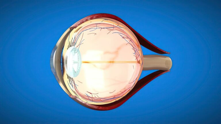
The vitreous is the transparent, gel-like structure located between the eye's lens and the retina that fills the back of the eye. It helps maintain the shape of the eye and allows light to reach the retina. It consists mainly of water and contains collagen fibres and hyaluronic acid. Ageing, trauma and certain eye diseases may cause changes in the structure of the vitreous. These changes may cause symptoms such as floaters, flashes of light or blurred vision. The need for treatment is determined according to the type of vitreous change and whether the retina has been affected.
What Is the Vitreous and What Does It Do?
The vitreous fills the large space at the back of the eyeball. Its transparent structure allows incoming light to reach the retina. The collagen fibres and hyaluronic acid it contains help maintain its gel-like consistency. Under healthy conditions, this structure does not contain blood vessels. Although it is in contact with the retina, it does not adhere with the same strength in every part of the eye. Age-related changes or certain diseases may affect this balanced structure of the vitreous.
Age-Related Vitreous Changes
At a young age, the vitreous has a more homogeneous and organised gel structure. With advancing age, this structure may partially liquefy, and the collagen fibres within it may clump together. The formation of small fluid-filled spaces within the gel may be regarded as part of the natural ageing process. Over time, the vitreous may shrink and begin to separate from its attachments at the back of the eye. Not all these changes necessarily cause symptoms. However, some people may notice moving spots or thread-like shapes in front of their eyes.
Age-related vitreous changes do not usually indicate a serious condition on their own. However, traction may develop in certain areas as the vitreous separates from the retina. The degree of traction on the retina determines the potential risks. Symptoms that begin suddenly require conditions such as a retinal tear to be ruled out. High myopia, previous eye surgery and eye trauma may also affect risk assessment in some patients. The time when the symptoms began and how they have changed are therefore considered during the examination.
Why Do Floaters Appear?
One of the most common symptoms of vitreous disorders is the appearance of floaters. These images are usually associated with the clumping of collagen fibres within the vitreous. The clumped fibres may cast small shadows in front of the light reaching the retina. These shadows are particularly noticeable when looking at light-coloured walls, the sky or bright screens. The images move as the eye moves and may continue to drift slowly when the gaze becomes fixed. The number and appearance of floaters may vary from person to person.
Visual symptoms described by patients include:
- Small black spots.
- Images resembling flying insects.
- A cobweb-like appearance.
- Transparent threads.
- Moving shadows.
- Flashes of light.
A small number of floaters that have remained unchanged for a long time may be associated with an age-related change in some people. However, a sudden increase in floaters requires a different assessment. The retina should be examined carefully, particularly if flashes of light or a curtain-like shadow in the visual field are also present. Sudden symptoms may be associated with vitreous separation or a retinal tear. Not every floater requires surgical treatment. The appropriate approach is determined according to the findings obtained during the fundus examination.
What Is Posterior Vitreous Detachment?
Posterior vitreous detachment is the separation of the vitreous gel from the back of the retina as it gradually shrinks. This condition becomes more common with advancing age. During the separation, the person may notice new floaters or flashes of light. Flashes may occur when traction exerted by the vitreous on the retina is perceived as a visual stimulus. In most patients, the separation is completed without causing a significant problem. However, a tear may develop in areas where the vitreous is firmly attached to the retina.
A fundus examination is important because a retinal tear may increase the risk of retinal detachment. During the examination, the retinal periphery is evaluated in detail. Even if no tear is found during the initial examination, an additional follow-up may be required depending on the course of the symptoms. A rapid increase in floaters or the development of a shadow in part of the visual field requires particular attention. The need for treatment is determined not only by vitreous separation but also by whether retinal damage is present. If a tear is detected, an appropriate intervention is planned without delay.
What Is Vitreomacular Traction?
The macula is the area at the centre of the retina responsible for detailed vision. As the vitreous separates from the retina, it may sometimes remain attached to the macula. As the gel continues to shrink, it may create traction on the macula. This condition is known as vitreomacular traction. Continuous pulling may cause changes in the natural structure of the macula and fluid accumulation. Patients may experience distorted straight lines, blurred central vision or image distortion.
The severity of vitreomacular traction is not the same in every patient. In some cases, the traction may decrease or resolve spontaneously over time. Regular monitoring may be appropriate if vision is not affected. However, treatment options are considered if there is a pronounced reduction in vision or progressive changes in the macular structure. Optical coherence tomography is one of the main examinations used to monitor the degree of traction. The treatment decision is made by evaluating the imaging findings and the patient's visual symptoms together.
How Does a Macular Hole Develop?
A macular hole is an opening in the retinal tissue at the centre of vision. Traction exerted by the vitreous on the macula may contribute to the development of this condition in some patients. When the tissue at the centre of the macula is affected, detailed vision decreases. The person may notice blurring, a dark spot or distortion in the centre of the area they are looking at. Reading, recognising faces and seeing fine details may become difficult. Peripheral vision is generally preserved better than central vision.
The size of the macular hole and how long it has been present are important for treatment planning. Monitoring may be considered appropriate in some small or early-stage cases. However, vitrectomy may be considered for patients with significant central vision loss. During surgery, traction on the macula is reduced, and a supportive gas may be used when necessary. Recovery after surgery varies according to the characteristics of the hole and the method used. The same visual outcome cannot be expected in every patient.
How Does an Epiretinal Membrane Develop?
An epiretinal membrane is a thin layer of tissue that forms on the surface of the macula. This membrane may develop in association with age-related changes, vitreous separation or certain retinal diseases. When the membrane contracts over time, it may cause traction on the underlying retinal tissue. This may cause straight lines to appear distorted and central vision to become blurred. Some patients may perceive objects as having a different size or shape than they actually do. The severity of the symptoms depends on the thickness of the membrane and its effect on the macula.
Not every epiretinal membrane requires surgery. Regular monitoring may be preferred if vision remains adequate and symptoms are mild. Optical coherence tomography helps evaluate the membrane's effect on the macula. Surgical treatment may be considered if visual quality decreases significantly. During vitrectomy, the membrane may be separated from the retinal surface using appropriate microsurgical instruments. Treatment is selected by considering the general condition of the eye and the effect of the symptoms on the patient's daily life.
What Is Vitreous Haemorrhage?
Vitreous haemorrhage occurs when blood from vessels enters the vitreous cavity. The haemorrhage may prevent light from reaching the retina and cause a sudden reduction in vision. Patients may describe dark spots, dense floaters or a curtain-like image in front of their eyes. Symptoms may be more limited when the amount of bleeding is small. In dense haemorrhages, examining the fundus may become difficult. Treatment is planned according to the source of the bleeding and the condition of the retina.
Conditions that may cause vitreous haemorrhage include:
- Diabetes-related retinal diseases.
- Retinal tears.
- Retinal vascular occlusions.
- Eye trauma.
- Abnormal blood vessel formation.
- Certain retinal diseases.
New blood vessels that develop due to diabetes may be more fragile and bleed easily. Retinal tears may also cause bleeding into the vitreous in some patients. Mild haemorrhages may resolve over time under certain conditions. However, the cause of the bleeding must be identified and the retina must be assessed. Vitrectomy may be considered for dense, recurrent or persistent haemorrhages that do not clear. If retinal detachment or a tear is suspected, the timing of intervention is determined according to the clinical findings.
How Are Vitreous Disorders Diagnosed?
The patient's symptoms and a detailed eye examination are considered together when evaluating vitreous disorders. A dilated fundus examination helps assess the retina. Optical coherence tomography may be used to examine traction and membrane formation over the macula. The retina may not be visible directly when there is dense bleeding inside the eye. In this situation, ocular ultrasonography may be used to assess the structures at the back of the eye. Imaging tests may be repeated at different times when necessary to monitor changes.
The main methods that may be used during diagnosis include:
- Fundus examination.
- Optical coherence tomography.
- Ocular ultrasonography.
- Retinal imaging systems.
- Special examination lenses.
Optical coherence tomography allows the tissue layers in the macular region to be examined in detail. Conditions such as vitreomacular traction, macular holes and epiretinal membranes may be assessed using this method. Ultrasonography may be helpful when the inside of the eye cannot be visualised because of bleeding or dense opacity. Imaging findings are important in patients with suspected retinal detachment. However, no imaging method is evaluated on its own independently of the clinical examination. The treatment plan is created by considering all findings together.
Non-Surgical Treatment Options
Treatment of vitreous disorders varies according to the type of problem and its effect on vision. Many patients do not require any additional intervention beyond regular monitoring. Observation may be sufficient, particularly in posterior vitreous detachments without a retinal tear. Floaters may become less noticeable over time. However, reassessment is necessary if visual symptoms become more pronounced. The treatment decision is based not only on the presence of symptoms but also on the fundus findings.
Laser procedures may be considered for certain vitreous opacities in selected patients. However, this method is not suitable for every floater. The distance of the opacity from the retina and the eye's lens is important for procedural safety. The expected benefits and potential risks of laser treatment should be assessed individually. Intraocular medication may also be considered in some cases of vitreomacular traction. The suitability of these options is determined according to the characteristics of the condition and the existing medical circumstances.
How Is Vitrectomy Surgery Performed?
Vitrectomy surgery is performed to remove the vitreous from inside the eye or relieve traction on the retina. This method may be considered for dense vitreous haemorrhage, macular holes, epiretinal membranes and certain retinal diseases. During surgery, specialised microsurgical instruments are inserted into the eye through small entry points. As the vitreous is removed in a controlled manner, an appropriate fluid is supplied to maintain the shape of the eye. When necessary, membranes on the retina are separated or retinal tears are treated. The scope of the procedure varies according to the underlying condition and intraocular findings.
The main stages that may be performed during vitrectomy include:
- Creating microsurgical entry points into the eye.
- Maintaining the intraocular fluid balance.
- Removing the vitreous gel.
- Removing membranes from the retina when necessary.
- Treating retinal tears.
- Using supportive materials in suitable patients.
The thickness of the surgical instruments and the procedures performed may not be the same for every patient. Some entry sites may close spontaneously, although sutures may be used when necessary. Retinal tears may be surrounded with laser treatment in suitable patients. Macular conditions may require more detailed microsurgical procedures. The duration of surgery varies according to the complexity of the condition and any additional procedures performed. The postoperative follow-up plan is prepared according to the intraocular findings.
Supportive Materials Placed Inside the Eye
After some vitrectomy procedures, a tamponade agent may be placed inside the eye to support the retina or macula. The type of material used is determined according to the nature of the condition and the surgical objective. Gas or silicone oil is not required in every vitrectomy procedure. In some patients, the eye is filled only with an appropriate fluid. In others, air, special gases or silicone oil may be preferred. The selected material also affects the postoperative follow-up process.
Supportive materials that may be used inside the eye include:
- Intraocular fluids.
- Sterile air.
- Short-acting medical gases.
- Long-acting medical gases.
- Silicone oil.
Air and gases may be absorbed spontaneously over time. The absorption period varies according to the type and amount of material used. Silicone oil is not absorbed spontaneously and may require additional surgery for removal at an appropriate time. The material placed inside the eye is selected by considering the location of the retinal tear or the characteristics of the macular condition. The choice of tamponade does not guarantee the treatment outcome on its own. Postoperative positioning recommendations and follow-up examinations are also important parts of the process.
Precautions After Vitrectomy
Recovery after vitrectomy varies according to the extent of the procedure and the material placed inside the eye. Some patients may be advised to maintain a specific head position. Particularly in operations involving gas, the required position is determined according to the treated area of the retina. Not every patient needs to lie face down. Using prescribed eye drops as recommended is important for monitoring the healing process. Intraocular pressure, retinal structure and the ocular surface are assessed during follow-up examinations.
Points that may require attention after surgery include:
- Maintaining the recommended head position.
- Using eye drops regularly.
- Attending follow-up appointments.
- Protecting the eye from impact.
- Following flight restrictions when necessary.
- Following hygiene recommendations.
Patients with gas inside the eye must not travel by air until the gas has been completely absorbed. Travelling to high-altitude areas may also cause the intraocular gas to expand and increase pressure. Whether the gas has completely disappeared should be assessed through examination findings. If anaesthesia is required for another reason, the healthcare team must be informed that gas is present inside the eye. Follow-up and travel recommendations may differ for patients with silicone oil. Severe pain, sudden loss of vision or pronounced redness requires assessment without delay.
Potential Risks of Vitrectomy
Vitrectomy is a surgical method used for certain retinal and vitreous disorders. As with every operation, this procedure carries certain risks. Infection, intraocular bleeding and changes in intraocular pressure may occur. Problems such as retinal tears or retinal detachment may also develop in some patients. Cataract progression may occur in patients who still have their natural lens. The level of risk varies according to the existing condition, the structure of the eye and the extent of the surgical procedure.
The time required for vision to recover after surgery is not the same for every patient. In macular conditions in particular, the level of vision is closely related to the tissue damage present before surgery. Some patients may require additional treatment or another surgical assessment. If silicone oil has been used, its removal may be planned at a later stage. The expected benefits and potential risks are considered together when making the treatment decision. Regular follow-up helps identify problems that may occur during recovery at an early stage.
What Is Asteroid Hyalosis?
Asteroid hyalosis is a condition characterised by small, bright particles within the vitreous. These particles are generally deposits associated with calcium and lipid components. They may create a distinctive appearance during a fundus examination. However, they do not significantly affect vision in many patients. Monitoring alone is therefore sufficient in most cases. The need for treatment is determined according to the patient's symptoms and whether the back of the eye can be adequately assessed.
Dense particles may make retinal examination difficult in some patients. Additional examinations may be performed if vision is significantly affected or assessing another retinal condition becomes difficult. Surgical intervention is considered only in selected cases. Asteroid hyalosis does not always mean that a retinal disease is present. However, it must be distinguished from other intraocular conditions that cause similar symptoms. This assessment is performed using a fundus examination and the necessary imaging methods.
Recommendations for Protecting Eye Health
There is no eye drop or method that can completely prevent age-related structural changes in the vitreous. However, regular monitoring of general health may help manage certain risks associated with eye diseases. In people with diabetes, blood sugar control is important for protecting the retinal blood vessels. Monitoring blood pressure is also among the factors that support vascular health. Wearing protective eyewear during high-risk sports or physical work may reduce the risk of trauma. Regular eye examinations may also help detect certain problems that do not cause symptoms at an early stage.
The main measures that support eye health include:
- Regular monitoring of diabetes.
- Keeping blood pressure under control.
- Protecting the eyes from physical impact.
- Attending regular eye examinations.
- Having newly developed visual changes assessed.
- Following general health recommendations.
Which Symptoms Require Urgent Assessment?
A sudden increase in floaters, newly developed flashes of light or a curtain-like shadow in the visual field are symptoms that require attention. Sudden vision loss or increasingly pronounced dark shadows may be associated with a retinal tear, retinal detachment or intraocular bleeding. It is not appropriate to wait for these symptoms to resolve on their own. An eye assessment should be performed on the same day when these symptoms occur. Early examination helps determine the condition of the retina and allows any necessary treatment to be planned in a timely manner. For non-urgent assessments, you can contact me using the details on my website to schedule an examination appointment.