Contents:
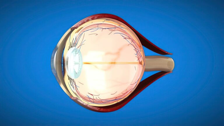
Retinal detachment occurs when the light-sensitive retinal layer separates from the supporting tissue underneath it. It may develop for different reasons, including a retinal tear, traction or fluid accumulation. Retinal detachment surgery costs may vary depending on the type and extent of the detachment and the surgical method to be used. The microsurgical instruments, choice of intraocular gas or silicone and anaesthesia method also affect the treatment plan. Since every patient has a different eye structure and degree of retinal damage, there is no standard surgical process. If sudden flashes of light, a new onset of numerous floaters or a curtain-like shadow in the field of vision occurs, an ophthalmologist should perform an assessment on the same day.
Why Does Retinal Detachment Require Urgent Assessment?
Retinal detachment is a serious eye condition that may cause permanent vision loss if left untreated. The timing of treatment is determined according to whether the detachment has affected the centre of vision and how quickly the condition is progressing. There may be significant differences in surgical planning between a recently developed detachment and one that has been present for a long time. Membranes and traction may develop on the retinal surface in delayed or complex cases. This may make it more difficult to reposition the retina and require additional surgical stages. Early assessment is important for preserving the existing visual potential.
Not every retinal detachment presents with the same symptoms or progresses at the same rate. Loss of the visual field may increase rapidly in some patients, while progression may be more limited in other cases. However, mild symptoms do not mean that the condition is unimportant. The location and extent of the detachment, number of tears and condition of the centre of vision are assessed during the examination. The surgical method is selected according to these findings. When preparing the treatment plan, the condition of the retina and preservation of vision should take priority over cost.
How Is the Surgical Method Determined?
The method used to treat retinal detachment is determined according to the cause of the separation and the anatomical characteristics of the eye. Treating the outer wall of the eye may be sufficient for some patients. In other cases, the eye must be entered to remove the vitreous tissue. In suitable cases, injecting gas into the eye to support closure of the tear may also be considered. One of these methods may be used on its own, or certain surgical techniques may be combined. The main methods considered for treating retinal detachment include:
- Vitrectomy.
- Scleral buckling.
- Pneumatic retinopexy.
- Combined surgical methods in suitable cases.
The choice of surgical method does not depend solely on the size of the detachment. The patient's age, condition of the natural lens and previous eye operations are also considered. The number and location of retinal tears may change the treatment approach. Additional procedures may be required when diabetes-related traction or intraocular haemorrhage is present. The duration of the operation and the materials used also vary according to the selected method. Therefore, retinal detachment surgery costs cannot be assessed individually without a detailed examination.
What Is Done During Vitrectomy?
Vitrectomy is a microsurgical method that provides access to the retina by removing the vitreous gel that fills the eye. The surgeon may access the inside of the eye through fine entry points and remove tissues that cause traction on the retina. Retinal tears may be surrounded with laser or freezing treatment when necessary. After the retina has been repositioned, air, gas or silicone oil may be placed inside the eye. The procedures performed vary depending on the type of retinal detachment and the findings obtained during surgery. The same surgical stages are not required in every vitrectomy.
Some of the instruments used during vitrectomy may be prepared for single use. Vitreous cutter probes, intraocular access cannulas, light sources and endolaser systems are used according to the surgical plan. These instruments are not robotic tips but microsurgical equipment controlled directly by the surgeon. The thickness and technical features of the systems are determined according to the patient's eye structure. Some operations may require membrane-peeling instruments, specialised fluids or additional retinal surgery materials. These differences affect the scope of retinal detachment operations involving vitrectomy.
When Is Scleral Buckling Used?
Scleral buckling is a retinal surgery method involving the placement of a silicone band or supporting material on the outer wall of the eye. The material supports the wall of the eye inward and helps reduce traction on the retinal tear. Freezing treatment or other supportive procedures may be applied to the tear when necessary. This method may be considered particularly for certain cases of retinal detachment caused by a tear. However, it may not be sufficient or suitable on its own for every patient. In some cases, it may need to be combined with vitrectomy.
Not entering the inside of the eye during scleral buckling does not mean that the method is always easier or carries less risk. Changes in the prescription, temporary double vision or discomfort around the eye may occur after surgery. In rare cases, problems such as infection or extrusion of the implanted material may develop. Therefore, the surgical method should not be selected solely according to the materials used. The condition of the retina, location of the tear and the patient's eye structure should be assessed together. Including scleral buckling in the treatment plan may change the scope of the operation.
Who May Be Suitable for Pneumatic Retinopexy?
Pneumatic retinopexy is a method that aims to support the retinal tear from within by injecting a gas bubble into the eye. It may generally be considered for patients whose number, size and location of retinal tears meet certain criteria. The patient may need to maintain a particular head position so that the gas bubble can be positioned over the tear. This method cannot be used for every retinal detachment. Suitability should be determined according to a dilated fundus examination and imaging findings. A shorter procedure time does not mean that this method is the most appropriate option for everyone.
If the retina does not settle into the expected position after pneumatic retinopexy, an additional surgical procedure may be required. Methods such as vitrectomy or scleral buckling may be considered in such cases. Maintaining the recommended head position may be an important part of treatment. However, the form and duration of the position should be determined according to the location of the tear. Air travel and travelling to high altitudes with gas inside the eye may cause a serious increase in intraocular pressure. If another operation or anaesthesia is required, the healthcare team must be informed that gas is present inside the eye.
Which Tests Are Performed Before Surgery?
Retinal detachment is usually diagnosed through a detailed retinal examination performed after dilating the pupil. Optical coherence tomography may be used to assess whether the detachment has affected the centre of vision. Ocular ultrasonography may be used if the retina cannot be visualised because of intraocular haemorrhage or a dense cataract. Ultra-widefield retinal imaging systems may help document the boundaries of the detachment in some cases. Not every imaging method is required for every patient. The main tests that may be considered before surgery include:
- Dilated retinal examination.
- Optical coherence tomography.
- Ocular ultrasonography when necessary.
- Ultra-widefield retinal imaging in suitable cases.
- Intraocular pressure measurement.
Fundus fluorescein angiography and specular microscopy are not routinely required in every case of retinal detachment. Different tests may be planned if another condition, such as a retinal vascular disease or cataract requiring surgery, is present. Imaging results help determine the surgical method and materials to be used. However, no test can predict every finding that may arise during surgery. The surgical plan is prepared by assessing the examination and imaging results together. The scope of the preoperative examinations may also be among the factors affecting retinal detachment surgery costs.
How Is Intraocular Tamponade Selected?
After the retina has been repositioned, some patients may require a supporting substance to be placed inside the eye. These substances help keep the retina in place for a certain period. The tamponade is selected according to the location of the tear, type of detachment and anatomical characteristics of the eye. The same tamponade is not used in every retinal detachment operation. Air or expansile medical gases may be sufficient in some cases. The main substances that may be used during treatment include:
- Sterile air.
- Expansile medical gases.
- Silicone oil.
Air and gases are naturally absorbed by the eye within a certain period. The absorption period varies according to the type and amount of substance used. Silicone oil does not disappear on its own and may be removed later in suitable patients. However, silicone oil does not need to be removed after the same period in every patient. The timing of removal is determined according to the condition of the retina and intraocular findings. Planning a second surgical procedure may change the scope of the treatment process and the materials required.
The use of gas or silicone requires different postoperative follow-up rules. Flight and high-altitude restrictions must be strictly followed when gas is present inside the eye. Some patients may be advised to maintain a face-down position or turn their head towards a particular side. In eyes containing silicone oil, intraocular pressure and the condition of the retina are assessed regularly. Each tamponade substance has a different purpose and follow-up process. Therefore, the tamponade should be selected according to the physician's surgical assessment.
Delayed and Complex Detachments
Membranes and traction may develop on the retinal surface in long-standing retinal detachments. This condition, known as proliferative vitreoretinopathy, may cause the retina to stiffen and contract. These membranes may need to be removed during surgery to release the retina again. These procedures may increase the duration of the operation and the range of microsurgical materials used. Silicone oil or other supportive surgical procedures may be used in some cases. The progression of the condition is one of the main factors considered when assessing retinal detachment surgery costs.
High myopia, previous cataract surgery and eye trauma are among the risk factors assessed for retinal detachment caused by a tear. In diabetes-related retinal diseases, tractional detachment may develop when membranes inside the eye pull on the retina. These cases may involve intraocular haemorrhage or extensive membrane formation. Additional equipment may be required during surgery to control bleeding, remove membranes and assess the retinal surface. However, not every patient with diabetes or high myopia will experience the same surgical difficulty. The treatment plan should be prepared according to the patient's examination findings.
Postoperative Follow-Up
Regular check-ups are required after retinal detachment surgery. The time required for vision to recover varies according to whether the detachment affected the centre of vision, the duration of the condition and the surgical method used. Anatomical success of the operation does not mean that vision will completely return to its previous level. Some patients may require additional laser treatment, removal of silicone oil or another surgical procedure. Intraocular pressure, the condition of the retinal tissue and the tamponade used are assessed during follow-up examinations. Eye drops and head-positioning instructions provided by the physician should be followed individually.
Retinal detachment surgery costs are assessed according to the type and extent of the detachment and the surgical method to be used. Whether vitrectomy, scleral buckling or pneumatic retinopexy will be used is determined after the examination. Placement of gas or silicone inside the eye and the need for additional surgical materials may change the scope of treatment. If sudden flashes of light, numerous floaters or a curtain-like shadow in the field of vision occurs, a routine appointment should not be awaited. If these symptoms are not present, you can contact me using the contact information on my website to arrange an examination. If urgent symptoms are present, an ophthalmologist or an appropriate ophthalmic emergency department should be consulted on the same day.