Contents:
Eye diseases include health problems affecting the eyeball, optic nerve, eyelids and tear ducts. Structures such as the cornea, eye lens, retina and optic nerve each perform different functions in vision. A disorder in any of these structures may affect visual quality or eye comfort. Blurred vision, eye pain, redness, sensitivity to light and changes in the visual field are common symptoms. Some conditions may progress without causing noticeable symptoms. Diagnosis, monitoring and treatment planning are therefore determined following a detailed eye examination.
Basic Anatomical Structures of the Eye
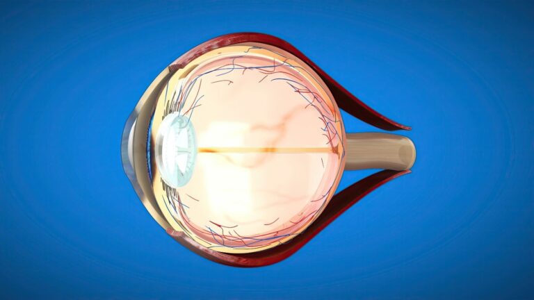
The eye performs its visual function through the coordinated work of multiple tissues with different roles. The sclera is the white outer layer that supports the structure of the eyeball. The cornea is the transparent tissue at the front of the eye and plays an important role in allowing light to enter the eye. The uvea forms the vascular middle layer of the eye. The retina is located at the back of the eye and converts incoming light into visual signals. These signals are transmitted to the visual centres of the brain through the optic nerve.
The main structures involved in vision include:
- Sclera.
- Cornea.
- Uvea.
- Eye lens.
- Retina.
- Optic nerve.
The eye lens helps focus light on the retina. The vitreous, which fills the back of the eye, allows light to reach the retina through its transparent structure. The eyelids and tear system are important for protecting the ocular surface. The eye muscles allow both eyes to move in coordination. A disorder affecting any of these structures may cause different symptoms. When evaluating eye diseases, the affected tissue is identified so that appropriate treatment can be planned.
How Are Eye Diseases Diagnosed?
Measuring visual acuity and performing a detailed eye examination are the primary methods used to diagnose eye diseases. Intraocular pressure measurement, corneal examination and fundus examination are added to the assessment when necessary. Different imaging methods may be used to evaluate changes in the retina and optic nerve. Regular examinations are important because some conditions do not cause noticeable symptoms during their early stages. The tests performed are determined according to the patient's age, symptoms and existing health conditions. Not every imaging method is required for every patient.
The main tests that may be used during diagnosis include:
- Optical coherence tomography.
- Computerised visual field testing.
- Corneal topography.
- Fundus imaging.
- Ocular ultrasonography.
- Visual evoked potentials.
- Electroretinography.
Optical coherence tomography helps evaluate the structural characteristics of the retina and optic nerve in detail. A visual field test may be used to examine whether peripheral vision has become restricted. Corneal topography shows the shape and regularity of the corneal surface. Electrophysiological tests may provide information about the function of the retina and visual pathways. In some cases, the pupils are dilated to examine the retina in greater detail. The diagnosis and monitoring plan is prepared by evaluating the examination and imaging findings together.
What Are Refractive Errors?
Refractive errors occur when light cannot focus on the correct point on the retina. In myopia, distant objects appear blurred, while nearby objects may be perceived more clearly. In hyperopia, near vision may be particularly difficult, and distance vision may also be affected in some people. Astigmatism is associated with the inability of the cornea or eye lens to focus light equally in different planes. This may cause blurred or shadowed vision at both near and far distances. The degree of the refractive error is determined through detailed eye measurements.
Glasses and contact lenses are frequently used to correct refractive errors. Laser surgery or intraocular lens procedures may also be considered in some patients. However, not every eye structure is suitable for every treatment method. Corneal thickness, changes in the prescription and the condition of the ocular surface must be evaluated together. Dry eye, retinal diseases and other structural characteristics may also affect the treatment decision. The appropriate method is determined according to the individual examination findings.
Laser Treatment of Refractive Errors
The purpose of laser treatment is to change the shape of the cornea in a controlled manner and help light focus on the retina. The method used is selected according to the prescription and corneal structure. In PRK and TransPRK, treatment is applied to the surface of the cornea. In LASIK and Femto-LASIK, a thin flap is created on the cornea and the underlying tissue is reshaped. In SMILE, a thin piece of tissue created within the cornea is removed through a small opening. Each method has different recovery processes and eligibility requirements.
Laser methods that may be used include:
- PRK.
- TransPRK.
- LASIK.
- Femto-LASIK.
- SMILE.
Corneal thickness, corneal mapping and tear structure are evaluated before laser treatment. Surface procedures may be suitable for some patients, but not everyone with a thin cornea is eligible for laser treatment. The selection of flap-based methods or small-incision procedures is also based on individual characteristics. It is also important for the prescription to have remained stable for a certain period. Dryness, sensitivity to light or fluctuations in vision may occur after surgery. The choice of method should therefore be based not only on the technology but also on the medical needs of the eye.
Age-Related Near Vision Problems
Presbyopia is difficulty focusing at close distances caused by the gradual loss of flexibility in the eye lens. It generally becomes more noticeable from middle age onwards. People may notice that they need to hold reading material farther away or require stronger lighting while reading. Eye strain may also occur when using computers and mobile phones. This is an age-related change and does not constitute an eye disease on its own. The degree of near vision difficulty is assessed through an eye examination.
Reading glasses or suitable contact lens options may be used to correct presbyopia. Intraocular lens procedures may also be considered in some patients. However, replacing the eye lens is a surgical procedure and is not suitable for every patient. The retinal structure, ocular surface and the person's visual expectations must be examined in detail. Factors such as night vision, light scatter and contrast perception are also included in the assessment. Surgical options are planned by considering the expected benefits and potential limitations together.
Intraocular Lens Options
Intraocular lenses may be used during cataract surgery or certain lens replacement procedures considered appropriate. Monofocal lenses are designed to provide focus at a specific distance. Bifocal and trifocal lenses may have multiple focal points. EDOF lenses have different optical characteristics designed to extend the depth of focus. Toric lenses may be considered for correcting astigmatism. The appropriate lens is determined according to the anatomical structure of the eye and the person's visual needs.
Types of intraocular lenses that may be used include:
- Monofocal lenses.
- Bifocal lenses.
- Trifocal lenses.
- EDOF lenses.
- Toric lenses.
Multifocal lenses may not be suitable for every patient. Some people may notice halos or glare around lights at night. Conditions such as retinal disease, pronounced dry eye or advanced glaucoma may affect lens selection. Astigmatism must be measured in detail before a toric lens can be used. Daily habits and occupational visual requirements are also evaluated when selecting a lens. No type of lens provides the same visual outcome in every patient.
What Is Cataract?
A cataract is the loss of transparency of the natural lens inside the eye. This change may make it difficult for light to reach the retina and cause blurred vision. Patients may notice that colours appear faded, lights scatter more or night vision becomes difficult. Cataracts most commonly develop with age. However, diabetes, trauma, long-term corticosteroid use and certain eye diseases may also affect their development. The degree of vision loss varies according to the density of the lens opacity.
Factors that may contribute to cataract development include:
- Advanced age.
- Diabetes.
- Eye trauma.
- Long-term corticosteroid use.
- Exposure to ultraviolet radiation.
- Certain congenital or systemic conditions.
The definitive treatment for cataract is surgical removal of the clouded natural lens. The timing of surgery is determined according to the patient's visual acuity and the effect on daily life. During phacoemulsification, the clouded lens is broken up and removed through a small incision. An artificial intraocular lens is implanted in its place in suitable patients. Recovery time and visual acuity after surgery vary according to the general health of the eye. Accompanying conditions such as retinal disease or glaucoma may affect the visual outcome.
What Is Glaucoma?
Glaucoma is an eye disease that may cause progressive damage to the optic nerve. Intraocular pressure is an important risk factor in most patients. However, glaucoma may also develop in people whose intraocular pressure is within the normal range. The disease may progress without causing noticeable symptoms, particularly during its early stages. Because visual field loss generally begins in the peripheral areas, it may not be noticed immediately. Regular assessment of intraocular pressure and the optic nerve is therefore important.
Factors considered in the diagnosis and monitoring of glaucoma include:
- Intraocular pressure.
- Corneal thickness.
- Structure of the optic nerve.
- Retinal nerve fibre layer.
- Computerised visual field.
- Intraocular fluid drainage angle.
The purpose of glaucoma treatment is to reduce intraocular pressure to a level appropriate for the condition of the optic nerve. Eye drops, laser procedures and surgical methods may be used for this purpose. Laser treatment to increase fluid outflow may be considered in some patients. Different surgical methods may be considered when medication is insufficient. Minimally invasive procedures, conventional glaucoma operations or drainage implants are selected according to the patient's condition. Because existing nerve damage cannot be reversed, early diagnosis and regular monitoring are important.
What Are Retinal Diseases?
Retinal diseases include various conditions affecting the light-sensitive layer at the back of the eye. Diabetic retinopathy, age-related macular degeneration, retinal tears and retinal detachment belong to this group. Macular holes and epiretinal membranes are also retinal conditions that may affect central vision. Symptoms may include blurred vision, flashes of light, floaters and image distortion. Some diseases progress slowly, while others may require intervention within a short time. The treatment approach is determined according to the affected area of the retina and the degree of damage.
A curtain, shadow or sudden darkening in the visual field may occur when a retinal tear or detachment develops. Newly developed flashes of light and a sudden increase in floaters are also symptoms requiring attention. Retinal assessment should not be delayed in these situations. Macular diseases may particularly make reading, recognising faces and seeing details more difficult. Early diagnosis helps ensure that appropriate treatment is planned in a timely manner. Not every retinal disease requires the same surgical method or monitoring period.
Diabetes-Related Retinal Problems
Diabetic retinopathy develops because of changes caused by diabetes in the retinal blood vessels. Persistently high blood sugar may affect the structure of the vessel walls. This may cause small retinal haemorrhages, fluid leakage and vascular occlusions. Fluid accumulation in the macular area may reduce central vision. In advanced stages, fragile new blood vessels may develop. Some patients may experience bleeding into the vitreous or traction on the retina.
Treatment of diabetic retinopathy is planned according to the stage of the disease. Blood sugar and blood pressure control are important parts of general monitoring. Intraocular medication injections or laser treatment may be used in suitable patients. Vitrectomy may be considered if there is dense bleeding or significant traction on the retina. The absence of noticeable eye symptoms does not mean that the retinal vessels have not been affected. People with diabetes should therefore have eye examinations at regular intervals.
Age-Related Macular Degeneration
Age-related macular degeneration is a disease affecting the central part of the retina. It may particularly cause problems with reading, recognising faces and seeing fine details. The disease has different forms known as dry and wet types. The dry type generally progresses more slowly. In the wet type, abnormal blood vessels developing beneath the macula may cause fluid leakage or bleeding. Distorted straight lines and impaired central vision are important symptoms.
The treatment approach varies according to the type of macular degeneration. Intraocular injections may be considered for suitable patients with the wet type. Monitoring and certain supportive approaches may be planned for the dry type according to the stage of the disease. Nutritional supplements are not necessary for every patient, and their suitability is determined through an individual assessment. Reviewing risk factors such as smoking is also important. Regular examinations help monitor changes in the macula.
Treatment Methods for Retinal Diseases
Treatments used for retinal diseases are determined according to the underlying problem and intraocular findings. Intraocular injections may be used for certain vascular diseases and macular oedema. Laser treatment may be considered for certain retinal tears or diabetes-related vascular changes. Vitrectomy is a surgical method used for intraocular bleeding and certain advanced retinal diseases. In some cases, implants that release medication inside the eye may also be preferred. The purpose of treatment is to control the progression of the existing disease and preserve visual function as much as possible.
Methods that may be used for retinal diseases include:
- Intraocular injections.
- Laser photocoagulation.
- Pars plana vitrectomy.
- Intraocular medication implants.
- Supportive treatments in suitable patients.
The frequency of treatment and duration of follow-up differ for each method. Some patients may require more than one treatment. The decision to operate is made by considering the extent of retinal damage and the person's level of vision. The visual outcome despite treatment may vary according to the initial stage of the disease. Control of accompanying diabetes or vascular disease also affects the process. Monitoring of retinal diseases is therefore planned individually.
Uveitis and Intraocular Inflammation
Uveitis is a group of diseases associated with inflammation of the uvea, the middle layer of the eye. It may affect only the front of the eye or cause inflammation in the back of the eye. Conditions involving the immune system accompany uveitis in some patients. Behçet's disease, rheumatic diseases and certain infections may contribute to its development. Severe redness, eye pain, sensitivity to light and blurred vision are common symptoms. If left untreated, changes in intraocular pressure, cataracts or retinal problems may develop.
Symptoms that may occur with uveitis include:
- Eye redness.
- Deep eye pain.
- Sensitivity to light.
- Blurred vision.
- Black floaters.
- Reduced visual acuity.
Treatment of uveitis is planned according to the location of the inflammation and the underlying cause. Corticosteroid eye drops may be used in some patients. Oral medication, injections or treatments that regulate the immune system may be considered in more extensive cases or those affecting the back of the eye. Different medications may be required for infection-related uveitis. Corticosteroid eye drops should therefore not be used without an examination. When necessary, treatment is managed together with rheumatology or other relevant specialties.
Eye Infections and Keratitis
Conjunctivitis is inflammation of the conjunctiva, which covers the white part of the eye and the inner surface of the eyelids. Viruses, bacteria or allergic causes may lead to this condition. Redness, watering, itching and discharge are common symptoms. Keratitis affects the cornea and may require closer assessment. Contact lens use, eye trauma and certain infections may increase the risk of keratitis. When the cornea is affected, pain, sensitivity to light and blurred vision may develop.
Treatment is determined according to the cause of the infection and the ocular surface findings. Not every case of redness requires antibiotic eye drops. Viral, bacterial and allergic conditions require different treatment approaches. Pain and redness developing in contact lens users should be evaluated more carefully. Using eye drops without medical guidance may cause additional problems, particularly when the medication contains corticosteroids. An examination should not be delayed if severe pain or reduced vision is present.
Eyelid and Tear Duct Disorders
The eyelids and tear ducts perform important functions in protecting the ocular surface. Drooping eyelids, inward or outward turning of the eyelids and tear duct obstructions may affect this area. Drooping of the eyelid, known as ptosis, may restrict the visual field in some patients. In entropion, the eyelid turns inward and causes the eyelashes to rub against the cornea. In ectropion, the eyelid turns outward, and dryness or watering of the ocular surface may develop. Problems affecting the eyelid glands, such as chalazia and styes, are also common.
Common eyelid and tear duct problems include:
- Ptosis.
- Entropion.
- Ectropion.
- Chalazion.
- Stye.
- Tear duct obstruction.
Persistent watering and discharge in babies may be associated with a congenital tear duct obstruction. Massage and monitoring may be recommended in suitable cases. Procedures such as probing may be considered in some patients when the obstruction persists. Different surgical methods may be used in adults according to the location of the obstruction. In some cases, a new drainage route may need to be created between the tear sac and the nasal cavity. The method selected is determined according to the anatomical characteristics of the eyelids and tear system.
Intraocular Tumours
Benign or malignant masses may develop inside the eye, on the eyelids or in the tissues surrounding the eye. Uveal melanoma in adults and retinoblastoma in children are among the intraocular tumours. Depending on the tumour's location, reduced vision, a change in pupil colour or different ocular symptoms may occur. A white reflection in the pupil of a child requires detailed examination. The patient's general health is considered a priority during treatment planning. Preserving the eye and visual function is also considered whenever possible.
Factors affecting the assessment of intraocular tumours include:
- Size of the tumour.
- Location.
- Cellular characteristics.
- Patient's age.
- Visual status of the eye.
- Extent of the disease.
Radioactive plaque treatment may be used for certain intraocular tumours. In this method, a radiation source suitable for the tumour's location is placed on the outer surface of the eye for a specified period. Not every tumour is suitable for this method. Different radiotherapy methods, medication or surgical options may be considered when necessary. Removal of the eye may also be medically necessary in some advanced cases. The treatment decision is determined according to the characteristics of the tumour and assessments by the relevant specialties.
Eye Diseases in Children
The visual system in children continues to develop after birth. Refractive errors, strabismus or drooping eyelids developing during this period may affect visual development. Amblyopia may develop when the image reaching the brain from one eye is not sufficiently clear. Because children cannot always express a problem with their vision, the symptoms may not be noticed by the family. Squinting, turning the head in a particular direction or eye misalignment require assessment. Early diagnosis helps ensure that treatments supporting visual development are applied in a timely manner.
Signs requiring attention in children include:
- Eye misalignment.
- Squinting.
- Constantly turning the head in the same direction.
- Frequent blinking.
- Persistent watering.
- A white reflection in the pupil.
- Viewing objects at close range.
Appropriate glasses and patching therapy may be considered when treating amblyopia. Different forms of visual support may also be used in some patients. If strabismus is caused by a refractive error, it may be controlled with glasses. Surgery may be required in cases involving a muscle imbalance. The response to treatment varies according to the child's age and the underlying problem. Regular follow-up allows visual development and the ability of the eyes to work together to be assessed.
Retinal Monitoring in Premature Babies
The development of retinal blood vessels may be incomplete in babies born prematurely. This may cause a condition known as retinopathy of prematurity. The risk of developing the condition is associated with the gestational age at birth, birth weight and the baby's general health. There may be no noticeable external signs during the early stages. Retinal screenings must therefore be performed at the appropriate time in babies within the risk group. Monitoring, laser treatment or intraocular medication may be considered according to the stage of the disease.
New Technologies in Eye Health
Research into new technologies for diagnosing and treating eye diseases is ongoing. Gene therapies are among the methods being evaluated for certain inherited retinal diseases. Artificial intelligence-assisted systems may help examine certain retinal images. Stem cell procedures and retinal prostheses are also being researched. However, not all these methods are suitable for every patient. Some are used in limited patient groups, while others remain at the research stage.
Technologies being researched or used in certain situations include:
- Gene therapies.
- Retinal prostheses.
- Stem cell procedures.
- Artificial intelligence-assisted image analysis.
- Wearable visual assistance systems.
The clinical use of new technologies varies according to the type of disease and the available scientific evidence. In inherited retinal diseases, genetic testing may help assess suitable treatment options. Artificial intelligence systems do not replace an examination but may support image assessment. Retinal prostheses and stem cell methods are not general solutions that can be used for every type of vision loss. A realistic assessment of treatment options is directly related to the patient's eye structure and current diagnosis. Scientific developments contribute to the investigation of new approaches in suitable patients.
When Is an Eye Examination Necessary?
An eye examination should be planned if blurred vision, eye pain, persistent redness or sensitivity to light develops. Symptoms such as sudden vision loss, flashes of light or a curtain-like shadow in the visual field may require urgent assessment. Follow-up plans for people with diabetes, a history of glaucoma or high myopia are determined according to their individual risks. Examination should not be delayed if eye misalignment, difficulty seeing or a white reflection in the pupil is noticed in a child. Regular examinations may help detect conditions that can progress without symptoms at an early stage. For non-urgent assessments, you can contact me using the details on my website to schedule an examination appointment.