
Contents:
Eye tomography is an imaging method used to obtain high-resolution cross-sectional images of specific tissues in the eye. In ophthalmology, the term eye tomography usually refers to Optical Coherence Tomography (OCT). OCT is particularly used for the structural evaluation of the retina, macula, and optic nerve head. Its non-contact application and the ability to compare the same area on different dates help with the diagnosis and monitoring of diseases.
OCT eye tomography does not establish a diagnosis on its own. The images are evaluated together with the person's symptoms, visual acuity, intraocular pressure, fundus examination, and any other tests considered necessary. Additional examinations may be required, such as a visual field test for glaucoma or fundus photography and angiography for retinal vascular diseases. The tests to be performed are determined according to the area of the eye being examined and the suspected disease.
What Is Eye Tomography?
The question what is eye tomography can be explained by examining how OCT images the tissues of the eye. OCT creates cross-sectional images by analyzing the reflection of low-coherence light from different tissue layers. Its operating principle can be compared to ultrasound, but it uses light instead of sound waves. This allows thin tissues such as the retina to be examined at the micrometer level.
One micrometer is equal to one-thousandth of a millimeter. Although it varies depending on the type of device used, OCT can provide cross-sectional resolution of approximately 10 to 15 micrometers in ocular tissues. The images show the layers of the retina and changes in the thickness or structure of these layers. These measurements help compare changes in disease findings over time.
How Does OCT Eye Tomography Work?
An OCT device sends near-infrared light into the eye and measures the light reflected from different tissue layers. The reflected light is compared with reference light and converted into interference data. Computer software processes this data into cross-sectional images, thickness maps, and numerical measurements. Depending on the area being examined, images of the retina, macula, optic nerve, or anterior segment can be created.
An OCT image is not a direct color photograph of the eye. The device's software displays the light-reflecting properties of tissues using different shades or colors. Structural changes such as fluid within the retina, tissue loss, thickening, thinning, or traction can be evaluated on the cross-sectional images. Although the device's automated measurements are useful, the results must be reviewed by an ophthalmologist together with the raw images.
How Is OCT Eye Tomography Performed?
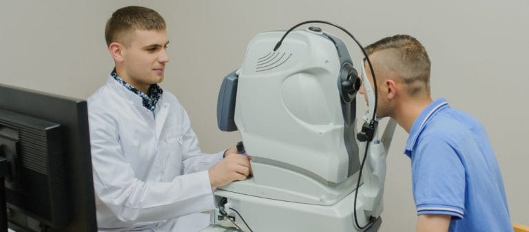
During the examination, the patient sits in front of the device and places their chin and forehead against the supports. The patient is asked to look at a target inside the device with one eye and remain as still as possible during the scan. The device captures images without touching the eye. The scan itself is brief, but examining different areas of both eyes and reviewing the images may extend the total duration.
Special preparation is generally not required. Pupil-dilating drops may be used if the pupil is small, if there is a dense cataract, or if a wider area of the retina needs to be evaluated. These drops may cause blurred vision and sensitivity to light lasting several hours. Whether the drops are used is determined according to the examination findings.
An OCT scan is generally painless and does not require physical contact with the eye. The images may need to be repeated in people who have difficulty keeping their head still or looking at a specific point. Frequent blinking and eye movements may cause lines or displacement in the scan. If the image quality is insufficient, the reliability of the results may decrease.
Which Eye Diseases Is OCT Used For?
OCT can be used for the structural evaluation of multiple areas of the eye. Its most common applications are retinal and macular diseases and glaucoma monitoring. Systems designed for the anterior segment can also image the cornea and anterior chamber structures. Whether the test is necessary is determined not only by the name of the disease but also by the clinical question that needs to be answered during the examination.
Retinal and Macular Diseases
The macula is the area at the center of the retina responsible for detailed vision. OCT can show fluid within the retina and changes in retinal thickness in diabetic macular edema and retinal vascular occlusions. In age-related macular degeneration, it helps evaluate findings such as drusen, changes in the pigment epithelium, and fluid within or beneath the retina. In patients receiving treatment, changes in the amount of fluid and retinal structure can be compared over time.
In conditions such as macular holes, epiretinal membranes, and vitreomacular traction, traction on the retinal surface and tissue integrity can be examined using cross-sectional images. OCT may also be used to monitor structural changes in uveitis and certain inherited retinal diseases. Standard OCT does not replace a fundus examination for ruling out peripheral retinal tears. People experiencing flashes of light, a sudden increase in floaters, or a curtain-like sensation must be evaluated with a dilated fundus examination.
Glaucoma and Optic Nerve Evaluation
Glaucoma is a group of diseases characterized by progressive optic nerve damage and is not defined solely by elevated intraocular pressure. OCT can measure the retinal nerve fiber layer around the optic nerve and the ganglion cell complex in the macula. Thinning in these layers helps evaluate suspected glaucoma and compare structural changes during follow-up. A single red or yellow measurement area does not establish a glaucoma diagnosis on its own.
Myopia, congenital differences in optic nerve anatomy, image quality, and the device's reference database may affect the measurements. During glaucoma monitoring, OCT results are interpreted together with intraocular pressure, optic nerve examination, and visual field testing. Structural changes and functional loss in the visual field may not always occur at the same time. Therefore, OCT is not a direct replacement for a visual field test.
Corneal and Anterior Segment Examination
Anterior segment OCT systems can provide cross-sectional information about the cornea, anterior chamber, iris, and drainage angle of the eye. Corneal thickness, certain corneal diseases, scars, and anatomical changes after surgery can be examined. Additional information may also be obtained about the position of intraocular lenses or certain structures in the anterior segment. The technical specifications of the device and the scanning protocol determine which areas can be imaged.
Anterior segment OCT and retinal OCT are not used for the same purpose. Devices optimized for the retina are intended to evaluate the back of the eye, while anterior segment systems assess its front section. Some newer devices may offer multiple modes. The type of scan is selected according to the examination findings.
What Are the Types of OCT?
OCT technology is divided into different types according to the image acquisition method, scanning speed, and depth of tissue penetration. Older systems known as time-domain OCT have largely been replaced by spectral-domain and swept-source devices. Newer systems may provide faster scanning and denser data acquisition. However, a newer device cannot automatically be considered more accurate in every clinical situation.
What Is the Difference Between SD-OCT and SS-OCT?
Spectral-domain OCT, abbreviated as SD-OCT, is one of the systems commonly used for retinal imaging. Swept-source OCT, abbreviated as SS-OCT, uses an adjustable light source with a longer wavelength and may provide advantages when imaging deeper tissues or wider areas. SS-OCT can make it easier to examine structures beneath the retina, such as the choroid, and may be less affected by certain media opacities. Image quality may still be affected by eye movement, cataract density, corneal transparency, and device settings.
Using the same device and the same scanning protocol whenever possible facilitates comparison during follow-up. The reference databases, segmentation methods, and measurement boundaries of different devices may not be identical. A retinal or nerve fiber thickness measurement from one device may not be directly comparable with a measurement from another device. These technical differences must be considered when analyzing changes.
What Is OCT Angiography?
OCT angiography, abbreviated as OCTA, obtains consecutive images of the same area and creates a vascular flow map based on the movement of blood cells. While standard OCT shows tissue structure, OCTA provides information about the vascular network in the retina and choroid. The procedure is generally performed without injecting dye into a vein. It may help evaluate certain vascular changes associated with diabetic retinopathy, retinal vascular occlusions, and macular degeneration.
OCTA is not a complete substitute for fluorescein angiography. Vessels with very slow blood flow may not produce a signal, and OCTA does not directly show dye leakage. Eye movement, blinking, projection artifacts, and segmentation errors may distort the vascular map. Depending on the clinical question, fluorescein angiography, indocyanine green angiography, or other imaging methods may also be required.
How Are OCT Results Interpreted?
An OCT report may include cross-sectional images, thickness maps, and numerical measurements for different areas. The color codes generally compare the measurement with the device's reference database for the same age group. Green usually represents the reference range, yellow may indicate a borderline value, and red may show a measurement outside the reference distribution. These colors alone do not determine whether the eye is healthy or diseased.
In addition to the automated thickness measurements, the physician examines the boundaries of the retinal layers and the quality of the image. In diseased tissues, the software may separate the layers incorrectly and create a segmentation error. When comparing the results with previous scans, changes in the device, displacement of the scan center, and signal quality are considered. If the result is inconsistent with the examination or other tests, the image may need to be repeated or confirmed using another method.
What Are the Limitations of OCT?
OCT relies on light reaching the ocular tissues and being reflected back. A dense cataract, corneal opacity, intraocular hemorrhage, or other media opacities may reduce the signal and lower image quality. Difficulty maintaining fixation, nystagmus, frequent blinking, and head movements may also affect the scan. In such cases, an inability to obtain an image does not mean that no disease is present.
OCT provides structural information and does not measure visual function on its own. Other tests may be more appropriate for retinal tears, intraocular tumors, the part of the optic nerve outside the eyeball, or certain vascular leakages. The device's automated analyses may produce false-positive or false-negative results. Therefore, an OCT report should not be interpreted solely by examining the color maps.
Does OCT Eye Tomography Involve Radiation?
OCT does not involve the ionizing radiation used in X-rays or computed tomography. It uses low-power near-infrared light for imaging, and no needle or other instrument is inserted into the eye during a standard examination. The test can be repeated on different dates when necessary. The frequency of repeated scans is determined according to the type of disease, the treatment plan, and previous findings.
When pupil-dilating drops are not used, OCT has no known serious side effects. If drops are required, temporary blurred vision, sensitivity to light, and, rarely, problems involving intraocular pressure may develop. The healthcare team should be informed before the drops are administered if the patient is pregnant or has a medication allergy, suspected narrow angles, or another special condition. The suitability of the test and the drops is evaluated according to the individual's circumstances.
What Is the Difference Between OCT and Ocular Ultrasound?
OCT uses light, while ocular ultrasound uses sound waves. OCT can provide finer structural details of the retinal layers and the area around the optic nerve. In contrast, when light cannot pass through because of conditions such as a dense cataract or intraocular hemorrhage, ultrasound can be used to evaluate the back of the eye. The two methods are not direct alternatives and answer different clinical questions.
Fundus photography shows the surface appearance of the retina, while OCT shows the cross-sectional structure of a selected area. Fluorescein angiography can dynamically evaluate vascular circulation and dye leakage, while OCTA creates a flow map without using dye. Some patients may require more than one of these methods. The examination plan is prepared according to the findings identified during the eye examination.










































