
Contents:
An eye examination is a medical evaluation that should be performed regularly to protect visual health and detect potential diseases at an early stage. Visual acuity, intraocular pressure and the internal structures of the eye are assessed in detail. This examination is important for both diagnosis and follow-up.
Methods used during an eye examination include visual acuity testing, biomicroscopy, fundus examination and optical coherence tomography. These tests allow structures such as the retina, optic nerve and cornea to be examined in detail. The scope of the examination may vary according to age group.
Eye examinations in children are critically important for detecting visual disorders at an early stage. Strabismus, amblyopia and refractive errors can be identified and treated early. For this reason, children should undergo an eye examination during the preschool period.
Regular eye examinations in adults help detect age-related eye conditions such as cataracts, glaucoma and macular degeneration at an early stage. More frequent examinations are particularly recommended for people with diabetes or hypertension.
| Purposes |
|---|
- Detecting visual disorders.
- Enabling the early diagnosis of eye diseases.
- Evaluating general eye health.
| Tests Performed |
|---|
- Visual Acuity Test: Measures the clarity of distance or near vision.
- Intraocular Pressure Measurement: Performed to evaluate the risk of glaucoma.
- Biomicroscopy: Detailed examination of the front part of the eye.
- Fundoscopy: Examination of the back of the eye, including the retina and optic nerve.
- Visual Field Test: Evaluates peripheral vision.
| Examination Duration | It usually takes 15-30 minutes. More comprehensive tests may take longer. |
|---|---|
| When Should It Be Performed? |
- When symptoms of a visual disorder are present.
- At regular intervals during childhood growth and development.
- Once a year after the age of 40, to screen for glaucoma and other age-related conditions.
| Conditions Diagnosed |
|---|
- Myopia, hyperopia and astigmatism.
- Cataracts.
- Glaucoma.
- Diabetic retinopathy.
- Macular degeneration.
- Infections and inflammation.
| Preparation |
|---|
- People who wear contact lenses may need to remove them before the examination.
- If dilating eye drops are used, blurred vision may occur for several hours after the examination.
| Interpretation of Results | If a visual defect or disease is diagnosed, a treatment plan is prepared, such as an eyeglass prescription or recommendation for surgical intervention. |
|---|---|
| Recommendations |
- Undergo regular eye examinations.
- Follow the 20-20-20 rule to prevent eye strain.
- Maintain appropriate posture and lighting while using screens.
Many people assume that no specific preparation is required before visiting an ophthalmologist. Although there is no extensive preparation process, paying attention to certain points can improve the quality and accuracy of the examination:
- Current Eyeglass or Contact Lens Prescription
If you have previously worn eyeglasses or contact lenses, it is helpful to bring your existing prescriptions with you. This allows us to compare previous measurements with current findings and monitor any necessary changes more effectively.
- Medical and Family History
It is very important to clearly disclose any chronic conditions such as diabetes or hypertension, medications you use regularly and any family history of serious eye diseases such as glaucoma, cataracts or macular degeneration. It should be remembered that genetic factors play a major role in eye health. For example, if glaucoma is common in the family, eye examinations should begin at a much younger age and be performed more frequently.
- Recommendations for Contact Lens Users
If you have worn contact lenses for an extended period before the examination, temporary changes may occur on the ocular surface. You may sometimes be asked to stop wearing lenses for several days. It is therefore helpful to consult your ophthalmologist before your appointment and ask how long before the examination you should remove your lenses.
- Pupil-Dilating Eye Drops
For a fundus examination, eye drops are applied to dilate the pupils. Temporary blurred vision, light sensitivity and sometimes a mild headache may occur after this procedure. It is therefore advisable to arrange transportation so that you do not need to drive or to have someone accompany you.
An eye examination consists of far more stages than is commonly assumed, and each stage serves a different purpose. The entire visual system is assessed using detailed techniques.
How Is Visual Acuity Measured?
One of the most basic tests is the visual acuity test, which measures how clearly we can see distant or nearby objects. People may say that they cannot see clearly at a distance or cannot read things up close. To determine the extent of these problems, they are asked to read letters from a specific distance using a Snellen chart or LogMAR chart.
- Snellen Chart: A standard chart viewed from a distance of 6 meters or 20 feet, with letters that become progressively smaller. In Türkiye, measurements are generally performed from a distance of 5 or 6 meters. The smallest line you can read determines your visual acuity.
- Near Vision Charts: Used to measure your performance at close distances, such as normal reading distance.
In broad terms, this test can be compared to checking a car's speedometer. Although visual acuity provides a basic indication of eye health, additional tests are required to understand any underlying mechanical or disease-related problems.
What Is a Refraction Test and Why Is It Performed?
If you have problems such as myopia, which causes blurred distance vision, hyperopia, which causes difficulty with near vision, astigmatism, which causes blurring along different axes of the visual field, or presbyopia, which causes age-related difficulty with near vision, their severity can be determined through a refraction test.
- Automated Refractometer: You look into a machine that automatically measures how your eye refracts light.
- Manual Reflex Test (Retinoscopy): Performed in a dark examination room by directing light into the eye and observing the reflection, known as the retinal reflex. It is particularly valuable for children or patients who have difficulty communicating.
- Subjective Refraction with a Phoropter or Trial Frame: The doctor presents different lens options and frequently asks, "Which one is clearer?" This helps identify the lens combination that provides the most comfortable and sharpest vision for the patient.
This test can be compared to adjusting the focus of a camera. When the lens is set correctly, clear photographs can be taken. Even a slight error in the setting, however, can completely eliminate sharpness. Accurately identifying a refractive error in the eye requires the same level of precision.
How Is Intraocular Pressure Measured?
Another important assessment is the measurement of intraocular pressure, known as tonometry. This is a critical test for the early diagnosis of glaucoma.
- Non-Contact Tonometry: Commonly known as the air-puff test. You place your chin on the device, a short puff of air is directed at your eye, and the device measures the eye's response to calculate the pressure.
- Applanation Tonometry: Performed with a biomicroscope, or slit lamp, using a measuring tip that gently touches the eye. It is a more precise and reliable method, but anesthetic eye drops are required to numb the corneal surface.
This pressure measurement can be compared to checking the pressure gauge in your home's plumbing system. Just as excessive pressure can cause serious problems such as a burst pipe, high intraocular pressure can cause permanent damage to the optic nerve over time.
How Is the Front Part of the Eye Evaluated?
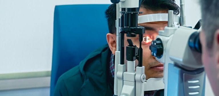
A slit-lamp examination is a microscopic method that allows ophthalmologists to examine anterior segment structures such as the cornea, iris, eyelids and lens in detail.
- Corneal Examination: The cornea is checked for scratches, wounds, infections or structural irregularities.
- Lens Evaluation: Opacities associated with cataracts and other structural changes can be detected.
- Eyelids and Eyelashes: Conditions such as infections and eyelid deformities, including entropion and ectropion, can be identified.
This can be compared to a jeweler examining a valuable gemstone with a magnifying glass. Certain details may not be visible to the naked eye, but even very small areas of damage or abnormal formations can be detected with specialized magnification and illumination.
How Is the Back of the Eye Examined?
Special eye drops are used to dilate the pupils so that the retina and optic nerve head can be examined in detail. The fundus is then examined using instruments such as an ophthalmoscope or binocular indirect ophthalmoscope.
- Direct Ophthalmoscope: A small handheld instrument that allows the doctor to examine the retina through the pupil.
- Indirect Ophthalmoscope: Uses a head-mounted lens and a powerful light source to provide a wider view of the retina, including its peripheral areas.
- Fundus Camera: Takes photographs of the retina and provides a permanent record.
This examination offers an advantage similar to being able to inspect the electrical wiring behind a wall without cutting into it. By directly observing the retinal blood vessels and nerve layer, an ophthalmologist can diagnose many diseases. Conditions such as diabetic retinopathy, hypertensive retinopathy, macular degeneration and intraocular tumors may be detected in this way.
- How Serious Is Glaucoma?
Glaucoma is an insidious disease that progresses when intraocular pressure reaches a level that damages the optic nerve. It usually does not cause obvious symptoms. It is recognized as one of the causes of irreversible blindness worldwide. If intraocular pressure is found to be elevated during routine examinations, additional tests such as visual field testing and nerve fiber layer thickness measurement are performed. These tests help determine whether glaucoma is present. Early diagnosis is critically important for preventing permanent vision loss in the future.
- What Exactly Is a Cataract?
A cataract occurs when the eye's natural lens loses its transparency and becomes cloudy. It can be compared to looking through a fogged or frosted window. Although aging is the most common cause, diabetes, corticosteroid use and trauma to the eye can also cause cataracts. A cataract can easily be detected by examining the lens with a slit lamp during an eye examination. Cataract surgery currently provides highly successful results, and early intervention restores visual quality.
- How Are Diabetic Retinopathy and Hypertensive Retinopathy Detected in the Eye?
Diabetes mainly affects the retinal layer of the eye. Diabetic retinopathy causes changes such as areas of bleeding, microaneurysms and the formation of new blood vessels in the fundus. Hypertension can cause narrowing of the retinal vessels, thickening of their walls and sometimes bleeding beneath the nerve fiber layer. These changes usually do not cause symptoms during the early stages. Detecting them during routine examinations is therefore highly valuable for monitoring both the patient's eye health and overall health.
- What Are the Symptoms of Macular Degeneration?
The macula is the central area of the retina responsible for the sharpest vision. Age-related macular degeneration causes this area to deteriorate over time. It is particularly common after the age of 60. People commonly report seeing straight lines as curved or noticing a dark spot in the center of their vision. It can be detected during an eye examination using methods such as fundus examination and Optical Coherence Tomography (OCT). Early diagnosis is extremely important for slowing its progression.
The visual field is a broad area that includes the peripheral images we perceive while looking straight ahead. In simple terms, it can be described as everything we can see out of the corners of our eyes while looking directly forward.
- Automated Perimetry: The patient sits in front of a large dome and presses a button whenever they see light stimuli presented at different points. A computer analyzes the responses and records blind spots, known as scotomas, or areas of visual field narrowing.
- Kinetic Perimetry (Goldmann Perimetry): Specialized optical equipment is used to detect hemifunctional visual field constrictions.
In addition to glaucoma, certain neurological diseases or mass lesions in the brain may cause characteristic visual field defects. Every defect detected through this test can indicate the location and severity of the underlying condition, much like marking a missing region on a map.
The recommended schedule for regular eye examinations varies according to age and risk factors:
- Children
Ages 0-2: The first eye evaluation should be performed at birth or, if possible, no later than 6-9 months of age. Early diagnosis of conditions such as congenital cataracts and glaucoma is vitally important.
Ages 3-5: An eye examination should be performed during the preschool years. This is especially important for detecting strabismus or amblyopia.
School Age (6-19 Years): Visual defects and the general development of the eyes should be monitored every year or every two years. A child who cannot see the board clearly may experience negative effects on both academic performance and social development.
- Adults (Ages 20-40)
If there are no obvious symptoms, an eye examination is recommended every 2-3 years. People who spend long periods at a computer, wear contact lenses or have a family history of eye disease may benefit from more frequent examinations.
- Middle and Older Age (Over 40)
An eye examination is recommended every 1-2 years between the ages of 40 and 64, and once a year from the age of 65 onward. The risk of diseases such as cataracts, glaucoma and macular degeneration increases in these age groups.
- High-Risk Patients
People with diabetes or hypertension should undergo an eye examination at least once a year, particularly if fundus damage has already begun. People at risk of glaucoma should also undergo intraocular pressure measurement and visual field testing at intervals considered appropriate by their doctor, such as twice a year.
The term "routine examination" refers to check-ups performed at regular intervals, similar to scheduled maintenance for a vehicle. Even when no obvious problem is present, these examinations may identify a minor issue at an early stage and allow it to be treated before it progresses.
With today's rapidly advancing technology, sophisticated imaging and measurement methods are also used during eye examinations. Some of these methods provide highly specific information and make diagnosis considerably easier.
- Optical Coherence Tomography (OCT)
OCT allows retinal tissue to be examined layer by layer. It provides valuable quantitative information about degeneration in the macular area, edema associated with diabetic retinopathy and thinning of the nerve fiber layer related to glaucoma.
This can be compared to an architect examining possible cracks in a building's foundation without cutting through a column. Because the internal structure of the eye is scanned layer by layer, even the earliest damage can be detected.
- Pachymetry (Corneal Thickness Measurement)
Corneal thickness is very important for accurately interpreting intraocular pressure and evaluating suitability for refractive procedures such as laser surgery. Pachymetry, performed using ultrasound or optical methods, can distinguish cases in which intraocular pressure appears low because of a thin cornea but is actually elevated.
- Gonioscopy
Gonioscopy evaluates the anterior chamber angle, which is the fluid drainage area between the iris and cornea. It is very important for determining glaucoma risk and identifying specific conditions such as angle-closure glaucoma. A special mirrored or prism lens is placed on the front of the eye, allowing direct examination of the angle structures that cannot be seen during a standard examination.
- Color Vision Tests
These tests are used particularly to diagnose color blindness and other color discrimination disorders. The Ishihara test is the standard method. Faint numbers or shapes are displayed within a graphic, and the patient is asked to identify them.
- Electrophysiological Tests
Tests such as Visual Evoked Potentials (VEP) and Electroretinography (ERG) measure the electrical signals generated in response to light stimulation of the retina or visual pathways. They allow us to objectively understand how the eyes respond in infants who cannot provide conscious feedback or in patients with special circumstances.
The eye drops used during an examination have the following primary purposes:
- Mydriatics Such as Tropicamide
The pupils must be dilated to examine the fundus in detail. This allows the retina to be observed as far as its peripheral areas. The disadvantages are temporary light sensitivity and blurred near vision.
- Alpha Agonists or Beta Blockers for Temporary Pressure Reduction
These may be used before or after tonometry to temporarily regulate intraocular pressure. This is not a routine practice and is decided according to clinical necessity.
- Anesthetic Eye Drops
These are used to numb the cornea during tonometry procedures that require contact with the eye, such as applanation tonometry, or during minor surgical interventions. They eliminate pain but do not directly affect visual quality.
The use of eye drops can be compared to a mechanic lubricating certain parts or opening the engine cover before examining a car engine. Each drop serves a different purpose and improves the precision of the procedure.
- Light Sensitivity: Because more light enters the eyes after the pupils are dilated, wearing sunglasses can provide considerable relief. You may also experience temporary difficulty when looking at a computer or phone screen. This is temporary.
- Temporary Blurred Vision: You may have difficulty with near tasks, such as reading or using a phone, and work requiring detail for several hours. If possible, it is therefore helpful to schedule the examination for a time when you do not have a demanding workload.
- Driving: Driving may be dangerous, especially after an examination involving dilating drops. It is therefore sensible to consider alternative transportation or arrange for someone else to drive you.
If a problem is detected during the examination, additional tests may be planned or treatment may be started. Early diagnosis is one of the most valuable steps for almost every eye disease.
- "I Don't Wear Glasses Because I Don't Want My Prescription to Increase"
This is a common but incorrect belief. Wearing glasses does not increase your prescription. Glasses sharpen your vision according to your existing refractive error.
- "Wearing Glasses Makes the Eyes Lazy"
On the contrary, amblyopia develops during childhood when the eye does not receive sufficient visual stimulation. Glasses help prevent amblyopia by making this visual input clearer.
- "Only Older People Develop Eye Diseases"
Although cataracts and macular degeneration are associated with older age, diseases such as glaucoma and keratoconus can also occur in younger people. Regular examinations are therefore important at every age.










































