
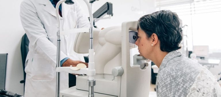
An eye check-up is a comprehensive examination performed to evaluate eye health and detect possible diseases at an early stage. Undergoing regular check-ups plays a critical role in preventing vision loss.
The examination includes stages such as measuring visual acuity, testing eye pressure, performing biomicroscopy, and examining the fundus. These evaluations allow all structures of the eye to be checked.
People in high-risk groups, particularly those with diabetes, hypertension, or a relevant family history, should undergo eye check-ups more frequently. Early diagnosis helps prevent permanent vision loss.
The findings obtained from a check-up enable a personalized treatment and follow-up plan to be created. This approach contributes to both the protection of eye health and an improvement in quality of life.
The first question about an eye check-up usually concerns its importance. Imagine a camera. If its lens, aperture, film or digital sensor, and background do not work perfectly, the resulting image will not be clear. The eye has a very similar structure. Components such as the cornea, lens, retina, and optic nerve must work together perfectly, just like the parts of a camera.
- Early diagnosis saves lives: Many eye diseases may not cause noticeable symptoms during their early stages. Glaucoma, for example, is commonly called the "silent thief" because patients may not notice it until the disease reaches an advanced stage.
- It improves quality of life: When refractive errors such as myopia, hyperopia, astigmatism, and presbyopia are corrected appropriately, daily life, work, reading, and social activities become much more enjoyable.
- It helps detect systemic diseases: The first signs of diabetes, hypertension, autoimmune diseases, and even some neurological conditions may appear in the eyes. From changes in blood vessels to certain details involving the optic nerve, the eyes may function like a mirror of the body.
An eye check-up is therefore not limited to saying, "I had my prescription measured, so everything is fine." The answer to the question "Why should I have an eye check-up?" is that it is vitally important for both your eye health and your overall health.
A patient's medical history is known as anamnesis in medical literature and is highly valuable. Just as a mechanic would want to know about a vehicle's previous faults and maintenance history before beginning work, an ophthalmologist needs to know your medical history and current complaints.
- Family history and genetic predisposition: The presence of conditions such as glaucoma, cataracts, macular degeneration, or diabetes in family members may increase the risk of similar eye problems.
- Existing diseases and medications: Systemic conditions such as diabetes and hypertension are closely associated with eye health. Certain medications may also cause eye problems as side effects.
- Eye-related complaints: Symptoms such as burning, stinging, blurred vision, flashes of light, or pain help guide the diagnostic process.
- Previous eye surgery or treatment: LASIK, cataract surgery, or retinal treatments provide important information when interpreting current examination findings.
Patients sometimes say, "I do not have any complaints at the moment," and assume the examination will be simple. However, many eye diseases may progress without symptoms. Sharing even the smallest details with the ophthalmologist is therefore critical for obtaining accurate results and maintaining proper follow-up.
An eye check-up is a multi-stage process. Patients may sometimes think, "The doctor just looked at my eyes, measured my prescription, and that was it," but many technical procedures are performed behind the scenes.
Visual Acuity Assessment
- Methods such as the Snellen chart, LogMAR chart, LEA symbols, or Teller cards are used.
- Just as an artist may produce incomplete work if unable to see details clearly, we identify even minor losses of visual acuity to determine whether the patient is unable to see clearly enough.
Refraction Test
- The process begins with objective tests such as an autorefractometer, which performs computerized eye measurements, and retinoscopy. These are followed by subjective tests requiring the patient's participation.
- This test is critical for determining the prescription for glasses. It may also identify subtle adjustments that fully reveal the problem.
Assessment of Eye Muscle Function
- Methods such as the cover test, EOM examination of the extraocular muscles, and the Parks-Bielschowsky three-step test may detect strabismus, muscle paralysis, or coordination problems.
- Imagine an orchestra. The eye muscles are like its instruments. If every muscle does not perform its task at the correct time, the eyes cannot move in synchronization and visual quality deteriorates.
Pupil Examination
- The pupils' responses to light and near targets, including the light reflex and near reflex, are examined.
- A unilateral light reflex abnormality may suggest an optic nerve problem or possible pathology involving the brain. A relative afferent pupillary defect, known as a Marcus Gunn pupil, may help ophthalmologists identify clues related to the brain.
Intraocular Pressure (IOP) Measurement
- Various methods are available, including Goldmann applanation tonometry, air-puff or noncontact tonometry, and the Schiøtz tonometer.
- This measurement is critical in diagnosing glaucoma. Elevated pressure should not be dismissed lightly. High intraocular pressure may permanently damage the optic nerve and lead to blindness.
Biomicroscopy (Slit-Lamp) Examination
- A high-powered microscope and narrow beam of light are used to examine the eye in detail, from the anterior segment to the posterior segment.
- This is similar to using a magnifying glass to examine the fine details of an ancient artifact before restoring it. Fine structures of the eye, including the cornea, iris, and lens, can be evaluated easily.
Retinal Assessment (Ophthalmoscopy and Fundus Examination)
- Direct and indirect ophthalmoscopy or fundus cameras are used to examine the retina, macula, and optic nerve head.
- The retina is like a strip of film on the back wall of the eye. Damage to even a small part of this strip may seriously affect vision.
Visual Field and Peripheral Vision Tests (Perimetry)
- Methods include automated static perimetry, Goldmann kinetic perimetry, Frequency Doubling Technology (FDT) perimetry, and Short-Wavelength Automated Perimetry (SWAP).
- Imagine a driver who cannot see dangers in the surrounding area and receives no information from the side mirrors about blind spots. Visual field tests perform the function of these side mirrors, allowing us to assess peripheral as well as central vision.
Additional Methods for Evaluating the Relationship Between the Eyes and Blood Vessels
- Specialized imaging methods such as fundus fluorescein angiography (FFA) and OCT angiography (OCTA) are used to detect retinal vascular changes, leakage, and blockages.
- Diabetes, hypertension, and even some autoimmune diseases may first produce signs in the blood vessels of the eyes. These methods map vascular abnormalities and provide information about systemic diseases.
Other Specialized Tests
- Methods such as electroretinography (ERG), visual evoked potentials (VEP), and microperimetry measure the functional capacity of retinal cells and the optic nerve.
- These tests may reveal the cause of otherwise unexplained vision loss or certain congenital retinal diseases.
As you can see, the methods used and tests included in an eye check-up form a long list. Each stage of the examination illuminates a different aspect of eye health. Every step is like a stage in constructing a building. Nothing from the foundation to the roof should be omitted.
Patients sometimes say, "Doctor, I do not have any eye problems, but I have diabetes and would like to have my eyes checked." Diabetes is not the only systemic disease that may produce signs in the eyes. Hypertension, rheumatic diseases, neurological conditions, and even certain blood disorders may also leave various clues.
- Retinal Findings in Diabetes: Abnormalities such as microaneurysms, hemorrhages, and hard exudates suggest diabetic retinopathy. Detecting these changes early is vital for preventing diabetes-related vision loss.
- Vascular Narrowing in Hypertension: People with high blood pressure may have narrowing of the retinal vessels, abnormal artery-vein crossings known as AV nicking, and sometimes optic disc swelling.
- Autoimmune Diseases such as Rheumatoid Arthritis and SLE: Inflammatory conditions such as scleritis, uveitis, and keratitis may affect the eyes. Red eyes, persistent pain, and light sensitivity may sometimes be the first signs of a rheumatic disease rather than a primary eye problem.
- Eye Findings in Neurological Diseases: Optic neuritis may develop in conditions such as multiple sclerosis. Initially, the patient may only experience mild blurred vision or pain behind the eye, but a detailed examination may detect changes in the optic nerve.
An eye check-up is therefore a window that indirectly reflects not only eye health but also the general health of the body. There have been many cases in which the first sign of undiagnosed diabetes or hypertension was observed in the eye, leading to a referral to internal medicine or endocrinology.
Cataracts and glaucoma are highly prevalent conditions commonly associated with advancing age. However, they may also occur in younger people.
Cataract
- A cataract is the loss of transparency in the natural lens of the eye.
- The opaque appearance of the lens is immediately noticeable during a biomicroscopic examination. Patients generally describe light scattering, impaired night vision, and faded colors.
- As with fogging on a camera lens, obtaining a clear image becomes difficult.
Glaucoma
- Elevated intraocular pressure damages the optic nerve.
- Measuring intraocular pressure through tonometry is one of the most critical tests. Increased cupping or excavation of the optic nerve head during a fundus examination may also indicate glaucoma.
- A visual field test may detect peripheral vision loss. During the early stage, the patient may not even notice that peripheral vision is narrowing, but the change can be identified through testing.
Early diagnosis is essential for preventing permanent vision loss in both conditions. Thinking, "There is nothing wrong with me because my eye does not hurt," can be misleading. Glaucoma often progresses without pain and may silently damage the optic nerve.
Children's eye health is particularly important for growth and learning. A child may have difficulty seeing the board at school without noticing the problem or being able to express it.
- LEA Symbols and Teller Cards: These specialized tests allow us to evaluate visual acuity and sometimes three-dimensional or stereo vision in children who cannot yet read.
- Strabismus and Amblyopia (Lazy Eye): These conditions may cause permanent vision loss if not diagnosed early. Simple methods such as the cover test may detect eye misalignment in children.
- Visual Development: A refractive error requiring glasses may adversely affect the development of neural connections between the child's eyes and brain. Regular examinations are therefore recommended at the ages of 1 and 3 and during the preschool period.
An important consideration is that children may have a limited ability to cooperate. Doctors try to make the examination entertaining by attracting their attention with games or colorful testing objects. The fundamental method is the same, but the testing materials and approach differ.
Fluctuating hormone levels during pregnancy may also cause changes in the eyes. The lens, corneal thickness, and tear volume may fluctuate. Diabetic retinopathy may progress rapidly in pregnant women with diabetes when blood sugar is not adequately controlled. Eye examinations should therefore not be omitted during pregnancy follow-up.
- The Glasses Prescription May Change: Some pregnant women may experience a slight change in their existing prescription. It usually returns to its previous level after childbirth, but permanent changes may occasionally occur.
- Blood Pressure Monitoring: Conditions such as preeclampsia may present with retinal hemorrhages or swelling.
- Anesthetic Drops and Medications: Many of the drops used during an eye check-up must be administered cautiously during pregnancy. The doctor performs the examination by considering the balance between benefits and risks.
Even when central vision is clear, the peripheral field may be narrowed without the person noticing for an extended period. The most typical finding of glaucoma, for example, is peripheral vision loss that may progress to tunnel vision.
- Automated Static Perimetry: Light stimuli appear in different areas within a bowl-shaped device in front of the patient, and the patient presses a button whenever a light is seen.
- Kinetic Perimetry (Goldmann Perimetry): A light source with a constant brightness and size is moved from the periphery toward the center. The patient responds when the light becomes visible.
- Frequency Doubling Technology (FDT) Perimetry: This method is particularly advantageous for detecting early glaucoma.
If you drive or require a wide field of vision for your occupation, such as security or traffic control, this test is vitally important. Including it in an eye check-up may prevent major problems later.
Like other areas of medicine, ophthalmology is a field in which technology develops rapidly. New devices improve patient comfort while enabling doctors to make earlier and more accurate diagnoses.
OCT (Optical Coherence Tomography)
- OCT produces layer-by-layer images of the retina at the micrometer level.
- In conditions such as macular degeneration, glaucoma, and diabetic retinopathy, it allows even the deepest structures of the eye to be examined, similar to inspecting the columns of a building.
OCTA (OCT Angiography)
- OCTA creates a three-dimensional map of blood vessels without injecting dye.
- It enables noninvasive examination of the vascular structure without requiring fluorescein or indocyanine dyes used in conventional angiography.
Digital Fundus Cameras and Artificial Intelligence Analysis
- High-resolution fundus photographs create a record of the patient's retina.
- Artificial intelligence algorithms may scan these images for early signs of diabetic retinopathy or macular diseases.
Ultrasound Biomicroscopy (UBM) and Other Ultrasound Techniques
- These methods are used to examine the anterior segment in detail or visualize the retina when opaque structures in the posterior segment, such as a dense cataract, prevent direct viewing.
These innovations make it possible to detect findings that previously required lengthy procedures or highly experienced eyes in a more systematic, rapid, and reliable manner.
Regular follow-up is more important than many contact lens users realize. Prolonged or incorrect use may cause corneal irritation, dryness, and even ulcers.
- Corneal Examination: Pressure exerted by the lens and reduced oxygen permeability may affect corneal health. The corneal surface is examined in detail with a biomicroscope.
- Lens Fit Test: The doctor evaluates whether the lens has the correct diameter and curvature for the eye. An incorrect fit may cause disruption similar to an aircraft landing on a runway and entering a pothole, potentially creating conditions for severe damage.
- Lens Cleaning and Care: The risk of microorganism growth is particularly high with soft contact lenses. During the examination, the doctor routinely answers questions about lens care and recommends different types of lenses when necessary.
Contact lenses should be used in a way that does not prevent the eye from "breathing," and their compatibility with the eye should be checked through regular examinations.
There is a common belief that "I do not wear glasses, so there is nothing wrong with my eyes." This is not entirely true. Some conditions are unrelated to a glasses prescription and may not produce any symptoms. Glaucoma and retinal tears may sometimes progress silently without causing a reduction in vision.
- Examinations at Specific Intervals: Routine eye examinations are recommended every 2-3 years beginning in a person's twenties and at least once a year after the age of 40.
- Age Factor: Presbyopia, or difficulty seeing at close distances, may develop during the forties or fifties. Even someone who has never required glasses may suddenly need reading glasses.
- Family History: People with a family history of serious eye conditions should be examined more frequently.
Even if you do not wear glasses, regular eye check-ups are invaluable for detecting many diseases at an early stage.
The duration of the examination varies according to the number and type of tests, the patient's condition, and any additional methods required. It may begin at approximately 20-30 minutes and last up to one hour. If additional examinations such as OCT or angiography are performed, it may take even longer.
- Eye Drop Administration: When pupil-dilating drops are used, blurred near vision and light sensitivity may continue for several hours. If you plan to drive, attending the examination using public transport or with a companion may be more appropriate.
- Findings Included in the Report: After the examination, the doctor provides detailed information about your eye health, any conditions detected, and the necessary treatment or follow-up plan. Recommendations concerning glasses, contact lenses, medication prescriptions, or surgical procedures are also provided at this stage.
Every patient's situation is different. Just as an architect prepares a different design for every building, an ophthalmologist creates an individualized examination and treatment plan for every patient.
Following the instructions given after the examination precisely is highly important for preventing future problems.
Regular Use of Prescribed Drops or Medications
- Drops prescribed for conditions such as glaucoma, dry eye, or allergic conjunctivitis should be used at the specified frequency and in the recommended manner.
Follow-Up Examination Appointments
- Some conditions cannot be resolved immediately and require regular monitoring. The ophthalmologist specifies when you should return. These are not random dates that patients determine independently. They are medical intervals established according to the nature of the condition.
Lifestyle Adjustments
- People who spend long periods in front of screens are advised to take regular visual breaks by looking at a distant point for 20 seconds every 20 minutes.
- Smoking and excessive alcohol consumption may damage the blood vessels and nerves of the eyes.
- Regular sleep and a balanced diet contribute to healthier function throughout the body, including the eyes.
Protective Measures
- Wearing sunglasses reduces the effect of ultraviolet light that may increase susceptibility to cataracts and macular degeneration.
- Entering a swimming pool while wearing contact lenses or leaving the eyes unprotected in dirty environments may increase the risk of infection.
A frequently asked question is: "How often should I have an eye check-up?"
- Ages 0-3: An initial examination at birth and examinations at approximately 6 months and 3 years are recommended.
- School Age (6-16 Years): Once a year or at least once every two years. Visual problems directly affect learning performance.
- Young and Middle-Aged Adults (20-40 Years): A general examination every 2-3 years.
- Over 40: Once a year because cataracts, glaucoma, and macular disorders occur more frequently during this period.
- People With Systemic Conditions Such as Diabetes or Hypertension: More frequent examinations according to the doctor's recommendation, for example every six months.
This schedule may vary between individuals. Family history, existing eye conditions, and occupational risks affect the recommended frequency of examinations.
Some patients make diagnosis and treatment more difficult through their actions before or after the examination:
- Attending While Wearing Contact Lenses: If tests such as refraction measurement or corneal topography will be performed, contact lens use may need to be discontinued at least one day before the examination. Otherwise, the corneal surface remains adapted to the shape of the lens, preventing accurate measurements.
- Failing to Report or Take All Symptoms Seriously: Thinking, "I have a headache and it affects my eyes, but it is not worth mentioning," is a serious mistake. Some headaches caused by conditions such as migraine, a brain tumor, or hypertension may produce findings during an eye examination.
- Avoiding Eye Drops Because of Fear: Drops that dilate or anesthetize the eye improve the quality of the examination. Patients may sometimes prevent a complete diagnosis by saying, "I do not want eye drops." A dilated pupil is particularly important for evaluating the retina.
- Discontinuing Prescribed Medication Without Medical Advice: Regular use of eye drops is essential, particularly in glaucoma treatment. Stopping treatment because "my symptoms have resolved" may cause the disease to flare up again.
An eye check-up is a much more comprehensive examination than commonly believed. It includes many stages, such as measuring the glasses prescription, checking intraocular pressure, examining the retina and nervous tissue, evaluating the eye muscles, and even detecting clues to systemic diseases. Every stage is like a piece of a puzzle and must be performed carefully to interpret the whole picture correctly.
The prescription provided after the examination may not be limited to glasses. Early signs of diabetes or hypertension, ocular symptoms of rheumatic diseases, and early stages of eye conditions such as glaucoma or cataracts may also be revealed through the check-up. These diseases may continue to exist even without noticeable eye symptoms.
"An eye check-up can sometimes be a doorway that saves your life." Many diseases can be identified through the eyes. When the necessary interventions are made promptly, both vision and general health can be protected. Comprehensive eye examinations performed at regular intervals help you and your loved ones look toward the future more clearly.










































