
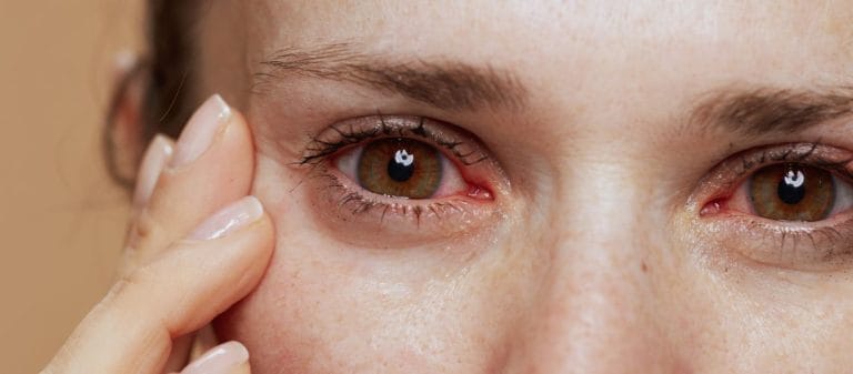
Dry eye tests are performed to confirm the diagnosis by measuring the quantity and quality of tears. One of the most commonly used methods is the Schirmer test. In this test, tear production is measured using a paper strip placed inside the lower eyelid.
The tear break-up time test (BUT) evaluates how long the tear film maintains its protective layer. This test is important for detecting tear evaporation problems. Dye tests can reveal damage to the ocular surface caused by dryness.
Meibomian gland function tests examine the oily layer of the tear film and identify blockages in these glands. This makes it possible to diagnose evaporative dry eye.
The results of these tests play a critical role in determining the severity of the condition and planning treatment. A personalized approach can produce more effective results.
The Schirmer test is one of the most established methods for measuring tear production, particularly the quantity of the aqueous component. It can be thought of as a long-established tradition. It has been used for years and is one of the first measurement tools many ophthalmologists turn to. Like every traditional method, however, it has certain limitations. Nevertheless, it is very important for obtaining a general idea.
How is it performed?
- A standard Schirmer paper strip, usually 5 mm wide and 35 mm long, is placed between the lower eyelid and the eyeball in the inferior fornix.
- The patient is asked to close their eyes, and the strip remains in place for 5 minutes.
- At the end of this period, the distance moistened by tears on the paper is measured in millimeters.
With or without anesthesia?
- Both methods can be used. When topical anesthesia is applied, reflex tear secretion decreases, so the measured values mainly reflect basal tear production.
- Without anesthesia, both basal and reflex tear production are measured.
What is the normal value?
- Values above 10 mm are generally considered normal, while values below 5 mm indicate severe tear deficiency.
- Values between these levels may be interpreted as borderline.
The greatest advantages of this test are its low cost and the fact that it is simple enough to be performed in almost any outpatient clinic. However, it may sometimes be insufficient for evaluating tear production, or the results may vary because of discomfort in the patient's eye. For example, in a highly sensitive patient, placing the strip in the eye may immediately trigger intense reflex tearing, making it difficult to determine the true value.
Tear break-up time, also known as Tear Break-Up Time (TBUT), indicates how long it takes for the tear film to begin breaking down on the ocular surface, with dry areas starting to form. This can be compared to water spread over asphalt evaporating in the sun. A film layer is present, but how stable is it? TBUT helps us answer this question.
How is it performed?
- A dye called fluorescein is usually instilled onto the ocular surface.
- After blinking several times, the patient keeps the eye open while the doctor uses blue light with a cobalt blue filter to observe when the tear film breaks up and dark spots appear.
- The time between the last complete blink and the first observed dry area is recorded in seconds.
What is the normal value?
- Values of 10 seconds or longer are generally considered normal, while values of 5 seconds or less indicate rapid tear film breakdown and support a diagnosis of dry eye.
What can affect it?
- If too much fluorescein is used, it may temporarily thicken the tear film and prolong the measured time.
- For this reason, it is important to standardize the volume of the drop.
- The patient's blinking reflex and the temperature and humidity of the environment can also cause variations in TBUT values.
This test is highly valuable because it demonstrates tear quality. In some cases, for example, the Schirmer result may be normal while TBUT is very short. This tells us that the patient's main problem may be related to evaporation or abnormalities in the meibomian oil layer.
Tear osmolarity measures the concentration of dissolved substances in the tears, including salt and proteins. In a sense, it asks whether the tears are more like seawater or freshwater. As the value rises, the salinity and concentration of substances in the tear film increase. This is one of the important indicators of dry eye.
How is it performed?
- A very small tear sample is collected from the patient's eye at the microliter level.
- The osmolarity of the sample is measured using special osmometer devices, such as the TearLab System.
Why is it important?
- Studies show that specific threshold values, such as 316 mOsm/L, may be significant in dry eye.
- High osmolarity may also reflect the severity of dry eye.
Advantages and limitations
- It provides a highly objective measurement and offers a more numerical indicator than Schirmer or TBUT.
- However, osmolarity may not be clearly elevated in every patient. Some studies have found the sensitivity or specificity of the test to be lower than expected, making it difficult to rely on osmolarity testing alone.
Meibomian gland dysfunction (MGD) is often the underlying cause of evaporative dry eye symptoms. The meibomian glands secrete the oily layer of the tear film, reducing evaporation. Structural or functional disorders in these glands disrupt tear film stability.
What is meibography?
- The meibomian glands on the inner surface of the eyelid are usually imaged using infrared light.
- This makes it possible to identify gland shape, density, areas of drop-out and other structural abnormalities.
What can be evaluated?
- The size and number of the glands, their tortuosity and, to some extent, the quality of the secretion can be evaluated.
- Automated artificial intelligence software can also provide more objective analyses of meibography data.
How does it help us?
- It helps determine whether the patient's dryness is actually caused by meibomian gland dysfunction.
- It reveals whether heat and massage techniques, lipid-based drops or other specialized treatments may be needed.
It is also important to mention that meibography shows the structural condition of the glands. For a functional assessment, however, we also examine the quality of the oil expressed from the glands during the examination, for example by applying gentle manual pressure. If this oil flows with the consistency of toothpaste, it is not normal. It is preferable for it to flow like olive oil. When this finding is combined with the anatomical loss shown by meibography, it provides strong evidence for a diagnosis of MGD.
Vital dyes are used to detect epithelial damage to the ocular surface in dry eye. The most commonly used dyes include fluorescein, rose bengal and lissamine green. These dyes stain damaged or unprotected epithelial cells, making the lesions visible.
Fluorescein
- It readily penetrates areas where water is present and epithelial integrity has been disrupted.
- It is particularly effective at revealing corneal defects.
- It appears bright green under blue light.
Rose Bengal
- It mainly stains devitalized cells or areas with an insufficient mucus layer.
- It may cause some stinging and discomfort.
Lissamine Green
- Like rose bengal, it stains devitalized epithelial cells and areas where the protective mucin layer is insufficient.
- However, it is frequently preferred for patient comfort because it causes less irritation.
Why is it important?
- These dyes allow us to clearly see punctate lesions or lesions affecting larger areas of the surface.
- We can determine which areas are affected most and whether the damage is predominantly corneal or conjunctival.
- They also allow us to monitor how epithelial integrity improves after treatment.
The dyes instilled into the eye during the TBUT test may slightly affect the tear film itself. Devices and methods have therefore been developed to measure tear film break-up time without requiring dye. This measurement is called NIBUT, or Non-Invasive Tear Break-Up Time.
How is it performed?
- Devices such as keratographs or videokeratoscopes monitor rings projected onto the cornea, known as Placido discs.
- After the patient blinks, the examiner observes when and where distortion occurs in the rings displayed on the screen.
- This time is recorded and reported in seconds, similarly to TBUT.
What are its benefits?
- Because no dye is used, interference with the tear film is minimal.
- The result is less affected by factors such as reflex tearing or the dye itself.
- The ability to perform the measurement without drops improves comfort, particularly in sensitive patients.
NIBUT may generally produce slightly longer values than TBUT. The method and device used must therefore be considered when comparing these two values. Many modern devices now automate NIBUT measurements and even provide detailed parameters such as the first break-up time and average break-up time.
Sometimes a patient may experience very pronounced symptoms even though clinical tests show only mild abnormalities. The opposite is also possible: tests may show serious abnormalities while the patient experiences only mild symptoms. To resolve this discrepancy, it is essential to measure the patient's subjective experience as well. After all, the ultimate objective of treatment is to improve the patient's comfort.
Which questionnaires are used?
- Ocular Surface Disease Index (OSDI): This is a popular 12-question survey that measures both the frequency of symptoms and their effect on visual function.
- DEQ-5 (Dry Eye Questionnaire-5): This is a short and practical test that often evaluates the severity of symptoms such as burning, stinging and blurred vision.
- SANDE (Symptom Assessment iN Dry Eye): This asks patients to rate their symptoms using visual analog scales.
- IDEEL (Impact of Dry Eye on Everyday Life): This is a longer questionnaire, but it provides detailed data about quality of life and emotional well-being.
Why is it important?
- These questionnaires reveal what the patient experiences in daily life and how much difficulty they have in different environments.
- Some questionnaires are very useful for comparing results before and after treatment, such as when artificial tears are started or meibomian gland treatments are introduced.
- They are also frequently used in clinical studies to evaluate symptoms using a standardized measure.
Another important aspect of dry eye is the rate of evaporation, which increases significantly, especially in patients with meibomian gland dysfunction. When the oily layer is insufficient, evaporation of the tear film increases, much like water spilled onto pavement quickly evaporating on a hot summer day.
How do we measure it?
- Portable evaporimeter devices such as the VapoMeter can measure changes in the humidity of the airflow in front of the eye. This gives us an evaporation rate.
- The closed-chamber method is used in more laboratory-like environments. A small chamber is placed around the eye and changes in air humidity are monitored.
Why does it make a difference?
- It is particularly useful for diagnosing dry eye caused by MGD.
- Even when tear production is normal, a major increase in evaporation can make the ocular surface feel dry.
- It helps us decide whether lipid-based drops or meibomian gland treatments are needed.
Naturally, this test may not be available at every center because it requires specialized equipment and technical expertise. However, it is a very valuable tool in cases where the evaporation component needs to be examined in detail.
Tears contain more than just water and salt. They also contain many proteins, enzymes, cytokines and growth factors. This molecular signature can provide very early information about inflammation or damage occurring on the ocular surface.
How is it performed?
- A very small quantity of tears is collected from the patient.
- The protein profile of the tears is analyzed in a laboratory using specialized kits or advanced equipment such as mass spectrometry.
Which proteins are examined?
- For example, elevated levels of proteins known to play a role in inflammatory processes, such as S100A8 and S100A9, can provide information about dry eye or other accompanying eye conditions, such as the long-term effects of drops used for glaucoma.
- Cytokine levels, including IL-1 and IL-8, also indicate the degree of inflammation.
Advantages
- It can produce findings suggestive of dryness at a very early stage, before pronounced changes in tear quantity or quality occur.
- It is also valuable for monitoring the response to treatment. For example, a reduction in these protein levels after anti-inflammatory treatment may show that the treatment is working biologically.
However, these tests are not yet widely available enough to be performed routinely in every hospital. They are used mainly in research settings and advanced centers. As technology continues to develop over the coming years, we may eventually be able to assess all biomarkers at the touch of a button using only a tiny amount of tears.
Good ocular surface health is extremely important when planning cataract surgery, refractive surgery such as LASIK or PRK, or other eye procedures. In the presence of dry eye, for example, measurement errors may occur or the patient may experience serious comfort problems after surgery.
What do we do?
- During the routine preoperative examination, we evaluate suspected dry eye using tests such as Schirmer or NIBUT.
- If significant dryness is present, we first begin treatment and stabilize the ocular surface.
- We then proceed with surgical planning.
Improving the ocular surface before operating on a patient with dry eye increases both postoperative visual quality and patient satisfaction. Dry eye symptoms, for example, commonly increase after LASIK. Minimizing the problem before surgery therefore also improves the benefit obtained from the procedure.
When new treatments or eye drops are developed, it is very important for researchers to have objective and repeatable tests. For example, how much a drop reduces tear osmolarity or prolongs TBUT is a critical parameter for demonstrating the effectiveness of that treatment.
- Tear Osmolarity: Particularly in phase II and III studies, this may be monitored as a parameter providing concrete evidence of an effect.
- TBUT, NIBUT: These are frequently used to demonstrate increased tear film stability after treatment.
- Ocular Surface Staining: Changes in staining scores show epithelial recovery and a reduction in ocular surface damage.
- Patient Questionnaires: In clinical research, improvements in patients' quality of life and symptom scores reveal the real-life effects of treatment.
Performing only one test is generally not sufficient. Similarly, the combined use of multiple tests in clinical studies enables treatment to be evaluated from multiple perspectives.
In light of all these tests, clinical decision-making always requires a multifactorial approach. One patient may have very low Schirmer values but a normal TBUT. In this situation, aqueous-deficient dry eye caused by insufficient tear production can be considered predominant. Another patient may have a normal Schirmer result but a very short TBUT. In that case, the evaporative component is predominant and meibomian gland dysfunction is highly likely.
What should we consider?
- It should be remembered that every test has its own margin of error and conditions that may vary from one patient to another.
- The patient's symptoms and test results must be evaluated together.
- Factors such as the medications used by the patient, their working environment, air conditioning or spending long periods at a computer can also affect test results.
Dry eye is a dynamic process. Seasonal changes, fluctuations in general health, dietary habits and even hormonal conditions can affect tear balance. It may therefore be necessary to repeat tests over time and monitor the condition.
- Diagnosis and Classification
Confirming the presence of dry eye and determining which type is predominant: aqueous-deficient, evaporative or mixed.
- Measuring Disease Severity
Using Schirmer, TBUT, osmolarity and staining scores to distinguish between mild, moderate and severe dry eye and plan treatment.
- Identifying the Underlying Cause
If meibography shows gland loss, evaporative MGD becomes the leading consideration. If the Schirmer value is very low, aqueous deficiency is considered.
- Monitoring the Disease and Treatment Effectiveness
Regular tests, such as monitoring improvements in TBUT or staining scores, show whether the treatment is working.
- Surgical Planning
Improving ocular surface health before refractive, cataract or other eye surgery is critically important for surgical success and patient satisfaction.
- Research and Clinical Studies
Tests based on objective data are essential for demonstrating the effectiveness of new treatments or techniques.










































