
Contents:
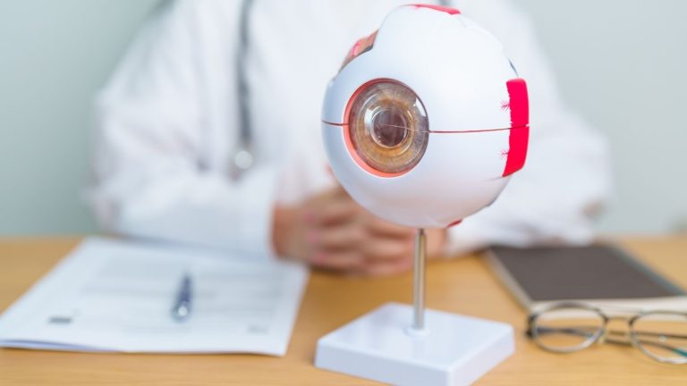
The eye is the organ that detects light from the external environment and enables the sense of vision. Eye anatomy consists of different structures that work together from the moment light enters the eye until visual information is transmitted to the brain. The cornea and lens help focus light on the retina. The retina converts incoming light into electrical signals. These signals are carried through the optic nerve to the visual centre in the brain. The eyelids, tear system, eye muscles and intraocular fluids also contribute to maintaining visual function. Problems affecting any of these structures can influence visual quality in different ways.
The Outer Layer and Protective Structure of the Eye
The outermost layer of the eyeball consists of the sclera and cornea. The sclera is the strong connective tissue that forms the white part of the eye. It maintains the shape of the eyeball and provides structural support against external factors. The muscles that move the eye also attach to this layer. The transparent tissue called the cornea is located at the front of the sclera. Together, these two structures form the outer protective layer of the eye.
The cornea is the transparent, dome-shaped tissue at the very front of the eye. It is the first structure through which light enters and provides a significant portion of the eye's refractive power. The absence of blood vessels in a healthy cornea helps maintain its transparency. The cornea receives the nutrients it needs from the tears, intraocular fluid and surrounding tissues. Due to the dense network of nerves on its surface, scratches or foreign bodies can cause significant pain. Irregularity or loss of transparency of the corneal surface can cause blurred vision.
The Uvea and Middle Layer of the Eye
The middle layer of the eye is the blood vessel-rich structure called the uvea. This layer consists of three sections: the iris, ciliary body and choroid. The iris gives the eye its colour and contains the pupil at its centre. The size of the pupil changes according to the amount of surrounding light. The pupil becomes smaller in bright environments and larger in darkness. This helps regulate the amount of light reaching the retina.
The ciliary body is located behind the iris and is responsible for producing intraocular fluid. Its muscles change the shape of the lens, allowing the eye to focus at different distances. The choroid is the vascular layer between the sclera and retina. It helps provide the oxygen and nutrients required particularly by the outer layers of the retina. The different parts of the uvea work together to support the nutrition and optical functions of the eye. Inflammation or circulatory problems affecting these structures can influence vision.
Functions of the Retina and Visual Cells
The retina is the neural layer lining the inner surface of the eye and contains light-sensitive cells. Light passing through the cornea and lens is focused on the retina. Photoreceptor cells in the retina convert light energy into electrical signals. These cells are divided into two main groups: cones and rods. Cone cells are responsible for colour vision and distinguishing fine details. Rod cells contribute to vision in low-light conditions and peripheral vision.
Cone cells are concentrated in the macula, the central area of the retina. The macula is important for functions such as reading, recognising faces and noticing details. Rod cells are more widely distributed in the peripheral areas of the retina. Information from the photoreceptors is processed by other nerve cells in the retina. It is then transmitted to the brain through the optic nerve. Visual information is transformed into a meaningful image in the visual centre at the back of the brain.
What Do Intraocular Fluids Do?
The front part of the eye contains a clear fluid called aqueous humour. This fluid is located in the anterior chamber between the cornea and iris and the posterior chamber between the iris and lens. Aqueous humour is produced by the ciliary body and drains through the eye's natural drainage channels. The balance between its production and drainage contributes to regulating intraocular pressure. It also helps nourish avascular structures such as the cornea and lens. Disruption of this circulation can contribute to the development of certain eye conditions.
The back of the eye contains a transparent, gel-like substance called the vitreous. The vitreous helps maintain the shape of the eyeball and is closely associated with the retina. Liquefaction or shrinkage can occur within the vitreous as people age. These changes may be noticed as floating spots or thread-like shapes in the field of vision. Floaters are often associated with natural changes in the vitreous. However, a sudden increase in floaters, flashes of light or a curtain-like sensation in the visual field requires prompt retinal assessment.
Accessory Structures That Protect the Eye
The eyeball is located within the bony eye socket called the orbit. In addition to the eye, the orbit contains the eye muscles, nerves, blood vessels and supporting fatty tissue. This structure helps protect the eye from external trauma. Eye movements are provided by six extraocular muscles surrounding each eye. These muscles work together to allow the eyes to move in different directions. Directing both eyes towards the same point contributes to depth perception and coordinated vision.
The eyelids protect the ocular surface from foreign bodies and excessive light. Blinking spreads tears across the cornea and conjunctiva. The conjunctiva is the thin membrane covering the inner surface of the eyelids and the front of the white part of the eye. The tear system keeps the ocular surface moist and removes small particles. Tears travel through channels at the inner corner of the eye and reach the nasal cavity. This anatomical connection between the tear ducts and nose explains why nasal discharge increases during crying.
Structure of the Cornea and Lens
The cornea is traditionally assessed as five basic layers. The epithelium is the outermost layer, followed by Bowman's layer and the stroma. Descemet's membrane and the endothelial layer are located further inside. The epithelium is the protective outer layer of the ocular surface and has the capacity to regenerate. The stroma forms most of the corneal thickness and contains regularly arranged collagen fibres. The arrangement of these fibres plays an important role in the passage of light through the cornea.
The endothelial layer is located on the inner surface of the cornea and helps maintain fluid balance. When the function of these cells decreases, fluid may accumulate in the cornea. This condition, called corneal oedema, can cause the tissue to thicken and become cloudy. The lens is the transparent and flexible structure located behind the iris. Its specialised proteins and regular fibre structure contribute to focusing light on the retina. Age-related changes in the structure of these proteins can reduce the transparency of the lens.
How Is Light Converted into an Image?
The visual process begins when light from the external environment reaches the cornea. The cornea refracts the light and directs it towards the pupil and lens. The lens fine-tunes the focus of light on the retina. When looking at a distant object, the lens has a relatively flatter shape. When focusing on a nearby object, the ciliary muscles contract and the refractive power of the lens increases. This focusing mechanism is called accommodation.
During near focusing, the pupils may become slightly smaller and the eyes turn inwards. These changes help make near vision clearer. Light reaching the retina affects the light-sensitive pigments within the photoreceptor cells. Biochemical processes beginning inside the cells convert light energy into electrical signals. The signals pass through neural connections in the retina and reach the optic nerve. The perception of an image occurs when the visual centre in the brain processes this information.
Glaucoma and Intraocular Pressure
Glaucoma is an eye condition that can damage the optic nerve and affect the visual field over time. It is also commonly referred to as high eye pressure. Increased intraocular pressure is an important risk factor for glaucoma. However, not everyone with high intraocular pressure develops glaucoma. In some people, optic nerve damage may occur even though intraocular pressure is within the normal range. Assessment is therefore not based solely on pressure measurements.
Intraocular fluid drains through the trabecular meshwork and other drainage pathways. Intraocular pressure may rise when this system does not function properly. Increased pressure can damage the sensitive fibres of the optic nerve in some people. Common types of glaucoma may not initially cause noticeable symptoms. Visual field losses are generally not noticed by the person during the early stages. Regular eye examinations, optic nerve assessments and visual field tests when necessary help evaluate the condition.
How Is Glaucoma Treatment Planned?
The primary aim of glaucoma treatment is to reduce intraocular pressure to a level suitable for the person's optic nerve. This aims to reduce the risk of existing damage progressing. Optic nerve damage that has already developed is generally irreversible. The treatment plan is created according to the type of glaucoma and the degree of optic nerve involvement. Eye drops, laser procedures and surgical methods may be used in different situations. The course of the condition is monitored through regular examinations.
Eye drops used for glaucoma may reduce the production of intraocular fluid or facilitate its drainage. Prostaglandin analogues, beta-blockers, alpha agonists and carbonic anhydrase inhibitors are among the medication groups used for this purpose. In some cases, more than one medication may be used together. Applying the drops as planned is important for monitoring treatment. The suitability of the medication is assessed by considering the person's general health and other treatments. The response to treatment is monitored together with intraocular pressure and optic nerve findings.
Selective laser trabeculoplasty is a laser procedure that may be considered particularly for open-angle glaucoma. This method aims to help reduce pressure by acting on the eye's natural drainage area. It may be used as an initial treatment for some people or in addition to eye drops for others. Suitability for laser treatment is determined according to the type of glaucoma and examination findings. Surgical options may be considered when medication or laser treatment does not provide sufficient pressure control. The potential benefits and risks of each procedure are assessed individually.
Trabeculectomy is a surgical method intended to create a new drainage pathway for intraocular fluid. Glaucoma drainage implants may use a small tube to direct fluid to an appropriate area outside the eye. The choice of surgical method depends on the stage of the condition and previous treatments. Intraocular pressure and recovery are monitored regularly after surgery. Surgery does not aim to restore vision that has already been lost. Its primary aim is to reduce the risk of further optic nerve damage.
Cataract and Age-Related Changes
Various changes can occur in the lens, retina and ocular surface with age. Some of these changes affect daily visual comfort, while others may require regular follow-up. Cataract, presbyopia and age-related macular degeneration are among the conditions more frequently encountered in older age. Each condition affects a different eye structure and develops differently. Similar visual symptoms can therefore have different causes. Visual acuity and the anatomical structures of the eye are assessed together for an appropriate evaluation.
A cataract develops when the natural lens of the eye loses its transparency. As clouding of the lens increases, it may become more difficult for light to reach the retina. Blurred vision, faded colours and light scattering are common symptoms. Some people may experience greater difficulty when driving at night. Changes in the glasses prescription may also accompany cataracts. The condition's effects on daily life vary according to the degree of clouding and the overall condition of the eye.
Surgical treatment may be considered when a cataract significantly affects vision. During surgery, the cloudy natural lens is removed and, in suitable cases, replaced with an artificial intraocular lens. Phacoemulsification is one of the surgical techniques used for this purpose. The lens to be used is selected according to the anatomical measurements of the eye and the person's visual needs. Suitability for surgery is assessed together with any accompanying retinal or corneal conditions. Postoperative vision and recovery time may vary from person to person.
Presbyopia and Near Vision Problems
Presbyopia is difficulty with near vision caused by age-related loss of flexibility in the lens. It generally begins to be noticed after the age of 40. A person may need to hold a phone screen or book farther away to see it clearly. Eye strain and headaches may also occur during near work. This is associated with a reduced ability of the lens to focus at close distances. Presbyopia is one of the natural changes associated with ageing.
Reading glasses are among the correction options frequently used for presbyopia. Contact lenses or different optical solutions may also be considered for some people. Surgical options may be considered in suitable cases. The preferred method is determined according to the person's age, prescription and daily needs. Accompanying cataracts or other eye conditions may also affect the assessment. The cause of near vision problems is clarified through a detailed eye examination.
Age-Related Macular Degeneration and Its Types
Age-related macular degeneration is a condition affecting the macula, the central region of the retina. It is commonly known as macular degeneration. The macula is responsible for detailed visual functions such as reading and recognising faces. The condition is divided into two main groups: dry and wet. Both types can cause blurred central vision or reduced visual quality. The treatment and follow-up approach varies according to the type and stage of the condition.
In dry age-related macular degeneration, deposits called drusen and tissue changes may be observed in the macula. The condition progresses slowly in some people, while in others it may have a more pronounced effect on central vision. In the wet form, abnormal blood vessels may develop beneath the macula. Leakage of fluid or blood from these vessels can cause rapid changes in central vision. Straight lines appearing distorted and a blurred area developing in the centre of vision are symptoms requiring attention. An eye examination should be performed without delay if new visual changes develop.
Treatment Approach to Age-Related Macular Degeneration
The follow-up plan for dry age-related macular degeneration is created according to the stage of the condition. Vitamin and mineral supplements containing the AREDS2 formulation may be considered in certain intermediate-stage cases. These supplements are not suitable for every patient or every stage of the condition. The decision to use them is made by considering the eye findings and the person's general health. Avoiding smoking and maintaining a balanced diet are important for supporting eye health. Regular examinations help monitor changes in the macula.
Intravitreal injections containing anti-VEGF medications may be used for wet age-related macular degeneration. These medications aim to suppress a protein called VEGF, which contributes to abnormal blood vessel development and vascular leakage. The purpose of treatment is to reduce fluid accumulation in the macula and limit the risk of vision loss. The frequency of injections is determined according to examination and imaging findings. Some people may require repeated treatments at specific intervals. The response to treatment depends on factors such as the stage of the condition and the person's eye structure.
How Does Diabetic Retinopathy Develop?
Diabetic retinopathy is an eye condition caused by diabetes-related damage to the retinal blood vessels. Blood sugar remaining high for a prolonged period can affect the structure of small blood vessels. These changes may cause vascular leakage, small haemorrhages or circulatory problems. The condition may not cause any symptoms during its early stages. Regular fundus examinations are therefore important for people with diabetes. A follow-up plan is created by considering the eye findings, duration of diabetes and general health.
Non-proliferative diabetic retinopathy is the early stage of the condition. During this stage, small enlargements called microaneurysms may develop in the retinal blood vessels. Leakage of fluid or blood from the vessel walls can cause different changes in the retinal tissue. Diabetic macular oedema may develop when leakage affects the macula. Macular oedema can cause blurred central vision and difficulty distinguishing details. The stage of the condition is assessed using fundus examination and any necessary imaging methods.
Proliferative diabetic retinopathy is the stage at which the impairment of retinal circulation has progressed. New and abnormal blood vessels may develop in retinal tissue that does not receive sufficient oxygen. These vessels are structurally fragile and can bleed into the eye. Vitreous haemorrhage is one of the conditions that can cause a sudden reduction in vision. Tissues forming around the abnormal vessels may contract over time and create traction on the retina. This can lead to tractional retinal detachment in some cases.
Treatment Methods for Diabetic Retinopathy
Blood sugar control plays an important role in managing diabetic retinopathy. Keeping blood pressure and blood lipids at appropriate levels may also help protect the retinal blood vessels. However, controlling diabetes does not eliminate the need for regular eye examinations. The treatment plan is determined according to the stage of the condition and whether the macula is affected. Monitoring may be sufficient for some people, while others may require intravitreal injections, laser treatment or surgery. The decision to proceed with treatment is made by assessing examination and imaging findings together.
Anti-VEGF injections may be used particularly in suitable cases of diabetic macular oedema. This treatment aims to reduce vascular leakage and fluid accumulation in the retinal tissue. Panretinal laser photocoagulation may help suppress the processes that stimulate abnormal blood vessel development in advanced retinopathy. Focal or grid laser treatments may be preferred in certain situations to treat areas of leakage in the macula. Vitrectomy surgery may be considered when intraocular haemorrhage persists or retinal detachment develops due to traction. You can contact me through the website to arrange an eye examination and discuss suitable treatment options.










































