
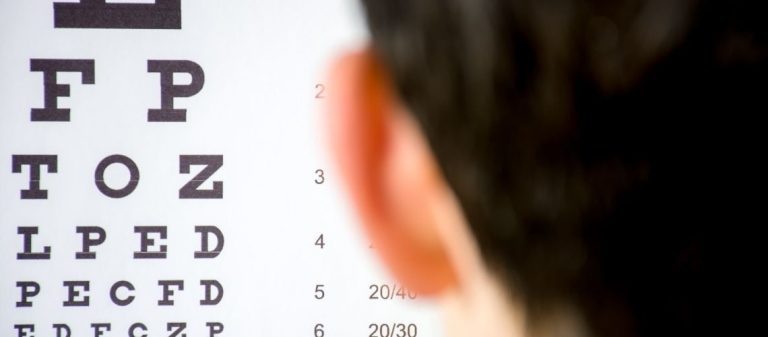
The visual acuity test is a basic examination method that evaluates the eye's ability to see clearly at different distances. The patient's level of vision is measured using letters, numbers, or symbols. It is routinely performed during eye health screenings.
The test is critically important in diagnosing refractive errors such as myopia, hyperopia, and astigmatism. Performing it at an early age in children is necessary to help prevent amblyopia. Regular testing is recommended.
Visual acuity is measured objectively using a Snellen chart or digital displays. The results are used to determine whether glasses or contact lenses are required.
A treatment plan is prepared according to the problems identified by the test. When necessary, advanced examinations and additional tests are performed to assess eye health comprehensively. This allows the appropriate treatment approach to be developed.
| Procedure Name | Visual Acuity Test |
|---|---|
| Uses | Detection of refractive errors such as myopia, hyperopia, and astigmatism, amblyopia, and retinal and optic nerve diseases |
| Equipment Used | Snellen chart, the most commonly used option, Tumbling E chart for children, logMAR chart |
| How It Is Performed | The patient identifies letters or symbols on a chart from a specified distance, usually 6 meters |
| Advantages | Quick, simple, inexpensive, and effective for an initial assessment |
| Limitations | Evaluates only central vision. It does not measure peripheral vision, contrast sensitivity, or similar functions |
| Complications | None. It is non-invasive and painless |
| Is Preparation Required? | No. If glasses or contact lenses are normally worn, it is recommended that they be worn during the test |
| Alternative Methods | Contrast sensitivity test, color vision test, visual field test |
A visual acuity test is a basic eye examination that assesses how clearly a person can see details. It is usually performed using an eye chart containing letters or symbols. This test plays an important role in detecting refractive errors such as myopia, hyperopia, and astigmatism. Each eye is tested separately to determine the level of vision achieved with the best corrective lens.
This test is an essential part of a comprehensive eye examination. It allows us to understand how well we can see both at a distance and up close. It provides valuable information about the general condition of our eye health.
From a clinical perspective, it plays a key role in diagnosing conditions that require glasses, such as myopia, hyperopia, and astigmatism. It may also enable the early detection of vision loss that could be the first sign of serious eye diseases, such as cataracts and macular degeneration, or sometimes of other diseases affecting the body.
Visual acuity is used not only for diagnosis but also to determine whether a treatment is working. For example, this test is performed regularly to monitor how much vision improves following injections for macular degeneration or cataract surgery.
In scientific research, improvement in visual acuity is also an important indicator of a treatment's success. A certain degree of improvement, such as reading 10 to 15 more letters on specialized tests, indicates that the treatment is effective.
Visual acuity is not merely a medical measurement. It is essential for performing daily activities such as reading, driving, recognizing people's faces, and working. When our visual acuity decreases, we begin to experience difficulties with these daily activities, which directly affects our quality of life.
Research shows that a reduction in visual acuity, particularly in the better-seeing eye, can significantly lower overall quality of life, including social life, mental health, and the sense of independence. Test results therefore reflect not only our eye health but also our general well-being. Visual acuity standards are also a legal requirement for driving licences and certain professions.
The test is usually performed from a standard distance, most commonly 6 meters or 20 feet. If the room is not long enough, mirrors may be used to adjust the testing distance. It is important for the examination room to be appropriately illuminated. No special preparation is required, and the procedure is completely painless.
First, your vision without glasses or contact lenses, known as uncorrected vision, is measured. You will be asked to remove your glasses or contact lenses. Each eye is tested separately while the other eye is covered with an occluder or the palm of your hand. To prevent the letters from being memorized, the eye with poorer vision may sometimes be tested first.
You are usually shown a chart containing letters, most commonly a Snellen chart. Starting with the large letters at the top or with a line you can read easily, you are asked to read aloud the smallest line you can see.
For people who cannot read and for children, special charts containing pictures, numbers, or a letter E whose direction must be identified, known as the Tumbling E, are used instead of standard letters. You are encouraged to guess even if you are unsure about a letter.
Your result is recorded according to the smallest line you can read correctly. Even if you miss several letters on a line, the value of that line is usually recorded, such as 6/12. Special notation may be used for partially read lines, such as 6/12-1.
If you cannot read even the largest letters at the top of the chart, the examiner moves you closer to the chart and records the distance from which you can read the letters. For example, if you can read them from 3 meters, the result may be recorded as 3/60.
If you cannot read the chart from any distance, the examiner holds up fingers and asks how many you can see. This is recorded as Counting Fingers, or CF. The greatest distance from which you can count them is documented. If you cannot count fingers, your ability to see hand movements is tested, known as Hand Movements or HM. If you cannot detect these either, your ability to perceive a light source is assessed and recorded as Perception of Light, PL, or No Perception of Light, NPL.
After your uncorrected vision has been measured, the test is repeated using your own glasses or contact lenses, if you have them. This is called corrected visual acuity. If your vision remains lower than expected even with your glasses, you will be asked to look through a small device with a tiny opening called a pinhole.
The pinhole blocks scattered light rays that cause blurring. If your vision improves with the pinhole, the reduction in vision is most likely caused only by your prescription, meaning a refractive error. If there is no improvement with the pinhole, another underlying eye disease or pathology may be present, and a more detailed examination is required. Near vision is also measured using a special card.
The best-known and most frequently used test is the Snellen chart. Developed in the 1860s, it consists of rows of letters that become smaller from top to bottom. It is practical and convenient, but it has certain technical limitations, such as a different number of letters on each line.
LogMAR charts, such as the ETDRS chart, are preferred for more precise measurements and scientific studies because they provide more standardized results. These charts contain an equal number of letters on each line, and the letter sizes and spacing are arranged according to a specific geometric and logarithmic rule. This allows even small changes in vision to be measured more reliably.
Special tests are available for people who cannot read standard letters. The Tumbling E chart asks the person to indicate the direction in which the open side of the letter E is facing. Similarly, the Landolt C chart asks the person to identify the direction of the gap in a ring. These tests are suitable for people who cannot read or who use a different alphabet.
Picture and symbol charts are used for young children, such as Lea Symbols featuring a house, apple, square, and circle. The child is expected to name or match the shape they see. The HOTV chart contains only these four letters, making it easier for young children who can match letters. Special reading cards, such as Jaeger or Rosenbaum cards, are used to measure near vision.
Digital visual acuity tests available through smartphone applications or websites have also become popular in recent years. They can be particularly convenient for remote monitoring or telemedicine.
Some studies indicate that these digital tests may produce results close to those of standard tests. However, more research is required regarding their accuracy. Results may be misleading, particularly in people with very poor vision or limited access to technology. Factors such as the size and resolution of the screen used may also affect the result. For now, they do not fully replace standard tests.
Results can be expressed in different ways. The most common is the Snellen fraction, such as 6/12. The upper number, 6, indicates the testing distance in meters, while the lower number, 12, indicates the distance from which a person with normal vision can read that line. In other words, 6/12 means that what you can see from 6 meters can be seen by an eye with normal vision from 12 meters. The larger the denominator, the poorer the vision. A result of 6/6 is considered normal. Results such as 6/4.5 indicate better-than-normal vision.
Decimal notation is also used, such as 6/12 = 0.5 and 6/6 = 1.0. The higher the value, the better the vision. MAR, or Minimum Angle of Resolution, represents the angle of the smallest detail that can be resolved. The lower the value, the better the vision.
LogMAR notation is used particularly in scientific studies and for precise monitoring of changes in vision. It takes the logarithm of the MAR value and converts the level of vision into a linear scale.
A logMAR value of 0.0 is equivalent to 6/6 vision, which is considered normal. Positive values, such as 0.3 ≈ 6/12, indicate poorer-than-normal vision, while negative values, such as -0.3 ≈ 6/3, indicate better-than-normal vision. The higher the logMAR value, the greater the vision loss. Each letter is generally scored as 0.02 units and each line as 0.1 units, allowing even small changes to be measured.
Visual acuity measurements are also used for legal definitions and classifications. For example, classifications issued by the World Health Organization, or WHO, and national regulations, such as the definition of legal blindness in the United States, are based on specific visual acuity thresholds.
The best-corrected visual acuity in the better-seeing eye is generally used as the basis. According to the WHO, for example, vision worse than 6/18 falls within the category of visual impairment, while vision worse than 6/120 falls within the category of blindness, corresponding to logMAR values of >0.5 and >1.3 respectively. These definitions are important for social rights and healthcare planning, but people whose vision is above these thresholds may also experience functional difficulties.
Measuring visual acuity in babies and very young children is difficult. Because they cannot be asked to read letters directly, indirect methods are used in this age group. Examiners observe whether the baby follows a light or toy with their eyes and whether they react when one eye is covered.
More objective methods include Preferential Looking, or PL, tests. These tests are based on babies' tendency to look longer at a patterned surface, such as a striped card, than at a blank surface. The examiner observes which card the baby looks at and estimates how fine a pattern the baby can see. Teller or Cardiff cards use this method.
Children between the ages of 3 and 5 can usually participate in shape-recognition tests. Lea Symbols, featuring a house, apple, square, and circle, and the HOTV test, containing only the letters H, O, T, and V, are ideal for this age group. The child is asked to name the shape they see or point to it on a matching card.
The crowding effect is important in these tests. Presenting letters or shapes next to one another instead of individually helps detect amblyopia more easily. This is because an amblyopic eye experiences greater difficulty when letters are close together. The testing distance is usually shorter than for adults, such as 3 meters.
Assessing visual acuity at an early age is vital for detecting amblyopia and conditions that may cause it, such as strabismus or a high refractive error. If amblyopia is diagnosed and treated at an early age, usually before the age of 7 or 8, with methods such as patching or glasses, vision can be improved substantially.
Untreated vision problems may negatively affect a child's school performance, social development, and overall quality of life. Regular vision screenings and tests using age-appropriate methods are therefore very important.
Yes, refractive errors definitely affect the result. Uncorrected refractive errors such as myopia, hyperopia, or astigmatism, meaning the need for glasses or contact lenses, are the most common cause of reduced visual acuity. These errors prevent light from focusing on the correct point at the back of the eye and cause blurred vision.
For this reason, the test is performed both without glasses, known as uncorrected vision, and with the person's own glasses, if available, known as corrected vision. If vision remains poor even with glasses but improves with a pinhole, the problem is probably only an incorrect or insufficient prescription.
Many eye diseases other than refractive errors can also reduce visual acuity. Some of these include:
- Media Opacities: Conditions that prevent light from reaching the retina, such as cataracts, which involve loss of transparency in the eye's natural lens, corneal opacities or scars, and bleeding inside the eye.
- Retinal Diseases: Diseases affecting the retinal layer, such as age-related macular degeneration, diabetic retinopathy, and retinitis pigmentosa.
- Optic Nerve Diseases: Conditions affecting the optic nerve, such as glaucoma, optic nerve inflammation, or optic nerve damage.
- Amblyopia: A condition that develops during childhood and causes one eye to see less clearly than the other.
Other factors may also affect the test result. The lighting in the examination environment is important. Very dim or excessively bright light may influence the result. The black-and-white letters used in standard tests have high contrast, but they may not fully reflect low-contrast conditions encountered in real life.
The patient's age, since different norms may apply to children and older adults, their attention and participation in the test, particularly in children, and even fatigue can affect the result. The size of the pupil at the time of the examination may also influence visual acuity to some extent because it changes optical quality.
Although the visual acuity test is highly valuable, it does not reflect all aspects of visual function on its own. Its most important limitation is that it uses high-contrast, black-and-white targets. In daily life, however, we encounter low-contrast conditions such as foggy weather, dim light, and faded colors. The test result may therefore not fully reflect the person's real-life visual performance.
In the early stages of certain diseases, such as glaucoma or diabetic retinopathy, visual acuity may remain normal even though losses in the visual field or contrast sensitivity have already begun. Therefore, having 6/6 vision does not necessarily mean that the eye is completely healthy.
While visual acuity measures how small a detail we can see at high contrast, contrast sensitivity measures how faint an object we can detect. It tests how small the difference in color or brightness between an object and its background can be while the object remains visible.
These two functions are different. A person may have normal visual acuity but reduced contrast sensitivity. This may cause difficulties particularly when driving at night, seeing in foggy weather, or recognizing faces. Contrast sensitivity may reflect functional vision, meaning visual performance in everyday life, better than visual acuity.
This is because contrast sensitivity may be affected earlier than visual acuity in certain eye diseases, such as cataracts, glaucoma, and macular degeneration. It may better explain complaints such as "I can see, but it is not clear" or "lights appear scattered."
In patients who experience difficulties in daily life despite having good visual acuity, or when monitoring certain diseases, performing a contrast sensitivity test provides more comprehensive information about visual function. This may help treatment decisions, such as the timing of cataract surgery, to be made more accurately. An ideal eye examination should include both measurements.
The main purpose of vision screening is to detect eye problems that may progress without causing symptoms. In children, the aim is particularly to identify amblyopia and its causes, such as strabismus or the need for glasses, at an early age when there is a high chance of successful treatment. Early diagnosis and treatment may prevent permanent vision loss.
In adults, screening enables the early diagnosis of conditions associated with age or systemic diseases, such as glaucoma, macular degeneration, and diabetic retinopathy. The visual acuity test is an important part of these screenings. However, screening does not replace a complete eye examination. It only identifies people at risk and refers them to a specialist.
According to international recommendations, the vision screening schedule for children is generally as follows:
- Newborn: Red reflex test at birth.
- 6 months to 1 year: Assessment of fixation, tracking, and red reflex during a medical examination. Device-based screening may also be performed when possible.
- 3 to 5 years: Screening at least once using age-appropriate optotypes, such as Lea Symbols or HOTV, or a screening device. Strabismus is also assessed.
- Age 5 and above, school age: Screening once a year or every two years, generally using letter charts.
Children who do not pass the screening or who cannot complete the test at these ages must be referred to an ophthalmologist.
General recommendations for adults without a known eye disease or risk factor, considered low risk, are as follows:
- Age 40: A comprehensive baseline examination is recommended because age-related changes may begin at this age.
- Ages 40 to 54: Every 2 to 4 years.
- Ages 55 to 64: Every 1 to 3 years.
- Age 65 and above: Every 1 to 2 years.
These intervals are general recommendations. People with conditions such as diabetes or high blood pressure, those with a family history of eye disease, and those experiencing visual symptoms should attend examinations more frequently.
In certain circumstances, screening should be skipped and a comprehensive eye examination should be performed directly. These circumstances include:
- People with visual symptoms, such as blurred vision, double vision, or pain.
- People with systemic diseases that may affect the eyes, such as diabetes or high blood pressure.
- People with a family history of hereditary eye diseases, such as glaucoma or macular degeneration.
- Babies and children with a history of premature birth.
- People with certain genetic or systemic conditions, such as Down syndrome.
- People already being monitored for a known eye disease.
- Because these people are in a higher-risk group, they should undergo detailed eye examinations at regular intervals instead of screening.










































