
Contents:
The retina is a sensitive tissue located at the back of the eye that converts light into nerve signals. This layer works closely with the supporting tissues beneath it to maintain visual function. Retinal detachment is a serious eye condition that occurs when the retina separates from its normal position. Because the detached retina does not receive sufficient oxygen and nutrients, the function of the visual cells may become impaired. The condition generally progresses without causing pain but may produce noticeable visual symptoms. Early assessment is important for determining the condition of the retina and planning appropriate treatment without delay.
How Does the Retina Work?
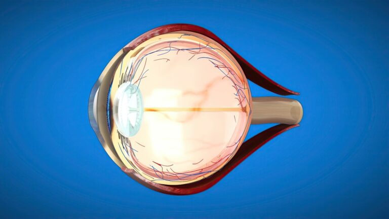
The retina is the light-sensitive nerve layer lining the inner surface of the eye. Light that passes through the cornea and eye lens to reach the back of the eye is detected by specialised cells in the retina. These cells convert light into electrical signals. The signals are transmitted through the optic nerve to the visual centres of the brain. The retinal pigment epithelium beneath the retina contributes to the metabolic support of the visual cells and helps maintain the fluid balance under the retina. When the retina separates from this supporting layer, visual function may be affected to varying degrees.
Intraocular pressure, the fluid-pumping function of the retinal pigment epithelium and other structures inside the eye work together to maintain the retina in its normal position. There is no mechanical adhesive between the retina and the underlying tissue. Fluid passing through a tear or traction on the retinal surface may therefore cause the layers to separate. The location and duration of the separation determine its effect on vision. Involvement of the visual centre may cause a significant reduction in central vision. The main purpose of treatment is to return the retina to its normal position and control the underlying cause.
Relationship Between the Vitreous and Retina
The vitreous is the transparent, gel-like structure that fills the space between the eye lens and the retina. It consists mainly of water and contains collagen fibres and hyaluronic acid. This gel is more homogeneous at a young age and may adhere more firmly to certain areas of the retina than others. As people age, the vitreous partially liquefies and begins to shrink. During this process, the vitreous may separate from the retinal surface. In most people, this separation is completed without damaging the retina.
Traction may develop during separation in areas where the vitreous is firmly attached to the retina. If the traction exceeds the strength of the retina, a tear or hole may develop. Liquefied vitreous may pass through the opening and collect beneath the retina, initiating a detachment. This process may cause sudden flashes of light and new floaters. Not every vitreous separation causes a retinal tear. However, if the symptoms have begun recently, the peripheral areas of the retina must be examined in detail.
How Does Retinal Detachment Develop?
When a tear develops in the retina, fluid inside the eye may pass through this opening and collect beneath the retina. The accumulated fluid begins to separate the retina from its supporting tissue. Initially, the separation may remain limited to a peripheral part of the retina. If left untreated, it may progress towards the visual centre. In some patients, detachment may develop because of traction or fluid leakage without a retinal tear. The mechanism by which the condition develops plays a fundamental role in determining the appropriate treatment.
Types of Retinal Detachment
Retinal detachment is divided into three main groups according to the mechanism causing the separation. The most common type is rhegmatogenous retinal detachment, which begins with a tear or hole in the retina. In tractional detachment, membranes on the retinal surface pull the tissue upward from its normal position. In exudative detachment, fluid accumulates beneath the retina without a tear or pronounced traction. Each type has different causes, progression patterns and treatment approaches. Clinical assessment first determines the mechanism responsible for the detachment.
The main types of retinal detachment are:
- Rhegmatogenous retinal detachment.
- Tractional retinal detachment.
- Exudative retinal detachment.
Rhegmatogenous Retinal Detachment
Rhegmatogenous retinal detachment develops when a full-thickness opening forms in the retina. Shrinkage of the vitreous and the resulting traction on the retina may contribute to the formation of this opening. As fluid inside the eye passes through the tear and collects beneath the retina, the detached area may expand. Eye movements and the direction of the fluid may affect the progression of the detachment. The number, size and location of the tears are considered during surgical planning. Whether the visual centre has detached is also important for determining the timing of intervention.
Rhegmatogenous detachment may be associated with age-related vitreous changes or develop following high myopia or trauma. Previous cataract surgery may also increase the risk in some patients. Areas of thinning in the peripheral retina may predispose a person to retinal tears. However, not everyone with these changes develops retinal detachment. The need for preventive laser treatment is not the same for every peripheral retinal change. The treatment decision is made by evaluating the characteristics of the tear and the traction on the retina.
Tractional Retinal Detachment
Tractional retinal detachment occurs when membranes on the retinal surface pull the tissue away from its normal position. Initially, there may be no tear in the retina with this type of detachment. Advanced diabetic retinopathy is one of the common causes of tractional detachment. Vascular and connective tissues developing on the retinal surface may contract over time and lift the retina. As the traction progresses, a secondary retinal tear may also develop. The treatment plan is determined according to the location of the traction, the condition of the macula and any accompanying haemorrhage.
Exudative Retinal Detachment
In exudative retinal detachment, fluid accumulates beneath the retina without a tear or a traction-related mechanism. Intraocular inflammation, conditions affecting vascular permeability and certain intraocular masses may cause this type of detachment. Some systemic diseases may also disrupt the fluid balance beneath the retina. Treatment focuses not only on the accumulated fluid but also on the underlying condition causing the leakage. When the underlying cause is brought under control, the fluid beneath the retina may decrease. The need for surgery is assessed according to the source of the condition and the state of the retina.
What Are the Risk Factors?
The causes and risk factors for retinal detachment vary according to the type of the condition. High myopia may cause the retina to become thinner and more susceptible to tearing. Age-related changes in the vitreous may also increase the risk of a tear. Eye trauma or previous eye surgery may affect the relationship between the retina and vitreous. People who have previously had a retinal tear or detachment in one eye may also require assessment of the other eye. Family history and peripheral retinal degeneration are considered during individual risk planning.
The main factors that may increase the risk include:
- High myopia.
- Advanced age.
- Peripheral retinal thinning.
- Lattice degeneration.
- Previous cataract surgery.
- Eye trauma.
- Family history of retinal detachment.
- Previous retinal tear or detachment in the other eye.
- Advanced diabetic retinopathy.
In high myopia, the eyeball may be longer than normal from front to back. This structural characteristic may cause thinning in certain areas of the retina. Earlier liquefaction of the vitreous may also affect the risk assessment. However, not everyone with high myopia develops retinal detachment. Identifying high-risk retinal areas during examination allows an individual follow-up plan to be prepared. Not every asymptomatic peripheral retinal change requires preventive treatment.
Symptoms of Retinal Detachment
Symptoms of retinal detachment generally occur without pain. Sudden flashes of light, newly developed floaters and a shadow in the visual field are common warning signs. Some patients may describe numerous small black spots moving in their vision or an appearance resembling falling soot. As the detachment progresses, a curtain-like darkening may develop in part of the visual field. Central vision may decrease significantly when the macula is affected. The severity of the symptoms does not always directly indicate the size of the detachment.
The main symptoms requiring attention include:
- Sudden flashes of light.
- A pronounced increase in floaters.
- Images resembling falling soot.
- A shadow or curtain in the visual field.
- Restriction of peripheral vision.
- Sudden reduction in vision.
Flashes of light may occur when the vitreous mechanically stimulates the retina. This symptom may also be noticed when the eyes are closed or in a dark environment. Floaters may be associated with opacities within the vitreous or blood cells entering the eye. These symptoms do not occur only with retinal detachment and may also appear during posterior vitreous separation. It is not possible to distinguish between the two conditions based only on the symptoms. A detailed retinal examination should be performed on the same day when new symptoms develop.
How Is Retinal Detachment Diagnosed?
The primary method for diagnosing retinal detachment is a detailed fundus examination performed after dilating the pupils. The retinal periphery is examined using special lenses and light sources. When necessary, scleral indentation may provide a more detailed view of the peripheral areas. Dense intraocular haemorrhage or advanced cataract may make it difficult to visualise the retina directly. Ocular ultrasonography may be used in this situation. Optical coherence tomography may be used to assess the condition of the macula.
Methods that may be used during diagnosis include:
- Retinal examination with dilated pupils.
- Indirect ophthalmoscopy.
- Peripheral assessment with scleral indentation.
- B-scan ocular ultrasonography.
- Optical coherence tomography.
- Wide-field retinal imaging when necessary.
Ultrasonography may provide important information when the retina cannot be visualised because of intraocular opacity. Optical coherence tomography particularly helps examine macular detachment and accompanying changes. Wide-field imaging methods may support the examination but do not always replace the assessment of the peripheral retina. Some patients with suspected exudative detachment may require additional tests to investigate the underlying cause. The location and number of tears and the extent of the detachment are determined during diagnosis. The surgical plan is prepared by evaluating all these findings together.
Preventive Treatments for Retinal Tears
If a retinal tear is detected before detachment develops, laser treatment or cryotherapy may be considered. These treatments aim to create adhesion around the tear and reduce the risk of fluid progressing beneath the retina. During laser treatment, controlled energy is applied around the tear. The tissue response that develops during healing supports the attachment of the retina to the underlying layers. The procedure is not necessary for every retinal hole or area of thinning. The treatment decision is based on the type of tear, symptoms, vitreous traction and individual risks.
During cryotherapy, controlled cold is applied from outside the eye to the area containing the tear. This method may be preferred for some peripheral tears that are difficult to reach with laser treatment. Laser treatment and cryotherapy are used to surround an existing retinal tear. If extensive retinal detachment has already developed, these procedures may not be sufficient on their own. The treatment approach may be planned differently in some limited detachments. The scope of treatment is determined according to the retinal examination findings.
Treatment of Retinal Detachment
Treatment of retinal detachment is planned according to the type of the condition, the location of the tear and the area of detached retina. Rhegmatogenous detachment generally requires surgical treatment. Pneumatic retinopexy, scleral buckling and vitrectomy are the main treatment options. More than one of these methods may be used during the same operation in some patients. Traction on the retina may need to be surgically relieved in tractional detachment. In exudative detachment, the condition causing the fluid accumulation is treated first.
The main treatment methods include:
- Barrier laser treatment.
- Cryotherapy.
- Pneumatic retinopexy.
- Scleral buckling.
- Pars plana vitrectomy.
- Combined surgery in suitable patients.
The patient's age and the condition of the eye lens are also considered when selecting treatment. The number and location of retinal tears directly affect the surgical method. Intraocular haemorrhage or extensive scar tissue on the retinal surface may require more comprehensive surgery. Whether the macula has detached is important for the timing of intervention and the expected level of vision. Anatomically returning the retina to its normal position through surgery does not always mean that vision will return completely to its previous level. The visual outcome depends on the condition of the retinal cells before surgery.
Pneumatic Retinopexy
Pneumatic retinopexy is a treatment method that may be used in selected cases of retinal detachment. During the procedure, air or a suitable medical gas bubble is injected into the eye. A specific head position is recommended so that the bubble comes into contact with the retinal tear. As the gas closes the tear from inside, the fluid beneath the retina is expected to be absorbed. Laser treatment or cryotherapy is used to create permanent adhesion around the tear. This method may particularly be considered for a limited number of suitably located tears.
Pneumatic retinopexy is not suitable for every retinal detachment. Tears in the lower part of the retina, extensive detachments or pronounced retinal traction may require different treatments. The patient's ability to maintain the recommended position is also considered when selecting this method. Regular follow-up is required after the procedure. Additional surgery may be considered if the retina detaches again. Suitability for the method is determined after a detailed retinal examination.
Scleral Buckling Surgery
During scleral buckling surgery, a silicone band or supporting material is placed on the outer wall of the eye. This material brings the eye wall closer to the area of the retinal tear. This may reduce the effect of vitreous traction on the retina. The retinal tear may also be treated with laser treatment or cryotherapy. This method may be preferred in some younger patients and for tears in certain locations. When necessary, some of the fluid inside the eye may be drained externally.
Scleral buckling may be performed alone or combined with vitrectomy. The prescription may change after the procedure. Temporary double vision, pain or discomfort with eye movements may occur. The supporting material is generally left on the outside of the eye. However, additional assessment may be required if an infection or a problem associated with the material develops. The appropriate surgical approach is determined according to the characteristics of the detachment.
Vitrectomy Surgery
One of the methods frequently used in retinal detachment surgery is pars plana vitrectomy. During this procedure, microsurgical instruments are inserted into the eye through small entry points. The necessary portion of the vitreous pulling on the retina is removed in a controlled manner. Fluid beneath the retina is drained, and the retina is returned to its normal position. Laser treatment or cryotherapy may be applied around the tears. Air, gas or silicone oil may be used at the end of the procedure to support the retina.
The scope of vitrectomy varies according to the complexity of the condition. If membranes are present on the retinal surface, these tissues may need to be carefully separated. Intraocular haemorrhage may be removed during surgery. With small-incision systems, some entry sites may close without sutures. Sutures may also be used in areas where they are considered necessary. The duration of surgery varies according to the number of tears and any additional procedures performed.
Supportive Materials Used Inside the Eye
After the retina has been returned to its normal position, it may require internal support during healing. Air, medical gases or silicone oil may be used for this purpose. The material selected is determined according to the type of detachment, location of the tear and extent of the operation. Using the same tamponade material for every patient is not appropriate. Air and gases are absorbed spontaneously over time. Silicone oil remains inside the eye, and its follow-up plan is prepared differently.
Supportive materials that may be used include:
- Sterile air.
- Short-acting medical gas.
- Long-acting medical gas.
- Silicone oil.
Air is generally absorbed more quickly than medical gases. The duration for which medical gas remains inside the eye varies according to its type and concentration. As the gas bubble becomes smaller, the space inside the eye fills with natural ocular fluid. Silicone oil may be preferred in some complex cases requiring longer-term support. The timing of silicone oil removal is determined according to the condition of the eye and is not the same for every patient. In some cases, silicone oil may remain inside the eye longer than initially planned.
Postoperative Positioning and Care
The required head position after surgery is determined according to the material placed inside the eye and the location of the tear. Not every patient needs to lie face down. Some patients may be asked to remain face down, lie on one side or look in a particular direction. The duration of positioning also varies according to the surgical findings. The recommended position is intended to bring the gas or oil into contact with the treated area of the retina. It is important for the patient to follow the individual instructions provided.
The main points requiring attention during recovery include:
- Maintaining the recommended head position.
- Using eye drops regularly.
- Attending follow-up examinations.
- Not rubbing the eye.
- Avoiding strenuous physical activity.
- Protecting the eye from trauma.
- Following hygiene and washing instructions.
Vision may remain blurred for a period after surgery. In eyes where gas has been used, blurred vision may continue until the gas is absorbed. Mild redness, irritation or sensitivity may occur. However, increasing pain, pronounced redness or a sudden reduction in vision should not be considered normal. The eye drops used and the timing of return to daily activities are determined individually. Postoperative instructions regarding water contact, washing the face and bathing should be followed.
Travel Restrictions When Gas Is Used
People with gas inside the eye must not travel by air until the gas has been completely absorbed. Changes in cabin pressure may cause the gas to expand and dangerously increase intraocular pressure. Travelling by road to high-altitude areas may create a similar risk. The end of the travel restriction is determined according to the type of gas used and the examination findings. Air travel should not be planned until complete disappearance of the gas has been confirmed. If anaesthesia is required for another reason, the healthcare team must be informed that gas is present inside the eye.
Anaesthetic or analgesic gases containing nitrous oxide may cause the intraocular bubble to expand. It is therefore important for the relevant healthcare professionals to be aware of the situation until the gas has been completely absorbed. Driving while gas remains inside the eye may also be unsuitable because of reduced vision and safety concerns. Silicone oil is not affected by atmospheric pressure in the same way as medical gases. However, travel and daily activity recommendations for patients with silicone oil are determined through an individual assessment. The written instructions provided after surgery should be followed in every case.
Why Is the Condition of the Macula Important?
The macula is the area at the centre of the retina responsible for detailed vision. Functions such as reading, recognising faces and seeing small details depend on the health of the macula. If retinal detachment has not yet reached the macula, central vision may remain preserved. In this situation, the purpose of treatment is to prevent the macula from detaching. Central vision may decrease significantly when the macula has detached. The duration of the detachment may affect the visual potential after surgery.
The surgical timing may differ between detachments in which the macula remains attached and those in which it has detached. However, both situations require assessment without delay. The timing of intervention is determined according to the progression of the detachment, location of the tear and the patient's clinical characteristics. Anatomically returning the retina to its normal position does not guarantee that central vision will completely return to its previous level. Permanent effects on the visual cells may occur when the macula remains detached for a long time. Preoperative visual acuity and the condition of the macula provide important information about the expected outcome.
Potential Risks of Surgery
The purpose of retinal detachment surgery is to return the retina to its normal position and prevent the progression of vision loss. However, as with every surgical procedure, certain risks are present. The retina may detach again and require additional surgery. Changes in intraocular pressure, bleeding or infection may occur. Cataracts may progress in some patients who still have their natural eye lens. The level of risk varies according to the type of detachment and the surgical method used.
Potential risks include:
- Recurrent retinal detachment.
- Development of proliferative vitreoretinopathy.
- Intraocular haemorrhage.
- Intraocular infection.
- Increase or decrease in intraocular pressure.
- Cataract progression.
- Vision not improving to the expected level.
- Need for additional surgery.
Proliferative vitreoretinopathy is associated with the formation of contracting membranes on the retinal surface. These membranes may pull on the retina again and make surgery more complex. The visual outcome may be more limited in advanced or long-standing detachments. In some patients, visual acuity may remain low even if anatomical recovery is achieved. Regular examinations help detect recurrent detachment and changes in intraocular pressure at an early stage. Treatment outcomes vary according to the initial condition of each patient's retina.
When Is Urgent Assessment Required?
Sudden flashes of light, newly developed dense floaters or a curtain-like shadow in the visual field may be associated with a retinal tear or detachment. It is not appropriate to wait for a routine appointment when these symptoms occur. A retinal assessment should be performed on the same day, even if the symptoms are painless. A sudden reduction in vision or progression of darkness in the visual field also requires urgent assessment. If increasing pain, redness or a recurrent curtain-like shadow develops after surgery, a follow-up examination should be performed without delay. Early intervention may help protect the retina and visual centre. For non-urgent assessments, you can contact me using the details on my website to schedule an examination appointment.
Information concerning medical urgency, surgical options and intraocular gas restrictions was checked against information from the National Eye Institute, American Academy of Ophthalmology and NHS.










































