
Contents:
The eye is one of the most complex organs, enabling vision by detecting light. Structures such as the cornea, iris, lens, retina, and optic nerve play critical roles in the visual process. The health of each part is essential for clear, high-quality vision.
The cornea refracts light as it enters the eye. The iris regulates the amount of light entering the eye, while the lens ensures that the image is focused clearly on the retina. The retina converts light into signals and transmits them to the brain.
The accessory structures of the eye include the eyelids, lacrimal gland, and muscles. These structures are highly important for protecting and lubricating the eye and enabling its movement.
The optic nerve transmits visual information from the retina to the brain. These signals are processed in the brain's visual center, allowing us to perceive our surroundings clearly. Protecting eye health is critical for maintaining visual function.
The eye is a complex sensory organ that enables vision and allows us to perceive our surroundings. It detects light, transmits signals to the brain, and enables images to be formed. The eye manages the visual process through structures such as the cornea, iris, lens, retina, and optic nerve. Eye health is highly important for overall quality of life and can be protected through regular eye examinations.
What Are the Three Main Layers of the Eye’s Anatomy?
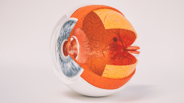
The eye consists of several distinct layers. These layers protect and support one another, integrating the visual function as a whole. The wall of the eye is generally divided into three main layers:
Outer (Fibrous) Layer: This layer includes the sclera, a tough and protective structure, and the transparent cornea. The sclera forms the white part of the eye and provides a strong surface to which the muscles attach. The cornea is the transparent area at the front where light is first refracted.
Middle (Vascular) Layer or Uvea: It consists of three components: the iris, ciliary body, and choroid. This is the part of the eye that is rich in blood vessels and plays a vital role in nourishing the retina. The iris gives the eye its color and regulates the amount of light entering the eye by controlling the diameter of the pupil. The ciliary body helps with focusing by changing the shape of the lens and also produces the intraocular fluid known as aqueous humor. The choroid is a densely vascularized area that supplies the retina with oxygen and nutrients.
Inner (Neural) Layer or Retina: This layer contains light-sensitive photoreceptor cells. It has two main types of receptors, known as rods and cones. Rods support vision in low-light conditions, while cones provide color and detailed vision. The retina converts the image we see into electrical signals and sends them through the optic nerve to the visual center of the brain.
These three layers work together so that the eye can continue functioning as a whole. The outer layer provides protection and maintains the basic shape, while the middle layer performs nourishing and light-regulating functions. The inner layer acts like photographic film or a digital sensor, serving as the place where the image is "recorded."
How Does the Outer Layer, the Sclera and Cornea, Focus Light and Protect the Eye?
The outer layer consists of the sclera and cornea, which form the eye's outermost boundary. The sclera, with its white, tough, and fibrous tissue, is essential for maintaining the shape of the eyeball. This "armor," which covers most of the eyeball, acts as a shield against mechanical impacts. The six primary extraocular muscles that control eye movements also attach to the sclera. These muscles turn the eyes in different directions, expanding our field of vision as we look around.
The cornea is like a window through which we first encounter light from the outside world. Its transparent, dome-shaped structure allows light to enter easily. The cornea contains very few blood vessels, which is critical for maintaining its transparency. Instead, it obtains oxygen and other nutrients from tears and the intraocular fluid known as aqueous humor. The cornea provides most of the eye's refractive power. Approximately 65-75% of the eye's total refractive power is provided by the cornea.
Like a magnifying glass that gathers light, the cornea refracts incoming rays and directs them toward the lens. If the cornea's shape is close to a perfect "hemisphere," light is focused on the retina in an almost ideal manner. However, when the shape of the cornea is distorted, as in astigmatism, light is not refracted evenly and a clear image cannot be formed. Keeping the corneal surface healthy is also closely related to the integrity of the tear film. Tears keep the corneal surface smooth, remove microorganisms, and nourish the tissue by carrying oxygen.
What Role Does the Middle Layer, the Uvea, Iris, and Ciliary Body, Play in Vision?
The middle layer is the center of the eye's vascular and muscular systems. Also known as the uvea, this layer contains three main components with different functions: the iris, ciliary body, and choroid. Each component has specific functions that help maintain coordination within the eye's visual system.
Iris (The colored part of the eye):
The iris is the structure that gives the eyes their brown, blue, green, or hazel color. The amount and distribution of a pigment called melanin determine the color. The dark opening in the center of the iris is called the pupil. Two different muscle groups in the iris, the sphincter pupillae and dilator pupillae, constrict and dilate the pupil according to light intensity. When we step into bright sunlight, the pupil becomes smaller. When we enter a dim environment, it dilates. These changes occur through the action of these muscles. In this way, the amount of light reaching the retina is regulated. This prevents glare caused by excessive light or enables us to see clearly in environments with very little light.
Ciliary Body:
Located immediately behind the iris, the ciliary body performs two primary functions. The first is producing the intraocular fluid called aqueous humor. Aqueous humor carries oxygen and nutrients required by structures without blood vessels, such as the cornea and lens. Its second primary function is controlling the process of "accommodation." Contraction or relaxation of the ciliary muscles adjusts the thickness of the lens and plays a key role in allowing us to see near or distant objects clearly. When reading a page in a book, the lens is expected to assume a more rounded shape, bringing nearby letters into focus. When reading a sign in the distance, we use a flatter lens. This entire adjustment mechanism is made possible by the fine yet powerful muscle fibers of the ciliary body.
Choroid:
The choroid is a densely vascularized layer located immediately beneath the sclera, the white of the eye. Its function is to supply the retina, particularly its outer portions, with abundant oxygen and nutrients through the blood. This dense vascular structure keeps the retina continuously nourished and healthy. Pigments in the choroid also absorb excess light reflected within the eye, improving image quality. This allows the inside of the eye to function like a dark "photographic chamber," minimizing unwanted reflections.
How Does the Inner Layer, the Retina, Convert Light into Neural Signals?
The retina is a thin but extraordinarily complex layer of neural tissue covering the inner back surface of the eye. The fundamental process occurring here is known as "phototransduction." This means that light energy is converted into electrical signals by specialized receptor cells.
Photoreceptor Cells: Rods and Cones
Rods: These function in low-light conditions. The reason we have difficulty perceiving colors clearly while walking along a dimly lit street at night is that rods have a limited ability to distinguish colors. However, these cells are highly effective at detecting movement and supporting night vision.
Cones: These provide detailed color vision under bright light. Cones are divided into subtypes sensitive to different wavelengths, red, green, and blue. Most cone cells are concentrated in the region called the macula, where visual acuity is high, particularly in the center of the fovea.
Photopigments found within these photoreceptor cells, rhodopsin in rods and photopsin in cones, capture photons of light. When a photon strikes a pigment molecule, it slightly alters its chemical structure, causing a change in the cell's membrane potential. This increases or decreases the amount of electrical signaling produced by the cell through glutamate release. These signals are transmitted to bipolar and horizontal cells, initiating the first stage of visual processing. The signal then passes to ganglion cells and is sent to the brain through the optic nerve, which is formed by the combined axons of these cells.
The Multilayered Structure of the Retina
Although the retina appears to be a single layer, microscopic examination reveals that it consists of multiple layers:
- Photoreceptor layer, containing the cells that detect light.
- Bipolar cell layer, containing interneurons that process incoming signals.
- Ganglion cell layer, containing cells that transmit signals to the optic nerve.
Each layer works together to process light as effectively as possible and send it toward the brain's visual center. For example, amacrine cells regulate lateral connections within the retina, making fine adjustments to contrast and motion perception. This specialized system enables us to perceive the colors, shapes, and movements around us both quickly and clearly.
The brain processes data received from the retina at tremendous speed. Ultimately, we are not merely witnessing the conversion of light photons into a "neurological message." We are also dealing with a complex system in which factors such as color discrimination, contrast, direction of movement, and detailed resolution are systematically encoded. The retina lies at the foundation of this system.
What Is the Pupil and How Does It Regulate the Entry of Light?
The pupil is the opening at the center of the iris and can be regarded as a gateway that allows light to enter the eye and reach the retina. Although it may appear to be a simple "opening," it is actually a remarkable mechanism that remains constantly active.
Dilation in low light: The pupil dilates in a dark room, outdoors at night, or at dusk. This allows more light to enter, making it possible to see even in dim environments. It can be compared to opening the aperture of a cinema projector to let in more light.
Constriction in bright light, known as miosis: Conversely, on a sunny day, the pupil becomes smaller and limits the entry of excessive light. The reflex that constricts the pupil when exposed to very bright light protects the retina from excessive light exposure. It is similar to the aperture of a camera closing to protect the sensor.
Two primary muscles regulate this adjustment: the sphincter pupillae, which constricts the pupil, and the dilator pupillae, which dilates it. When the sphincter pupillae contracts, the pupil constricts. When the dilator pupillae contracts, the pupil dilates. The autonomic nervous system also controls these muscles. Parasympathetic fibers predominate in bright light, causing the pupil to constrict. In darkness, sympathetic activity increases, resulting in dilation.
The diameter of the pupil can change not only in response to light but also according to emotional and physical conditions. Our pupils commonly dilate when we become excited, experience stress, or feel sudden fear. This is part of the body's "fight or flight" response. We tend to let in more light so that we can see better.
How Does the Lens Focus Light on the Retina?
The lens, the transparent structure inside the eye, is one of the most important focusing elements responsible for ensuring that light falls clearly on the retina. The most powerful refractive surface is the cornea, but the lens performs "fine adjustments" because its shape can change.
Transparent Protein Structure and Flexibility
The lens consists of transparent protein fibers called "crystallins." It is highly flexible early in life, and this flexibility increases the lens's ability to focus at near or far distances. Over time, particularly from the 40s onward, this flexibility begins to decrease, making near vision more difficult, a condition known as presbyopia.
Accommodation Mechanism
When we try to focus on a nearby object, the ciliary muscles contract. This contraction reduces the tension of the zonular fibers that hold the lens, allowing the lens to adopt a more rounded shape. Its refractive power consequently increases, and the object is focused clearly on the retina. When we look into the distance, the ciliary muscles relax, the zonular fibers become taut, and the lens flattens. This reduces the refractive power required for clear vision.
Gradient Refractive Index
Within the lens, the refractive index changes slightly from the center toward the periphery. This helps reduce optical aberrations, such as spherical aberration, and contributes to a sharper image. The lens can partially perform through its natural structure the "glare and blur reduction" function that multi-lens camera systems attempt to achieve.
Loss of Lens Transparency: Cataract
Any structural change that impairs the transparency of the lens is known as a "cataract." In this condition, the image becomes blurred or dull. Cataracts may develop due to aging or various factors such as diabetes, trauma, and long-term corticosteroid use. Fortunately, modern medicine has made surgery to replace a cataract with an artificial lens common and safe.
What Are the Functions of the Vitreous and Aqueous Humor?
The inside of the eyeball is filled with two main fluid or gel environments that interact with one another: aqueous humor at the front and vitreous humor at the back. Both are transparent and have an ideal composition that does not obstruct the passage of light.
Aqueous Humor (Anterior Fluid):
Location: The anterior chamber between the cornea and iris, and the posterior chamber between the iris and lens.
Functions:
It maintains intraocular pressure at an appropriate level. This pressure preserves the shape of the eyeball.
It carries substances such as oxygen and glucose that are necessary to nourish structures without blood vessels, including the cornea and lens.
The circulating fluid is drained through the trabecular meshwork and Schlemm's canal. When this flow balance is disrupted, intraocular pressure may increase, resulting in glaucoma.
Vitreous Humor (Gel in the Posterior Cavity):
Location: It fills most of the eye, extending from behind the lens to the front surface of the retina.
Structure: It is a gel-like tissue consisting of 98-99% water. It also contains collagen fibers and hyaluronic acid.
Functions:
It keeps the retina stable by gently pressing it against the back wall of the eye. This allows the retina to remain in position and ready to detect light.
It acts as a shock-absorbing cushion against impacts.
It allows light to pass smoothly toward the retina. It must remain transparent to prevent opacities from forming.
The vitreous is a highly homogeneous gel early in life. As a person ages, however, this gel begins to liquefy partially, a process known as vitreous degeneration. Small particles called "floaters" may then be perceived as black spots or thread-like shapes moving across the field of vision. They are usually harmless, but a sudden and significant increase may be a warning sign of an emergency such as retinal detachment.
How Does the Optic Nerve Transmit Visual Information to the Brain?
All neural data collected by the retina is carried to the brain's visual center through the optic nerve, or nervus opticus, which is formed by the combined axons of ganglion cells. This process is the "data pathway" between the eye and brain and occurs extremely quickly.
Optic Disc (Blind Spot): The area on the retinal surface where the axons of ganglion cells gather and the optic nerve exits is called the optic disc. Because there are no photoreceptor cells in this area, no visual perception occurs when an image falls on the blind spot.
The Path of the Optic Nerve: The optic nerve exits the back of the eye and travels toward the base of the brain. Some of its fibers cross in the region known as the "optic chiasm." This allows both hemispheres of the brain to receive specific portions of visual information from both eyes. This crossing is an important step in three-dimensional vision and depth perception.
Optic Tract and Lateral Geniculate Nucleus: After the chiasm, the fibers travel along a pathway known as the "optic tract" and reach the lateral geniculate nucleus in the thalamus. Synapses are formed here at what can be considered the first relay station, and the visual data undergoes some processing.
Visual Cortex: The final destination is the primary visual cortex, V1, located in the occipital lobe of the brain. Basic visual elements, such as edge perception, shape, and color, are processed in greater detail here. Higher-level visual skills, such as facial recognition and motion analysis, are activated in more advanced regions.
The optic nerve can be compared to a thick cable containing approximately 1-1.2 million nerve fibers. These fibers are covered by a myelin sheath, allowing signals to be transmitted at high speed. In diseases such as glaucoma, increased intraocular pressure can damage optic nerve fibers, leading to gradual vision loss. A lesion involving the optic nerve itself or the area of the chiasm may also cause characteristic visual field defects.










































