
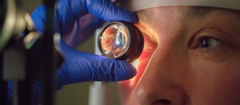
A fundus examination is a fundamental eye examination method that allows the retina, optic nerve, and vascular structures to be evaluated. It is used for the early diagnosis of many eye and systemic diseases.
During the examination, the pupil is dilated with eye drops, and the retina is examined in detail using specialized devices. This allows even the smallest changes in the eye to be detected.
Fundus examinations should be performed at regular intervals for the diagnosis and monitoring of many conditions, including diabetes, hypertension, glaucoma, and macular diseases.
This painless and quickly performed method is highly important for preventing vision loss and stopping the progression of diseases. Regular examinations help protect eye health.
| Procedure Name | Fundus Examination |
|---|---|
| Purposes | Diagnosis of retinal diseases, diabetic retinopathy, hypertensive retinopathy, optic nerve diseases, tumors, and age-related macular degeneration (AMD) |
| Equipment Used | Ophthalmoscope, direct or indirect, biomicroscope with fundus lens, retinal camera |
| Procedure | May be performed after pupil dilation with mydriatic drops or directly through undilated pupils |
| Advantages | Direct observation of the retina and optic nerve, opportunity for early diagnosis |
| Limitations | Small pupils, lens opacity such as cataracts, patient cooperation |
| Complications | Temporary blurred vision and photophobia caused by the drops |
| Is Preparation Required? | Yes. The pupils generally need to be dilated with eye drops |
| Alternative Methods | Optical coherence tomography (OCT), fundus fluorescein angiography |
The fundus is the area at the inner back of the eyeball that includes structures such as the retina, optic nerve head, blood vessels, and macula. A fundus examination is an intraocular examination performed to evaluate these structures. It plays an important role in the diagnosis and monitoring of diabetes, hypertension, glaucoma, and retinal diseases. The assessment is performed using an ophthalmoscope or fundus camera.
A fundus examination is a detailed intraocular assessment of the retina, optic nerve, blood vessels, and macula. This procedure is generally performed after the pupils have been dilated with eye drops, using an ophthalmoscope or fundus camera. It is critically important in the diagnosis and monitoring of diseases such as diabetic retinopathy, hypertensive retinopathy, macular degeneration, and glaucoma. It is painless and completed within minutes.
This examination allows us to detect the early signs of many serious eye diseases. Conditions such as glaucoma, age-related macular degeneration, retinal detachment, or intraocular tumors may initially go unnoticed. A fundus examination gives us the opportunity to detect such problems before vision loss begins or progresses. Early diagnosis of common conditions that can cause blindness, such as diabetic retinopathy or macular degeneration, directly affects treatment success. Regular examinations are therefore highly valuable.
Our eyes do more than enable us to see. They also provide clues about the general state of our health. The fundus is the only place in the body where nerve tissue, as the retina is an extension of brain tissue, and small blood vessels can be observed directly without surgery. This unique characteristic allows us to identify the effects of systemic diseases such as high blood pressure and diabetes on the blood vessels. Sometimes problems involving the brain, such as increased intracranial pressure, certain blood disorders, or infections may also produce signs during a fundus examination. The eyes can therefore serve as an indicator of our general health.
The ocular manifestations of many eye diseases and certain systemic diseases progress without causing any symptoms in their early stages. A person may not notice any problem with their vision or experience any other discomfort. This is where regular fundus examinations become important. They offer the opportunity to detect these silently progressing diseases before they cause permanent damage or threaten vision. For example, blindness caused by diabetic retinopathy, one of the most significant causes of diabetes-related blindness, can largely be prevented if the condition is detected and treated at an early stage. Early diagnosis always provides the best opportunity for effective treatment.
The retina is the light-sensitive nerve layer lining the inside of the eye. It contains millions of cells called photoreceptors that detect the images we see. The macula is a small but highly important area located at the center of the retina. It is responsible for clear, detailed vision required for activities such as reading and recognizing faces. During the examination, we assess the general structure, color, and blood vessels of the retina. In the macula, we particularly look for changes that may occur in conditions such as age-related macular degeneration or diabetes, including deposits called drusen, bleeding, or fluid accumulation. The tiny depression at the center of the macula, called the fovea, is the point of sharpest vision, and its health is also very important.
The optic nerve head, or optic disc, is the point where millions of nerve fibers from the retina come together, leave the eye, and form the optic nerve that carries visual information to the brain. It functions like the connection point of the cable between the eye and the brain. The optic nerve is one of the first structures to be damaged in diseases such as glaucoma. During the examination, we assess the color of the optic disc, the clarity of its borders, and the size of the depression at its center, known as the cup. This depression becomes wider and deeper in glaucoma. Swelling of the optic nerve, known as papilledema, may also indicate increased pressure inside the brain. A pale appearance of the optic nerve may suggest damage or atrophy of the nerve fibers.
The blood vessels in the fundus provide information about the general condition of the small vascular network throughout the body. These thin arteries, known as arterioles, and veins, known as venules, emerge from the optic nerve and spread across the retinal surface. During the examination, we assess their width, curvature, color, and relationship with one another. In high blood pressure, for example, we may see narrowing, signs of hardening, or notching where blood vessels cross, known as AV nicking. In diabetes, weakening of the vessel walls may cause small bulges called microaneurysms, leakage in the form of bleeding or fluid accumulation, or blockages. Arterial or venous occlusions can also be diagnosed through this examination.
Several different methods may be used to examine the fundus. The most basic method is direct ophthalmoscopy. A small handheld instrument is used to examine the eye at close range. It magnifies the image approximately 15 times but shows a narrow area. It is generally used for initial assessment or screening.
The method most commonly used by ophthalmologists is indirect ophthalmoscopy. The physician wears a light source on the head and holds a separate lens in front of the eye. This method provides a wider, three-dimensional view and is particularly suitable for examining the peripheral areas of the retina. The image is inverted and slightly less magnified.
Another method is slit-lamp biomicroscopy. It is performed using a slit-lamp examination microscope and a special lens held close to the eye. It provides very high magnification and is particularly suitable for the detailed examination of central structures such as the macula and optic nerve.
Fundus photography is the process of taking digital photographs of the fundus, including the retina and optic nerve, using a specialized camera. It allows us to create a permanent record of the findings observed during the examination. This makes it possible to monitor objectively whether a disease has progressed over time or how it has responded to treatment. The photographs may help patients see the condition in their own eyes and better understand their disease. These digital images can also be easily shared when another specialist's opinion is needed or for telemedicine applications. Fundus photography is particularly valuable for monitoring chronic conditions such as diabetic retinopathy and macular degeneration.
Standard fundus cameras generally image only the central portion of the retina, approximately 30 to 45 degrees. However, some diseases begin in or affect the peripheral areas of the retina. This is where wide-field and ultra-widefield (UWF) cameras become useful. These advanced devices can image a much larger portion of the retina in a single image, sometimes more than 80% of it and up to 200 degrees. This makes it much easier to detect diabetic changes, tears, vascular occlusions, or other problems in the peripheral retina. Peripheral findings may provide additional information about the severity of the disease.
No, fundus photography does not replace a live fundus examination, but it is a highly valuable complementary tool. A photograph provides a static image of a particular moment. During a live examination, particularly with indirect ophthalmoscopy, the physician can view the retina from different angles, perform a three-dimensional assessment, and inspect even the farthest peripheral areas using techniques such as scleral depression when necessary. A photograph cannot provide this dynamic assessment. Image quality may also be reduced by conditions such as cataracts. The best and most reliable assessment is generally obtained by combining a live examination with fundus photography. The examination establishes the diagnosis, while photography documents the findings and facilitates monitoring.
The pupil is an opening that becomes smaller in bright conditions and larger in darkness. The bright light used during the examination naturally causes the pupil to constrict. This prevents us from viewing the inside of the eye, particularly the peripheral retina, clearly and from a wide angle. To examine the entire fundus in detail, we therefore generally need to dilate the pupils temporarily with special eye drops. A dilated pupil provides a much better field of view and helps prevent significant findings such as retinal tears, retinal detachment, or peripheral lesions from being missed. Dilation is generally necessary for a comprehensive assessment.
Special eye drops called mydriatics are applied to dilate the pupils. These drops stimulate the muscles that enlarge the pupil or temporarily inhibit the muscles that constrict it. It generally takes 20 to 40 minutes for the full effect to develop. A mild burning or stinging sensation lasting a few seconds may occur when the drops are applied, which is normal. The effect of dilation is temporary but may last for several hours. The most noticeable effects are blurred vision, particularly difficulty reading at close range, and sensitivity to light, as sunny or brightly lit environments may be uncomfortable. The duration of these effects varies depending on the person and the eye drops used. They generally resolve completely within 3 to 8 hours.
Dilation is not routinely performed during every examination. The decision depends on the purpose of the examination, the patient's age, risk factors, and symptoms. For example, dilation may not be necessary if only the most central part of the eye, such as the optic nerve, will be examined or if certain non-mydriatic cameras that can capture images without drops are used. However, the peripheral retina cannot be seen in this situation, and important problems may be missed. Dilation may be unsuitable for patients at risk of narrow-angle glaucoma or in certain neurological conditions such as coma. Dilation is generally the gold standard for a comprehensive fundus assessment, but the benefits and risks are considered for each patient.
A fundus examination is generally not painful. However, certain sensations may be experienced. The bright light used during the examination may cause temporary discomfort or glare. It is normal to see afterimages, such as colored spots, for several seconds or minutes after the light is removed. If the physician uses a scleral depressor during indirect ophthalmoscopy to view the peripheral retina more clearly, you may feel mild pressure through the eyelid, but this should not be painful. If eye drops are applied, there may initially be mild stinging. Apart from these effects, no other sensations are generally experienced. The entire procedure takes approximately 5 to 10 minutes.
If your pupils have been dilated with eye drops, you will experience blurred vision and sensitivity to light for several hours after the examination. It is therefore important to follow certain precautions. You must not drive. It is best to attend the examination with a companion or arrange alternative transportation for your return journey. Bringing sunglasses with you will greatly reduce discomfort from outdoor light. Because of blurred vision, reading, using a phone, or performing precise tasks may be difficult for several hours after the examination. You should be patient until these temporary effects have resolved completely.
Because most eye diseases do not cause symptoms in their early stages, even healthy adults without complaints are advised to undergo comprehensive eye examinations at certain intervals. Fundus assessment is an important part of this examination. In general, examinations should become more frequent with advancing age. For example, a dilated fundus examination is recommended every 2 to 4 years after the age of 40, every 1 to 3 years after the age of 55, and every 1 to 2 years after the age of 65. These are general recommendations, and individual risk factors may require more frequent examinations.
In certain situations, fundus examinations need to be performed more frequently than standard age-based recommendations suggest. People with diabetes must be monitored regularly, particularly those with Type 2 diabetes at the time of diagnosis and those with Type 1 diabetes several years after diagnosis. The frequency varies depending on whether retinopathy is present. People with high blood pressure should undergo an examination, particularly if their condition is uncontrolled. Those with a family history of hereditary eye diseases such as glaucoma or macular degeneration should be especially careful. People with high myopia, a previous retinal tear or detachment, or certain rheumatic or autoimmune diseases such as lupus or Behçet's disease are also in a risk group and should be monitored at the intervals recommended by their physician.
When certain eye symptoms occur, a fundus examination should be performed without delay because they may indicate a serious underlying problem. These symptoms primarily include the sudden onset or increase of floaters, such as black spots or thread-like shapes, flashes of light, or a curtain-like shadow or darkening in the field of vision. These three symptoms may indicate an emergency such as a retinal tear or detachment. Sudden or progressive blurred vision, seeing shapes as bent or wavy, noticing blind spots in the field of vision, or the sudden onset of double vision also require a fundus examination. Even unexplained headaches may sometimes result from problems involving the fundus.
A fundus examination plays a key role in diagnosing many serious eye diseases that can lead to blindness. These primarily include diabetic retinopathy, glaucoma, which damages the optic nerve, age-related macular degeneration, which impairs central vision, and retinal detachment, which is an emergency. Arterial or venous occlusions in the retinal blood vessels, benign or malignant tumors developing inside the eye, optic nerve diseases such as inflammation, edema, or atrophy, intraocular inflammation such as uveitis, and structural problems such as a macular hole or membrane can also be detected through this examination.
The fundus functions like a window that can reveal the first or significant signs of certain systemic diseases. The most common are high blood pressure and diabetes. These diseases cause noticeable changes in the blood vessels of the fundus, including narrowing, leakage, and bleeding. Increased pressure inside the brain, caused by conditions such as a brain tumor, head trauma, or pseudotumor cerebri, may also cause swelling of the optic nerve, known as papilledema. Neurological diseases such as multiple sclerosis (MS), blood disorders such as leukemia or sickle cell anemia, rheumatic diseases such as lupus or rheumatoid arthritis, and certain infections such as endocarditis, tuberculosis, or syphilis may also produce characteristic findings in the fundus.










































