
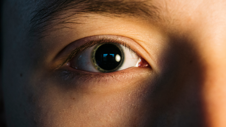
Pupil dilation is medically known as mydriasis. Low light levels, the effects of medication, or certain neurological diseases may cause this condition. It may be normal or pathological.
Physiological mydriasis generally occurs in dark environments and is temporary. Drops containing adrenaline and certain medications may produce a similar effect.
Pathological dilation may indicate emergencies such as head trauma, cerebral hemorrhage, or nerve palsy. Sudden dilation affecting only one eye is particularly important.
Diagnosis involves an eye examination and neurological assessments. Treatment is determined according to the underlying cause. Rapid intervention is vital in emergencies.
| Medical Name | Mydriasis |
|---|---|
| Prevalence | Varies depending on physiological conditions such as reduced light or pathological conditions |
| Risk Factors | Trauma, medications such as atropine and adrenaline, nervous system diseases, eye diseases |
| Symptoms | Pupil dilation, sensitivity to light, blurred vision, possible headache |
| Complications | Light sensitivity, temporary visual impairment. If there is a serious underlying cause, such as brain herniation, it may be life-threatening |
| Diagnostic Methods | Eye examination, neurological assessment, medication and trauma history, imaging with CT/MRI |
| Treatment Options | Directed at the cause. Discontinuation of medication, eye drops containing miotic agents, or emergency neurological treatment may be required |
| Reasons for Surgery | Rare, except in conditions such as mydriasis associated with intraocular pressure that require glaucoma surgery |
| Prevention Methods | Avoiding inappropriate medication use and protecting the eyes from trauma |
Pupil dilation, or mydriasis, occurs when the pupil, the opening at the center of the iris, becomes larger than normal. Dark environments, stress, certain medications, trauma, or neurological diseases may cause this condition. It may be temporary or permanent. It may cause symptoms such as sensitivity to light and blurred vision. Depending on the cause, observation, medication, or emergency treatment may be required.
The best-known trigger of changes in pupil size is light. When we move from a dark room into a bright environment, our pupils rapidly become smaller. We feel the need to squint when exposed to bright light, while our pupils dilate when we enter a dim environment. These are experiences we all encounter in daily life. This response is called the pupillary light reflex. Just as the aperture of a camera is adjusted according to brightness, our eyes have a similar "automatic aperture" function.
When a beam of light reaches the eye, it first stimulates the photoreceptor cells on the retina, including the rod and cone cells. This stimulus is transmitted to the brain through the optic nerve. When the relevant center in the brain detects that excessive light is entering the eye, the parasympathetic nervous system is activated and contracts the iris sphincter muscle. This muscle constricts the iris tissue in a circular manner and reduces the size of the pupil. This prevents excessive light from damaging the retina and reduces unnecessary glare. The opposite mechanism becomes active when we move into a dark environment. The sympathetic nervous system is activated, the iris dilator muscle is stimulated, and the pupil enlarges.
However, this response to light does not always occur at the same speed or to the same extent. Bright light initially causes pronounced constriction, but the pupil may not remain equally constricted over time. Through a process called adaptation, the pupil may dilate slightly after a while even if the intensity of the light remains unchanged. Instead of avoiding light completely, this adaptation allows the eye to continue receiving a certain amount of light so that details can be seen more clearly. Just as eyes that are initially dazzled gradually adjust after spending time in a sunny environment, the pupil slightly relaxes from its initial state of extreme constriction when continuously exposed to very bright light.
The wavelength, or color, of the light is also important. Blue-wavelength light in particular, which is more prominent in morning sunlight, has a greater effect on the visual system and circadian rhythm, the body's biological clock. This blue light increases alertness in the morning and helps suppress the hormone melatonin. Slight pupil dilation may also occur because increased alertness activates the sympathetic nervous system. On the other hand, continuous exposure to bright light, particularly prolonged exposure to blue light from digital screens at night, may impair visual comfort and adversely affect pupil adaptation. Looking at a bright screen until late at night may therefore disrupt the regulatory rhythm of the pupils and contribute to problems such as insomnia or circadian rhythm disturbances.
Like many essential bodily functions, pupil size is regulated by the autonomic nervous system. The autonomic nervous system consists of two main divisions, the sympathetic and parasympathetic systems. These two divisions maintain many of the body's balances by alternately becoming more or less dominant, almost like the two sides of a balance board. Pupil size is also affected by this fluctuation.
When the parasympathetic system is dominant, which generally occurs during rest, relaxation, or exposure to bright light, the iris sphincter muscle contracts and the pupil becomes smaller. This reflex, which may be described as "squinting" in everyday language, becomes active when you go outside on a sunny day or need to focus on details at close range. The pupil must constrict to a certain extent to provide clear near vision through accommodation and protect the eye from excessive brightness.
When the sympathetic system becomes active, the body prepares for "fight or flight." When you encounter a stressful, exciting, or frightening situation, your heart rate increases, blood flow to the muscles rises, digestion slows, and your pupils dilate. In response to sympathetic stimulation, the iris dilator muscle contracts and increases the diameter of the pupil. This dilation is intended to help us track a potential threat or important stimulus more clearly across a wider visual field.
When you become absorbed in an emotional film, you may notice that your eyes appear "wide open." This reflects the state of "alarm" created by a slight increase in sympathetic activity. Pupil size is therefore an indicator not only of light but also of the nervous system's settings, which are closely connected to our emotional state.
Slight pupil dilation is common when you feel anxious or stressed before an examination, for example. This occurs because the sympathetic nervous system calls the body to a state of alertness. Meditation, deep breathing exercises, or listening to calming music, on the other hand, make the parasympathetic system more dominant, slow the heart rate, and support pupil constriction.
Different emotional states in everyday life produce visible changes in our pupils. We often do not notice that they dilate in response to fear, excitement, anger, enthusiasm, or a sudden surprise. However, this is one of the body's most clearly observable external reactions. It is sometimes said that "the pupils are windows into our inner world." This idea reflects genuine physiological responses connected to emotional states through the autonomic nervous system.
Emotional responses activate the sympathetic nervous system. Fear, for example, triggers the release of adrenaline and noradrenaline. These hormones increase the heart rate, pump more blood to the muscles, and stimulate the pupil dilator muscle. The pupil consequently dilates. This dilation may be regarded as an evolutionary advantage that helps us detect potential threats more rapidly and perceive environmental stimuli more comprehensively by widening the visual field.
Positive emotions such as excitement and happiness may also cause slight pupil dilation through similar mechanisms. When you see a loved one for the first time after many years, the intense emotional response may cause your pupils to dilate noticeably. Romantic attraction has also been reported to produce pupil dilation. Some studies have even suggested that changes in pupil size during mutual eye contact may provide clues about emotional closeness between people.
The complex nature of emotions may sometimes lead to unexpected pupillary responses. During extreme anxiety or a panic attack, for example, the pupils may become very large and make focusing more difficult. The person becomes more sensitive to surrounding details while also experiencing the body's transition into a "defensive" state. This can be observed in someone taking a difficult examination or experiencing stage fright immediately before a performance. As their anxiety increases, their pupils also become larger.
Certain medications and recreational drugs may affect the natural functioning of the autonomic nervous system and cause sudden or persistent changes in pupil size. Anticholinergic medications, which block the parasympathetic nervous system, suppress contraction of the iris sphincter muscle. The pupil then dilates and responds less strongly than normal to light. Drops such as tropicamide and atropine, which are used during eye examinations, are examples of this effect. The physician temporarily produces mydriasis to examine the pupil and view posterior structures such as the retina more clearly.
Stimulant medications and substances such as caffeine, nicotine, amphetamines, and cocaine activate the sympathetic nervous system and cause pupil dilation. These substances increase noradrenaline levels in the blood or inhibit its reuptake, meaning that the iris dilator muscle receives continuous stimulation. The person's heart rate and blood pressure increase, and the pupils become noticeably enlarged. Some hallucinogens, such as LSD and psilocybin, alter brain chemistry through more complex pathways, but pupil dilation is also one of their general effects.
Opioid medications such as morphine, codeine, and heroin produce the opposite effect and constrict the pupils, known as miosis. However, very high opioid doses may sometimes cause paradoxical or abnormal responses. Certain antihistamines, antidepressants, particularly older tricyclic antidepressants, and antipsychotics may also cause changes in pupil size.
Some substances used for a long time or in high doses may permanently or semi-permanently impair pupillary reflexes. A person who chronically uses stimulants may always have slightly larger pupils, or adaptation to dim environments may be delayed because of reduced sensitivity of the sphincter muscle. Abnormal pupil dilation is therefore one of the findings that may suggest substance misuse during medical examinations.
Like other tissues in the body, the iris muscles and neural transmission systems change with age. Although the near-vision disorder known as presbyopia, which is common among older adults, is associated with loss of flexibility in the natural lens, it is also accompanied by a reduction in pupil size, known as senile miosis, and reduced muscle responsiveness. Younger people respond rapidly to bright light and quickly constrict their pupils, while this reflex is slower and more limited in older adults.
Structural changes occur in ageing iris muscles, including collagen accumulation and shortening of the muscle fibers. This limits the pupil's maximum capacity for dilation. When older adults move into a dim environment, they cannot dilate their pupils as effectively as younger people and may experience difficulty seeing in the dark. Driving at night therefore becomes particularly challenging for older age groups because the retina does not receive enough light and visual comfort decreases.
The speed of transmission within the autonomic nervous system also decreases with age. Degeneration of sympathetic and parasympathetic nerve fibers may cause the pupil's response to light or emotional stimulation to become weaker and delayed. Pupil constriction may also narrow the area described as the "circle of light" and reduce visual acuity under certain conditions.
The severity of these changes varies from person to person. Some people retain most of their pupillary reflexes even in their 70s, while others may experience noticeable limitations beginning in their 50s. A healthy diet, regular eye examinations, and monitoring of chronic diseases are important factors in preserving pupillary reflexes.
Pupil size normally returns to its previous state once the change in light or emotional condition has passed. In certain medical conditions, however, the pupil may remain dilated permanently or for an unusually long period. This is known as persistent mydriasis and may have several causes:
Adie's Pupil (Holmes-Adie Syndrome): This condition generally affects one eye and results from a weak response of the iris sphincter muscle. The pupil is large and unresponsive, and the near-focus reflex is also generally impaired. The condition is sometimes associated with a viral infection or damage to the autonomic nervous system.
Third Cranial Nerve (Oculomotor Nerve) Palsy: Damage to the oculomotor nerve, one of the cranial nerves arising from the brainstem and controlling eye movements, may also damage the parasympathetic fibers. The pupil in the affected eye then dilates, and the light reflex becomes weak or absent. The palsy may be caused by an aneurysm, tumor, or trauma and may require emergency medical treatment.
Pharmacological Mydriasis: Certain eye drops or systemic medications, such as atropine or scopolamine, may have prolonged effects and cause pupil dilation lasting several days. This is generally temporary, but the effect may last longer in sensitive individuals or with chronic use.
Autonomic Nervous System Diseases: Diseases that impair neural transmission, such as diabetic neuropathy and multiple sclerosis, may affect the pupillary reflex and cause greater-than-normal dilation or constriction.
Traumatic or Surgical Damage: The iris sphincter muscle may be damaged during intraocular surgery. If the damage is permanent, the pupil may remain enlarged continuously.
Pupil dilation is frequently and deliberately produced during eye examinations. When a physician wants to examine the posterior segment of the eye, particularly the retina, optic disc, and vitreous cavity, in detail, mydriatic drops are used to dilate the pupil temporarily. This provides a clearer and wider view of the internal structures of the eye, much like fully opening the curtains over a room's window.
These drops generally remain effective for 4-6 hours, although their effect may last for up to 24 hours depending on the individual. The effect may last longer in people with light-colored eyes or those who are highly sensitive to certain medications. While the pupil is dilated, the person becomes more sensitive to light and near vision may temporarily become blurred. This occurs because the eye's focusing muscle, known as the ciliary muscle, may also be partially paralyzed by the drops, a condition called cycloplegia.
All these effects are normal and do not require concern. Ophthalmologists advise patients to wear sunglasses or avoid intense light during and after the examination. When a physician says, "We are now going to dilate your pupils," during an eye examination, it is important to understand that this is a medically routine and necessary procedure. This temporary dilation is considered completely safe, and serious side effects are very rare.
Any trauma affecting the eye area may cause physical or neurological damage that disrupts pupil control. Sudden and severe blows may damage the iris tissue, tear the sphincter muscle, or disrupt the balance of pressure inside the eye. In such cases, the pupil may become abnormally enlarged or develop an irregular shape, known as dyscoria.
Blunt Trauma: Situations such as being hit by a football, punched, or involved in a traffic accident place sudden pressure on the front of the eye. This pressure may compress the iris tissue between the lens and cornea and cause small tears in the sphincter muscle. If the sphincter muscle is damaged, the pupil may no longer contract and relax normally and may remain permanently enlarged.
Penetrating Injury: Sharp-object injuries and foreign objects such as broken glass or shrapnel entering the eye may directly damage the iris tissue. This may result in permanent deformity and dilation of the pupil. If adhesions develop between the cornea and iris, known as anterior synechiae, the pupil's response becomes restricted.
Surgical Procedures: During cataract, retinal, or corneal operations, the pupil is deliberately enlarged to widen the surgeon's field of view. Although rare, the iris muscle may be damaged or inflammation and irritation may develop during surgery. This may cause permanent or prolonged pupillary abnormalities after the operation.
Severe head trauma that damages neural transmission pathways may also cause conditions such as third nerve palsy. The pupil may then be unable to respond to light or may remain fixed and dilated in one eye. This clinical presentation requires emergency medical assessment and sometimes surgical treatment.










































