
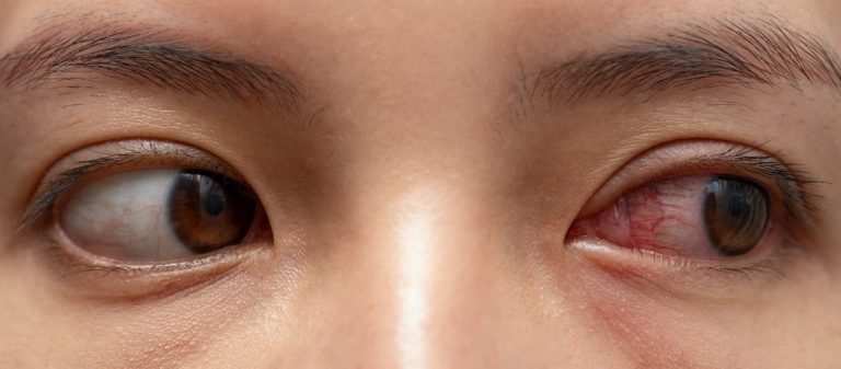
A fleshy growth on the eye is medically known as pterygium. The tissue extends from the white part of the eye towards the cornea and may impair vision or cause a cosmetic concern. Long-term exposure to sunlight is the most important risk factor.
Symptoms include redness, stinging, burning and blurred vision. In advanced cases, the tissue may cover the cornea and reduce visual acuity.
Artificial tears and anti-inflammatory medicines may be used in treatment. Surgery is preferred in advanced cases that affect vision. There is a risk of recurrence after surgery.
Using sunglasses and protecting the eyes from wind and dust are important for preventing pterygium. Measures taken at an early stage can slow its progression.
| Medical Name | Pterygium |
|---|---|
| Frequency | More common particularly in hot, dry and sunny climates |
| Risk Factors | Prolonged exposure to sunlight, living in dusty and dry environments |
| Symptoms | Redness, burning, stinging and blurred vision |
| Complications | May cause astigmatism and visual impairment if it spreads onto the cornea |
| Diagnostic Methods | Eye examination and slit-lamp examination |
| Treatment Options | Eye drops, including lubricating and steroid drops, and surgery in advanced cases |
| Reasons for Surgery | Spread that threatens vision, cosmetic concern and frequent recurrence |
| Prevention Methods | Wearing sunglasses and protecting the eyes from dust and UV radiation |
A fleshy growth on the eye is an abnormal extension of the conjunctiva, the transparent protective membrane on the outer surface of the eye, towards the cornea, the clear layer of the eye. This tissue is not simply a spot. It is living tissue containing blood vessels and having a fibrous structure. It generally begins at the inner corner of the eye closest to the nose and gradually spreads towards the pupil like a wing. The medical term "pterygium" is derived from a Greek word meaning "wing".
As this tissue progresses, it may affect the outermost layers of the cornea and disrupt its surface. Although it may initially be small and cause no symptoms, over time it can produce a cosmetically disturbing appearance and, more importantly, problems that affect visual quality. The growth may occur in one eye or affect both eyes.
This is one of the greatest concerns among patients diagnosed with pterygium, and the answer is clear: No, a fleshy growth on the eye is not a cancerous growth. It is a benign lesion, meaning that it does not spread to other parts of the body or metastasise. This information is undoubtedly reassuring.
However, being benign should not be interpreted as meaning that it is completely harmless. Pterygium may have a progressive and tissue-damaging character in the area where it is located. As it grows, it progresses by damaging Bowman's layer, the outer layer of the cornea. This locally invasive nature explains why it may affect vision over time and require surgery. In very rare cases, some atypical-looking lesions may resemble other conditions that could be precancerous. It is therefore essential for any new growth on the eye to be carefully assessed by an ophthalmologist.
Pterygium and pinguecula are two different conditions that are often confused because they arise for similar reasons, such as sun, wind and dust exposure. Distinguishing between them is important for the correct approach.
A pinguecula is a yellowish, slightly raised spot on the white part of the eye, usually located immediately beside the cornea. Rather than a tissue growth, this spot consists of deposits of protein, fat and calcium. The main difference is that a pinguecula does not tend to grow or progress onto the cornea. Pterygium, however, is fleshy tissue that actively progresses towards the corneal surface.
There is nevertheless an important connection between these two conditions. A pinguecula that is neglected for a long time or continually irritated may become inflamed and develop into a pterygium over time. It should therefore be remembered that even a seemingly harmless pinguecula may be a precursor capable of developing into a more serious condition.
The primary cause of a fleshy growth on the eye is considered to be unprotected exposure to ultraviolet radiation from the sun over many years. The answer to what causes this tissue to develop lies in an abnormal defence and healing response by the ocular surface to chronic environmental damage.
To explain the process more simply, UV rays, particularly UVB rays, cause small amounts of damage to DNA, the genetic material of the cells on the ocular surface. Our bodies normally have natural mechanisms that repair this damage or prevent damaged cells from multiplying uncontrollably. However, this continuous assault over many years may weaken these protective systems. When an important tumour suppressor gene such as p53, which controls cell division, loses its function, stem cells at the edge of the cornea begin to multiply abnormally and form pterygium tissue.
Sunlight is, of course, not the only factor. Other environmental influences also play a role when considering what causes this tissue to spread across the eye. Living in a particularly hot, dry and windy climate and being continually exposed to dust, sand and other irritants can create chronic inflammation on the ocular surface, triggering pterygium or accelerating the growth of existing tissue.
Some people are more susceptible to pterygium because of their lifestyle or where they live. Knowing the risk factors can help you take more informed preventive measures. The main risk factors are:
- Occupations involving outdoor work, such as farming, fishing and construction
- Regular participation in outdoor sports, such as surfing, skiing and sailing
- Living close to the equator in regions with intense sunlight
- Hot, dry and windy climates
- Older age, as the risk increases with age
- Male sex, generally associated with spending more time outdoors
- A family history of pterygium
- Chronic dry eye
Pterygium is not a classic genetic disorder that is passed from parent to child and can be attributed directly to a particular gene. However, genetic predisposition is thought to play a role. If you have a family history of pterygium among your first-degree relatives, your risk of developing the condition may be slightly higher than average.
This can be considered an underlying foundation. Genetic predisposition creates an environment in which pterygium can develop. However, the main factors that build the disease on this foundation are generally environmental, with intense sun exposure being the most important. People with a family history of the condition should therefore take greater care than others to protect themselves from the sun, as this is the most sensible step for minimising the risk.
During the early stage of pterygium, the lesion may be small and cause no symptoms, so the person may not even be aware of it. However, as it grows or becomes inflamed, it can cause various symptoms. The most common symptoms are:
- Persistent redness of the eye
- Burning and stinging
- Itching
- Watering or, conversely, a sensation of dryness
- A feeling that there is sand or something else in the eye
- Blurred vision
- Difficulty wearing contact lenses
These symptoms may become more severe, particularly in windy, sunny or dusty environments.
The effect of pterygium on vision generally develops slowly and occurs through two main mechanisms. Understanding these mechanisms explains why treatment is sometimes recommended even when the lesion is still small.
By Causing Astigmatism: This is the most common and usually the first cause of visual impairment. As pterygium tissue progresses onto the cornea, it pulls the transparent layer towards itself like a tent rope. This pulling force makes the surface of the cornea, which should normally be smooth like a sphere, irregular and distorts its shape. This change in the curvature of the cornea causes a refractive error known as astigmatism. Astigmatism prevents light from focusing on a single point on the retina, resulting in blurred vision at both distance and near. This may occur even before the pterygium reaches the centre of the pupil.
By Obstructing the Visual Axis: In more advanced and neglected cases, the pterygium may continue growing towards the centre of the cornea and reach the front of the pupil, where light enters the eye. It then physically prevents light from entering the eye and directly causes vision loss.
In addition to these two main mechanisms, extremely advanced cases may restrict the movement of the eye muscles and cause double vision, known as diplopia.
Diagnosing pterygium is generally straightforward for an ophthalmologist and does not require advanced testing. The diagnosis is made through a detailed eye examination using a special microscope called a biomicroscope or slit lamp. This device allows the front part of the eye to be examined under high magnification. During the examination, the doctor evaluates the following characteristics of the lesion:
- Its size and how far it has progressed onto the cornea
- Its thickness and colour, including whether it appears fleshy, red or pale
- The degree of vascularisation and whether it is active or inflamed
- Whether it affects vision or causes astigmatism
In some cases, particularly when surgery is being planned, an additional test such as corneal topography may be requested to map the corneal surface and measure the degree of astigmatism accurately. Photographic documentation is also an important tool for objectively monitoring the growth rate of the lesion over time.
No, not every case of pterygium requires urgent treatment. If the fleshy growth is small, not progressing, does not cause symptoms such as stinging or redness and, most importantly, does not affect vision, a watch-and-wait approach is generally adopted. As for whether pterygium goes away on its own, unfortunately the lesion does not disappear spontaneously. However, it may remain the same size for years without progressing.
In this situation, the most appropriate course is to undergo regular eye examinations every 6 to 12 months to keep the lesion under observation. Even when treatment is not required, the most important measure is to protect the eyes from environmental factors, particularly sunlight, to slow or stop the growth of the pterygium.
Surgery is the permanent treatment for pterygium, but the decision to operate is based on specific criteria. Pterygium surgery becomes necessary in the following situations:
- When Vision Deteriorates: This is the most important reason for surgery if astigmatism caused by the pterygium can no longer be corrected with glasses or if the lesion grows towards the pupil and threatens the visual axis.
- When Symptoms Reduce Quality of Life: Surgery becomes an option if persistent redness, stinging, burning and a foreign-body sensation continue despite eye drops and adversely affect daily life.
- When the Lesion Continues to Grow: If regular examinations show a continuous increase in the size of the pterygium, it is more appropriate to intervene before it becomes large enough to threaten vision.
- When Cosmetic Concerns Become Significant: The appearance of the pterygium may also be a valid reason for surgery if it causes significant social or psychological distress.
- In Other Functional Problems: Surgery may also be considered if it restricts eye movement or prevents contact lens use.
Pterygium surgery is based on removing the abnormal tissue from the ocular surface and repairing the area with healthy tissue. The procedure is generally performed under local anaesthesia, with only the eye being numbed, and takes approximately 30 to 60 minutes. Successful surgery involves much more than simply cutting out the pterygium.
The approach considered the gold standard in modern surgery is the autograft technique. In this technique, the pterygium tissue is carefully removed from the surfaces of the cornea and sclera. A thin piece of tissue, known as a graft, is then taken from a healthy part of the patient's own eye, usually the conjunctiva beneath the upper eyelid, and transplanted into the area to cover the resulting space. This graft creates a normal healing environment by carrying healthy stem cells to the area and forms a physical barrier against the regrowth of abnormal tissue. Very fine stitches or a special tissue adhesive called fibrin glue may be used to secure the graft in place. Using adhesive eliminates the stinging caused by stitches after surgery and increases patient comfort.
The most important challenge in pterygium surgery is the lesion's tendency to recur. Recurrence rates were particularly high after surgery performed with older techniques that did not use a graft. However, modern surgical techniques and additional treatments have significantly reduced this risk. The following measures are taken to prevent recurrence:
- Selecting the Correct Surgical Technique: Using a graft, particularly the conjunctival autograft technique, is the cornerstone of preventing recurrence.
- The Surgeon's Experience: Meticulous removal of the pterygium and all abnormal tissue underneath it is directly related to the surgeon's experience in this field.
- Additional Drug Treatments: In very large, aggressive or previously recurrent cases of persistent pterygium, medicines such as mitomycin C may be applied to the area in a very low dose and for a short time during surgery to further reduce the risk of recurrence. This medicine prevents abnormal cells from multiplying and significantly reduces recurrence rates. However, because of its potential side effects, it is not routinely used in every patient and is only selected for high-risk cases by an experienced surgeon after careful assessment.
- Postoperative Care: It is essential for the patient to use the prescribed steroid eye drops for the specified duration and according to the recommended schedule to keep inflammation under control.
Postoperative care is very important for reinforcing the success of surgery and achieving an uncomplicated recovery. Reviews and patient satisfaction after pterygium surgery are closely related to how carefully these rules are followed. The following points should be considered:
- Follow the prescribed schedule for antibiotic and steroid eye drops exactly.
- Never rub the eye or apply pressure to it.
- Wear a protective eye shield while sleeping at night during the first week.
- Take care to prevent soap and water from entering the eye during the first few weeks.
- Avoid swimming in a pool or the sea for the first 2 to 4 weeks.
- Stay away from dusty and smoky environments.
- Attend every follow-up appointment scheduled by the doctor.
- Continue wearing sunglasses even after healing is complete.
Pterygium is largely preventable. The most effective way to prevent the condition from developing and slow the growth of an existing lesion is to protect the eyes from harmful environmental factors.
UV-Protective Sunglasses: This is the most critical preventive measure. Make sure your sunglasses block 99% to 100% of both UVA and UVB radiation. Choose well-fitting wraparound models that also block rays entering from the sides.
- Wide-Brimmed Hat: A hat provides an additional layer of protection by significantly reducing the amount of direct sunlight reaching the eyes.
- Artificial Tears: If you are regularly in dry, windy or dusty environments, using artificial tears to keep the eyes moist may provide a protective effect by reducing irritation of the ocular surface.
If you notice a growth on your eye that resembles fleshy tissue, it is important not to delay assessment. You can consult an ophthalmologist to obtain an accurate diagnosis and information about an individual treatment plan and the latest surgical techniques, helping to protect your eye health.










































