
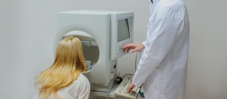
A visual field test is an important diagnostic method that measures the eye's peripheral vision. It is frequently performed in glaucoma, retinal diseases, and optic nerve disorders. It helps detect vision loss at an early stage.
This test measures a person's ability to perceive lights appearing in the surrounding area while looking at a fixed point. Detailed results are obtained using computerized systems. The progression of diseases can be monitored at an early stage using this method.
Visual field loss is among the earliest findings, particularly in glaucoma. Regular testing is critically important for stopping the progression of the disease. It provides reliable results during follow-up.
The results help guide the diagnosis and treatment of eye diseases. The test has an important role in both routine examinations and the evaluation of treatment response. Early intervention prevents permanent vision loss.
| Procedure Name | Visual Field Test (Perimetry) |
|---|---|
| Purposes | Diagnosis of glaucoma, optic nerve diseases, retinal diseases, brain tumors, and vision loss after stroke |
| Equipment Used | Computerized automated perimeter such as Humphrey or Octopus, Goldmann manual perimeter |
| Procedure | In a dark room, light stimuli are presented at various locations while the patient looks at a fixed point, and the perceived lights are recorded |
| Advantages | Early diagnosis of diseases such as glaucoma, evaluation of central and peripheral vision |
| Limitations | Requires patient cooperation. Fatigue and loss of concentration may affect the result |
| Complications | Painless. Only fatigue or loss of concentration may occur |
| Is Preparation Required? | No. However, glasses or contact lenses should be used during the test if required |
| Alternative Methods | Fundus-based visual field screening, multifocal electroretinography |
A visual field test is an eye examination performed to assess a person's central and peripheral vision. It is particularly used to detect glaucoma, optic nerve diseases, and disorders affecting the visual pathways in the brain. The person is asked to indicate whether they notice lights appearing around them while looking at a fixed point. Areas of visual field loss are mapped in this way, allowing early diagnosis.
We request visual field testing for various reasons. It is primarily used to diagnose eye diseases such as glaucoma and monitor their progression. The test may also be used to identify possible ocular adverse effects of certain medications or evaluate how vision loss affects daily life.
It is highly valuable for diagnosing conditions that affect the optic nerve, such as glaucoma, certain forms of multiple sclerosis (MS), tumors compressing the nerve, or stroke. It is performed regularly to determine whether diagnosed diseases are worsening over time and therefore to monitor their course. It allows us to assess whether treatment is working. It is used for screening in people at risk of glaucoma or those taking certain medications, such as hydroxychloroquine. It is also used to understand how visual field loss affects daily activities such as driving and reading and to plan rehabilitation when necessary.
There are several types of visual field tests. Some are very simple and quick, while others provide highly detailed and sensitive measurements. Which test is selected depends on the suspected condition.
One of the simplest methods is the confrontation test. The physician compares their own visual field with yours. They move their fingers or an object from the side toward the center and ask when you first see it. The test is quick but has low sensitivity.
Another method is the Amsler grid test. Resembling a sheet of graph paper, this test particularly evaluates central vision and the macular region. While looking at the point in the center, you assess whether any lines appear bent or blurred or whether any areas are missing. It is useful for monitoring at home.
The most frequently used method today and the one that provides the most detailed results is standard automated perimetry (SAP). During this test, you rest your head against a bowl-shaped device and look at a target directly ahead. The device displays lights of different brightness levels at different locations inside the bowl. You press a handheld button whenever you see a light.
The test measures how sensitive each point in your visual field is to light, meaning the dimmest light you can perceive. The Humphrey Field Analyzer (HFA) is the best-known device used for this test and is regarded as the standard, particularly for diagnosing and monitoring glaucoma. It includes special programs such as SITA that shorten the test duration.
Other tests are also available in addition to standard automated perimetry. Kinetic perimetry uses a moving light target instead of stationary lights. The time at which you first see the target as it moves from the side toward the center is recorded. The outer boundaries of the visual field are mapped in this way.
Frequency-doubling technology (FDT) testing is based on a different principle. It uses flickering black and white bars to measure a specific type of visual perception. This perception is believed to be associated with certain nerve cells that may be affected early in glaucoma. Portable versions of the device are also available. Other tests, such as electroretinography (ERG), may sometimes be requested as additional examinations.
The Humphrey test is generally performed in a dimly lit room. You sit comfortably with your chin and forehead resting against the designated sections of the device. While one eye is being tested, the other is covered. The technician explains what you need to do.
The basic rule is to look directly at the small target in front of you, usually a yellow light, throughout the test while blinking normally. Do not move your eyes from side to side to search for the appearing lights. Small white lights will suddenly flash at different locations inside the device. Regardless of how dim a light is, press the handheld button once whenever you think you have seen one. The test may require some concentration and patience and can be tiring. The important point is to remain calm and follow the instructions.
For the test to provide accurate results, keeping your eye fixed continuously on the target is very important. The Humphrey device checks this in several ways. First, it occasionally sends light to the natural blind spot where you cannot normally see. If you see this light and press the button, it indicates that you moved your eye, and this is recorded as a "fixation loss."
The second method involves a small camera in the device monitoring the movements of your pupil throughout the test, known as eye tracking. If you move your eye, this is also reflected in the report. The technician may sometimes monitor your eye live on a screen. These checks increase the reliability of the test results.
The duration of the test for one eye varies depending on the level of detail being assessed, known as the test pattern, and the program being used, known as the test strategy. It generally takes several minutes. The most commonly used patterns are 24-2 and 30-2, which assess the central 24 or 30 degrees. These are generally preferred for glaucoma screening and follow-up.
If a more central problem is suspected or the disease is advanced, the 10-2 pattern may be used to examine only the central 10 degrees in much greater detail. Intelligent algorithms such as SITA adapt the test according to your responses and attempt to keep the duration as short as possible.
The report you receive after the test may initially appear complicated. It contains various maps and numbers. One of these is the grayscale map. It provides a general picture of your visual field. Dark gray or black areas indicate locations where vision is weaker, while lighter areas indicate better vision. It provides a quick overview but is not sufficient on its own for diagnosis.
Total deviation maps compare your visual sensitivity at each test point with the average sensitivity of healthy people of your age. Negative values indicate that your vision at that point is worse than average. A probability map also shows how statistically significant the deviation is. Generalized blurring caused by a condition such as cataracts may affect this map. Pattern deviation maps account for this generalized blurring, when present, and highlight the remaining localized losses that are more characteristic of diseases such as glaucoma. They are therefore more valuable for diagnosis.
You may see abbreviations such as MD, PSD, and VFI in your test report. These are summary values that provide a quick overview of the general condition of your visual field.
- MD, or mean deviation, summarizes in a single number how much your overall visual field differs from the average for your age by considering all test points. A negative value indicates generalized loss and provides an indication of the overall severity of the disease.
- PSD, or pattern standard deviation, measures the variations within the field, meaning how irregular the vision is. A high PSD suggests that localized losses may be present, with some regions being noticeably weaker than others. This may be an early sign, particularly in diseases such as glaucoma.
- VFI, or visual field index, is a more recent measurement showing what percentage of your visual field remains healthy. A value of 100% indicates a completely healthy field, while 0% indicates complete loss. VFI assigns greater importance to central vision and is useful for monitoring progression over time.
You may see the result of the glaucoma hemifield test (GHT) in one section of the report. This analysis was developed specifically for glaucoma. It compares the sensitivity of particular regions in the upper half of the visual field with the corresponding symmetrical regions in the lower half.
This comparison is important because glaucoma generally causes unequal damage in the upper and lower halves of the visual field. The GHT result is usually reported as "Within Normal Limits," "Borderline," or "Outside Normal Limits." A result of "Outside Normal Limits" strongly suggests that glaucomatous damage may be present.
The test must be performed reliably for the results to be accurate. An unreliable result may be misleading. Test reliability depends on both the patient and how the test is performed.
Patient-related factors include fatigue and inattention, which may cause lights to be missed, not fully understanding the test, being unable to keep the eye fixed on the target, known as fixation loss, pressing the button when no light is present, known as a false positive, or failing to press the button when a light is seen, known as a false negative. A "learning effect" may also occur in people taking the test for the first time, meaning their results may improve slightly as they become accustomed to it.
Test-related factors include whether the testing environment is quiet, the technician's experience, the clarity of the instructions, and whether any corrective lenses used during the test are clean and correctly positioned. The edge of the lens or a mark on it may create an artificial defect.
Glaucoma can create certain highly characteristic patterns of visual field damage. Identifying these patterns during the test supports the diagnosis. One of the most common is an arcuate scotoma. This is an area of loss that begins at the blind spot and extends in an arc upward or downward, generally without crossing the horizontal midline.
Another pattern is the nasal step. This refers to a step-like difference in sensitivity between the upper and lower portions of the visual field on the nasal side. It is also an early finding of glaucoma. In advanced glaucoma, the visual field may become generally constricted from the periphery toward the center. An enlarged blind spot, in which only the natural blind spot is larger than normal, may sometimes be observed.
Visual field defects are not caused only by glaucoma. Problems involving the brain or optic nerves may also produce specific patterns. Hemianopia, for example, is the loss of an entire half of the visual field. If the same side, right or left, is lost in both eyes, this is called homonymous hemianopia and generally indicates a problem behind the optic chiasm, the point where the nerves cross, such as a stroke or brain tumor.
If the outer temporal halves of both eyes are lost, this is called bitemporal hemianopia and frequently indicates a pituitary tumor compressing the optic chiasm. An altitudinal defect is the complete loss of the upper or lower half of the visual field and is generally seen with blood supply problems affecting the optic nerve, such as anterior ischemic optic neuropathy (AION). Central or cecocentral scotomas involving both the central field and the blind spot are more frequently encountered in optic nerve inflammation, known as optic neuritis, or certain medication toxicities.
Glaucoma cannot be diagnosed solely by measuring intraocular pressure. Glaucoma may sometimes be present even when the pressure is normal, known as normal-tension glaucoma, or the pressure may be elevated before damage has begun. A visual field test shows whether structural damage to the optic nerve has affected visual function, meaning your actual vision.
Detecting visual field losses typical of glaucoma, such as an arcuate scotoma or nasal step, is a very important step in confirming the diagnosis. Particularly in suspicious cases, it allows us to detect early damage that has not yet caused other symptoms. Visual field testing is therefore one of the cornerstones of glaucoma diagnosis.
Glaucoma is a chronic, lifelong, and generally progressive disease. Regular follow-up is therefore essential. Visual field testing is used to understand how the disease changes over time. It shows whether the disease remains stable or is progressing and, if it is progressing, how quickly this is happening.
This monitoring allows us to assess whether the treatment being used, generally medications, laser treatment, or surgery to reduce intraocular pressure, is sufficient. If the visual field continues to worsen, the treatment is insufficient, and it may be necessary to use stronger treatment or reduce the target intraocular pressure further. If the field remains stable, the treatment is working. Regular visual field monitoring is therefore an integral part of glaucoma management.
One challenge in glaucoma follow-up is determining whether small changes between tests represent genuine deterioration or normal fluctuation. This is where guided progression analysis (GPA) is used. GPA is specialized software in the Humphrey device.
GPA statistically analyzes all visual field tests performed over time. It determines whether deterioration at particular points is random or significant. If recurring deterioration is detected in consecutive tests, the report marks it with special symbols, such as triangles. This is called event analysis. It also estimates the rate of disease progression by calculating how quickly values such as VFI or MD decrease over time. This is called trend analysis. This objective analysis provides more reliable information about whether glaucoma is progressing and supports treatment decisions. For GPA to work correctly, the tests being compared must have been performed with the same program, such as SITA Fast 24-2.
Visual field testing is also important in diagnosing and monitoring many diseases that directly affect the optic nerve connecting the eye to the brain. For example, in optic neuritis, which is inflammation of the optic nerve that generally affects young people and may be associated with MS, it is used to determine the degree and type of vision loss, generally central loss, and monitor recovery.
The test also supports the diagnosis of anterior ischemic optic neuropathy (AION), which causes sudden vision loss due to vascular problems affecting the optic nerve, and shows typical patterns of loss, usually involving the upper or lower half of the field. It is also used to assess visual field changes in hereditary optic nerve diseases such as Leber hereditary optic neuropathy (LHON) or optic nerve damage caused by trauma or compression.
Problems affecting the visual pathways in the brain may also appear during visual field testing. Pituitary gland tumors can compress the optic chiasm, where the optic nerves cross, and cause loss of the outer halves of the visual field in both eyes, known as bitemporal hemianopia. Visual field testing is highly valuable for detecting this condition, determining the degree of compression, and monitoring recovery after surgery.
Conditions such as stroke, brain tumors, or head trauma that affect the visual pathways behind the optic chiasm generally cause loss on the same side of the visual field in both eyes, known as homonymous hemianopia. Visual field testing may help us estimate the location of damage in the brain. In conditions involving increased intracranial pressure, such as idiopathic intracranial hypertension (IIH), the optic nerve head becomes swollen, known as papilledema, and this may cause an enlarged blind spot or other visual field losses. The test is also used to monitor this condition.
There is no single answer to how frequently visual field testing should be performed. The testing frequency is determined individually. This decision depends on many factors, including your age, risk factors, any existing eye disease, the severity of the condition, and its rate of progression.
Particularly with glaucoma, more frequent testing, such as two or three times a year, may be recommended during the first few years after diagnosis to determine the rate of progression. Once the disease is stable and intraocular pressure is controlled, the interval may be extended, for example to once a year. However, more frequent monitoring is required if the disease is progressing or the risk is high. People with suspected glaucoma may initially be monitored every 6 to 12 months, with longer intervals if the condition remains stable. Annual monitoring is generally recommended for people using hydroxychloroquine. Your physician will determine the most appropriate follow-up frequency for you.










































