
Contents:
The pupil is the structure located at the center of the iris that regulates the amount of light entering the eye. It dilates or constricts according to light intensity and helps provide clear vision. It has reflexes that are directly connected to the brain.
The diameter of the pupil changes according to ambient light. It dilates in darkness to allow more light to enter and constricts in bright conditions to protect the eye. This mechanism is necessary for the continuity of visual function.
Anatomically, the pupil is controlled through the contraction and relaxation of the iris muscles. The parasympathetic and sympathetic nervous systems regulate this process.
Pupil functions provide important clues during neurological examinations. Asymmetrical or unresponsive pupils may indicate serious health problems.
| Medical Name | Pupilla |
|---|---|
| Anatomical Structure | A circular opening at the center of the iris that allows light to reach the retina |
| Function | Regulates the amount of light reaching the retina by dilating and constricting according to environmental conditions |
| Size Control | Controlled by the parasympathetic system, which causes constriction or miosis, and the sympathetic system, which causes dilation or mydriasis |
| Normal Diameter | Varies between 2 and 8 mm depending on ambient light |
| Clinical Significance | Evaluated through the light reflex during neurological examinations. Anisocoria, meaning asymmetrical pupils, and non-reactive pupils may be pathological |
| Associated Conditions | Uveitis, glaucoma, Horner syndrome, Adie pupil, medication effects, head trauma |
| Diagnostic Methods | Light reflex test, pupillometry, neurological assessment |
What Is the Pupil and How Is It Defined in Ophthalmology?
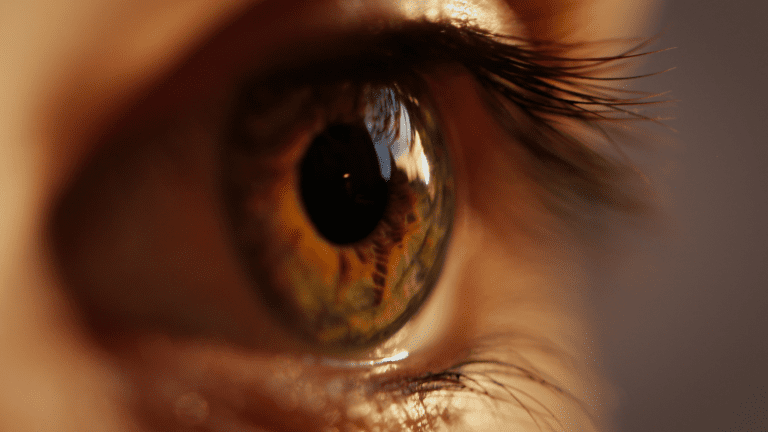
The pupil is the circular opening at the exact center of the iris, the colored part of the eye. In ophthalmology, it is known as the dynamic opening formed by the iris that controls the amount of light entering the eye. Some sources also describe it as the dark space within the iris. However, the black appearance we see is actually caused by the darkness inside the eye. In a sense, it is like looking into a darkened theater through a small opening in the stage curtain. Because the interior appears dark, the pupil also looks black.
This opening is not an actual piece of tissue. It is more like a "window" created by the muscles of the iris. The muscles surrounding the iris contract and relax to enlarge or reduce the pupil. In ophthalmology, examining the pupil's diameter, symmetry, and response speed provides important information about eye health and the function of the brain and spinal nervous system. Pupils that constrict in bright light, for example, indicate a normal reflex. If the expected response does not occur or one pupil is noticeably different from the other, a condition known as anisocoria, an underlying neurological or eye-related problem may need to be considered.
Pupil assessment is frequently referred to as a pupillary examination in ophthalmology. During both routine examinations and emergencies, it provides physicians with clues about many conditions, ranging from changes in intraocular pressure to brain injuries. In short, the pupil is like a "control point" that manages the body's interaction with the outside world through a highly sensitive mechanism while also reflecting the body's state of health.
Where Is the Pupil Located in the Anatomy of the Eye?
The pupil is located in the part of the eye known as the anterior segment, between the transparent cornea and the natural lens. When viewed from the outside, it is the black area at the center of the iris, the colored part of the eye. Anatomically, it has the cornea in front of it and the lens and vitreous behind it. This location is ideal for allowing light to enter the eye and creating a clear image on the retina. Like a ceiling light positioned to illuminate a room effectively, the pupil's central location allows light to pass directly through the eye.
The iris itself is a muscular structure containing circular muscle fibers, known as the sphincter pupillae, and radial muscle fibers, known as the dilator pupillae. The pupil is the opening at the center of these muscles. Its anatomical boundaries are determined by the movement of the iris muscles. This functions in almost the same way in everyone. However, congenital or acquired factors, such as trauma, may sometimes cause slight displacement or deformity of the pupil.
The pupil's fixed anatomical location allows light to reach the eye's optical axis as efficiently as possible. If the center of the pupil were not aligned with the axis of the cornea, for example if the pupil were significantly displaced to one side, light might not be refracted correctly. This could result in focusing problems, blurred vision, or double vision. In summary, the pupil remains centrally positioned and guides light toward the "correct point."
How Does the Pupil Function as the Eye’s Aperture?
The pupil functions as the eye's "aperture." Just as the aperture of a camera regulates the amount of light reaching the sensor, the pupil controls the light reaching the retina. People familiar with cameras know that when the aperture is narrowed, less light enters, but the image becomes sharper and the depth of field increases. When it opens widely, more light enters, but the area in focus becomes narrower and unwanted glare may occur. The eye works in a very similar way.
When light levels are low, the radial muscles within the iris, known as the dilator pupillae, contract and enlarge the pupil. This allows us to see better in low-light conditions. For example, the pupils dilate noticeably when we enter a dim room or go outside at night. Conversely, when we open our eyes in bright sunlight, the circular muscles, known as the sphincter pupillae, contract and constrict the pupil. This prevents excessive light from flooding the sensitive retinal layer at the back of the eye.
Another important aspect of this aperture function is that a constricted pupil reduces optical imperfections. When the pupil becomes smaller, the beam of light entering the eye follows a more central path. This reduces the possibility of light deviating from the correct path and increases visual sharpness. In dim environments, the pupil dilates to allow as much light as possible to reach the retina. In short, the pupil constantly attempts to establish the ideal balance across varying levels of light, from brightness to darkness.
Why Is the Pupil Important for Light to Enter the Eye?
The pupil is critically important because it regulates the amount of light required by the retina. Excessive light may damage retinal cells through long-term wear on the photoreceptors and may also cause immediate glare or dazzling. Insufficient light prevents a clear image from forming and may worsen difficulty seeing in low-light conditions, such as night blindness. The pupil is the primary structure responsible for maintaining this balance.
When we suddenly step outside into daylight, for example, our eyes are dazzled for a few seconds. The pupils then constrict rapidly and reduce this glare. Without this reflex, we would need to squint significantly and could even experience retinal damage over the long term. Similarly, our pupils are dilated while watching a film in a dark cinema. If this mechanism did not activate, too little light would enter the eyes for us to see what was happening on the screen.
The pupil also influences light not only in terms of quantity but also in terms of direction and focus. When it constricts, incoming rays pass through a more central path and create a sharper image on the retina. In daily life, this allows us to see details more clearly in bright environments and minimize glare caused by external factors. All these reasons show that the pupil is not simply a "hole." It is an active regulator that directly affects the quality of vision.
How Do the Pupils Adapt to Changes in Light?
The response of the pupils to changes in light is explained by a mechanism known as the pupillary light reflex. This reflex is a rapid and involuntary nervous system response. Photoreceptor cells in the eye, particularly rod and cone cells as well as specialized ganglion cells containing melanopsin, detect the amount of light and send signals to the brain indicating whether light is present. Relevant centers in the brain, particularly the pretectal nucleus in the midbrain, receive this signal and send instructions to the iris muscles through the oculomotor nerve, the third cranial nerve.
When bright light is detected, this signaling mechanism causes the sphincter pupillae muscles of the iris to contract and the pupil to constrict, known as miosis. In darkness or low-light conditions, the sympathetic pathway becomes active and the dilator pupillae muscles contract, causing the pupil to dilate, known as mydriasis. This occurs within only a few seconds. If the headlights of another vehicle suddenly shine into our eyes while driving at night, for example, our pupils immediately constrict. They dilate again after the headlights pass.
Another interesting feature is that this reflex affects both eyes together. Even if light is directed into only one eye, the pupils of both eyes constrict. This is called the consensual light reflex. It has considerable clinical importance because whether this reflex functions properly provides clues about various health indicators, from nerve conduction within the visual pathways to the function of certain areas of the brain. If this reflex is impaired, for example if one pupil does not constrict in response to bright light while the other responds normally, investigations may be performed because of suspected nerve damage or a biochemical disorder.
What Role Does the Iris Play in Regulating Pupil Size?
The iris is a colored and muscular diaphragm that surrounds the pupil. Its color is mainly determined by genetic factors, resulting in colors such as blue, brown, or green. Its most important feature, however, is the presence of two groups of muscles: circular muscle fibers, known as the sphincter pupillae, and radial muscle fibers, known as the dilator pupillae.
Sphincter Pupillae: This muscle forms a ring near the center of the iris. When stimulated by the parasympathetic system, it contracts and reduces the size of the pupil. The activation of this muscle causes our pupils to become smaller while walking in bright daylight.
Dilator Pupillae: This group of muscles is positioned radially in the outer parts of the iris. When stimulated by the sympathetic nervous system, the muscle fibers dilate the pupil through a movement that effectively "pulls the pupil outward." This muscle becomes active and enlarges the pupil when we enter a dim environment or suddenly experience fear or excitement.
The coordination of these muscle groups allows the pupil to adapt instantly to its environment. The structure of the iris is also important because it must be flexible enough to contract and relax. In some people or certain conditions, such as age-related stiffening of the iris or eye trauma, this flexibility decreases and the pupil cannot demonstrate a complete normal light reflex. This may cause significant problems with visual quality. In summary, the iris works like a gatekeeper and has full control over the amount of light entering the eye. This control is a critical mechanism that both protects eye health and regulates visual clarity.
How Does the Pupil Allow Light to Reach the Retina?
The pupil regulates the passage of light to the retina like a "traffic officer." Light first passes through the cornea and enters the eye. It then passes through the pupil and reaches the lens. The lens refracts the light and helps focus it on the retina. If the pupil is too large, the lens may have difficulty focusing after a large amount of light enters. Excessive light may cause glare on the retina and reduce visual comfort. If the pupil constricts excessively, insufficient light enters and vision becomes poorer in dark environments.
The angle from which light arrives is as important as its quantity. Rays passing close to the center are focused more clearly on the retina, while rays entering from the edges may cause scattering or refractive imperfections. The pupil's constriction in bright conditions therefore does more than reduce excessive light. It also improves image quality by allowing more centrally positioned rays to pass through. The optical precision of the eye is made possible in part by this "central passage."
Dilation and constriction of the pupil also support the adaptation of the retinal photoreceptors, the rod and cone cells. In dim conditions, for example, rod cells become active, and a dilated pupil makes it easier for enough photons to reach them. In bright environments, cone cells need to process large numbers of photons. A constricted pupil prevents them from being exposed to an unnecessarily intense amount of light. All these processes are managed through highly coordinated, immediate reflexes and neural feedback.
What Triggers the Pupil to Dilate or Constrict?
The dilation and constriction of the pupil are primarily triggered by the autonomic nervous system. This system has two main divisions: the sympathetic and parasympathetic systems. Bright light activates the parasympathetic system to constrict the pupil. Conversely, low light levels or sudden emotions such as excitement or fear activate the sympathetic system and dilate the pupil.
Not only light and emotional states but also various medications can change pupil diameter. Medications such as atropine, which block parasympathetic nerve transmission, dilate the pupil and cause mydriasis. Ophthalmologists therefore use these types of drops to dilate the pupil temporarily during a fundus examination. Medications such as pilocarpine, on the other hand, constrict the pupil and cause miosis. They are used as treatment in certain conditions involving elevated intraocular pressure, particularly glaucoma.
Lesions in certain areas of the brain and nerve damage may also affect pupil diameter. Damage to the brainstem, for example, may stop or delay the pupil's response to light. Certain systemic diseases, such as diabetes, may cause long-term nerve damage and weaken pupillary reflexes. Powerful painkillers, narcotics, or stimulant substances such as morphine, cocaine, and amphetamines may also cause the pupils to constrict or dilate abnormally. All these factors show how many different influences control the pupillary response and reveal that, despite appearing simple, the pupil is part of a highly complex system.
Which Eye Conditions Are Associated with Pupil Abnormalities?
Pupil abnormalities may indicate many different diseases. Some are directly related to the eye, while others are associated with systemic or neurological problems:
Anisocoria (Pupils of Different Sizes): A slight difference in size between the two pupils may normally be present. However, marked asymmetry may indicate conditions such as Horner syndrome, which involves damage to the sympathetic nerve pathway, third cranial nerve palsy, or iris damage following trauma.
Adie Syndrome (Holmes-Adie Syndrome): This condition is generally characterized by a large pupil in one eye that responds poorly to light. Reduced deep tendon reflexes may also occur. It may be more common in young adults and generally involves neurological dysfunction whose exact cause is not fully understood.
Argyll Robertson Pupil: The response to light is weak or absent, but the pupil constricts when focusing at near, known as accommodation. It is a classic finding particularly associated with neurosyphilis, although it may also occur in other conditions such as diabetes.
Third Cranial Nerve Palsy: This nerve carries the parasympathetic fibers responsible for eye movements and pupil constriction. When it is paralyzed, the pupil dilates and does not respond to light. Abnormalities of eye movement are also observed.
Iritis (Anterior Uveitis): This is inflammation of the iris. It may develop after eye trauma or as a result of autoimmune diseases or infections. Adhesions may form around the edges of the pupil, its shape may become irregular, and severe pain in response to light may occur.
Glaucoma: Particularly during acute angle-closure glaucoma, the pupil may be moderately dilated, fixed, and painful. The person may present to hospital with sudden vision loss, nausea, and vomiting.
Traumatic Mydriasis: Trauma to the eye or head may damage the iris muscles and cause temporary or permanent dilation. Similar conditions may also occur during surgery.
How Does the Pupillary Reflex Protect the Retina from Damage?
The pupillary reflex protects the retina from exposure to excessive light during periods of intense illumination. When we look at a bright source, such as sunlight, the pupil constricts within fractions of a second. This prevents excessive light from reaching the retina and reduces the risk of damage to photoreceptor cells. Understanding how this reflex works demonstrates the highly effective protective mechanisms developed by the body.
This protective function is more complex than a simple instruction to "constrict when light is present." Light-sensitive retinal ganglion cells, particularly those containing melanopsin, continuously send signals to the brain. In response to these signals, the brain activates the parasympathetic system and instructs the sphincter muscles of the iris to contract. This happens not only in bright light but also during sudden bursts of light, such as a camera flash. The reflex also activates rapidly when we move suddenly from a dark environment into daylight and experience temporary glare.
Protecting the retina from damage does not involve only preventing long-term light damage. The reflex also prevents immediate excessive brightness and glare, providing clearer and more comfortable vision during daily life. It is particularly important in snowy environments with high levels of reflection or by the sea under intense reflected sunlight. If this reflex mechanism is impaired, the person loses visual comfort and may also face a long-term risk of retinal damage.










































