
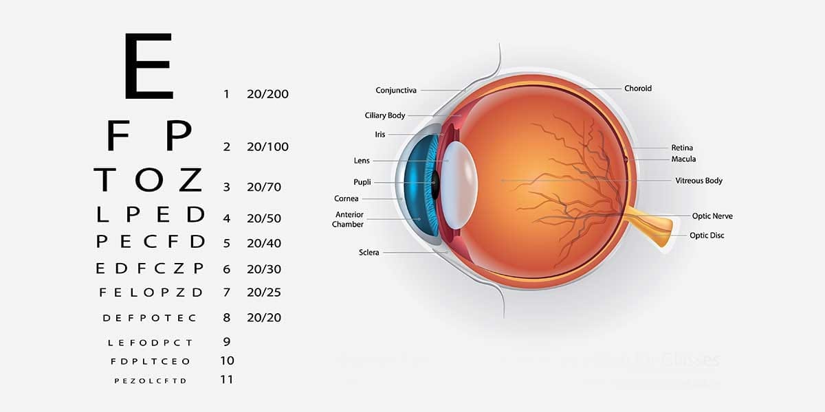
Diabetic retinopathy is an eye disease that occurs when long-term or uncontrolled diabetes damages the blood vessels of the retina at the back of the eye. Diabetic retinopathy may progress without symptoms at first and can eventually cause vascular leakage, bleeding inside the eye or swelling at the centre of vision. Commonly described as diabetes affecting the eyes, this condition can occur in people with both type 1 and type 2 diabetes. The treatment approach is determined according to the stage of the disease, the level of vision and the nature of the retinal damage. Control of blood glucose, blood pressure and cholesterol forms an important part of the treatment plan at every stage. Intravitreal medication injections, retinal laser treatment or vitrectomy surgery are considered only when medically necessary. Early diagnosis and regular follow-up may help reduce the risk of permanent vision loss.
What Causes Diabetic Retinopathy?
Diabetic retinopathy develops when high blood glucose levels damage the structure of the small blood vessels in the retina over time. When the vessel walls weaken, small dilations, haemorrhages and fluid leakage may occur. Blockage of some vessels prevents the retinal tissue from receiving sufficient oxygen. As oxygen deprivation increases, the eye begins to produce certain substances that stimulate the formation of new blood vessels. These new vessels are more fragile than healthy vessels and can bleed easily. When vascular leakage affects the centre of vision, it may cause central vision loss due to macular oedema. High blood pressure, kidney disease and cholesterol problems may contribute to the acceleration of this process. Retinal damage does not progress at the same rate in every patient, and individual risks may differ.
Risk Factors for Diabetic Retinopathy
The likelihood of damage to the retinal blood vessels increases as the duration of diabetes becomes longer. Blood glucose levels remaining above the target range for an extended period are an important risk factor. High blood pressure and abnormal blood lipid levels may also negatively affect the vascular structure of the retina. Diabetic kidney disease, anaemia and certain systemic conditions may require closer monitoring of the disease. Pregnancy may accelerate the progression of existing retinopathy, particularly in people who had diabetes before pregnancy. Smoking may impair overall vascular health and make eye-related complications more difficult to control. Rapid changes in blood glucose treatment may be associated with temporary worsening in some patients with advanced retinopathy. However, the presence of a single risk factor does not mean that a person will necessarily experience vision loss.
What Are the Symptoms of Diabetic Retinopathy?
Diabetic retinopathy may not cause pain or noticeable visual symptoms in its early stages. Normal vision does not definitively indicate that there is no damage to the retinal blood vessels. As the disease progresses, blurred vision, fluctuating vision or difficulty reading may occur. Black floaters, thread-like shadows or dark spots in the field of vision may be associated with bleeding inside the eye. People with macular oedema may have difficulty recognising faces, reading text and seeing details. Some patients may perceive colours as more faded or notice changes in night vision. A sudden reduction in vision, flashes of light or a curtain-like shadow may require urgent assessment. The severity of symptoms does not always correspond directly to the extent of retinal damage.
What Are the Stages of Diabetic Retinopathy?
Diabetic retinopathy is generally assessed in two main stages, non-proliferative and proliferative. In the non-proliferative stage, small dilations, haemorrhages and fluid leakage occur in the retinal blood vessels. As vascular blockages increase, this stage may progress from mild to moderate or severe. In the proliferative stage, retinal tissue deprived of oxygen triggers the formation of new and abnormal blood vessels. Bleeding from the new vessels or traction forming around them may cause more serious visual problems. Diabetic macular oedema may occur at different stages of the disease, not only in its advanced stage. Staging is performed using a fundus examination and necessary imaging methods. Determining the stage of the disease is important for establishing the frequency of follow-up and the treatment to be applied.
What Is Non-Proliferative Diabetic Retinopathy?
Non-proliferative diabetic retinopathy refers to early or intermediate retinal damage in which abnormal new blood vessels have not yet formed. Microaneurysms, small retinal haemorrhages and deposits caused by vascular leakage may be seen during examination. Some patients may also develop white spots indicating that the retinal tissue is not receiving adequate nourishment. Intravitreal injections or laser treatment may not be immediately necessary in mild cases. However, the treatment plan may change if macular oedema develops or the disease progresses to a severe stage. Regulating blood glucose, blood pressure and cholesterol control is particularly important at this stage. Regular retinal examinations should not be neglected simply because there are no symptoms in the eye. The rate of disease progression varies according to the duration of diabetes and other systemic risk factors.
What Is Proliferative Diabetic Retinopathy?
Proliferative diabetic retinopathy is the advanced stage in which abnormal new blood vessels develop on the retina or around the optic nerve. Because these vessels are structurally fragile, they may bleed into the gel-like tissue inside the eye. Connective tissues accompanying the new vessels may contract over time and pull on the retina. This traction may lead to tractional retinal detachment and seriously affect vision. In some patients, new vessels may form in the front of the eye and cause intraocular pressure to rise. Panretinal laser treatment, intravitreal anti-VEGF injections or vitrectomy when necessary may be considered. The method selected is determined according to the patient's ability to attend follow-up and the complications that have developed in the retina. Regular monitoring is important because proliferative disease does not always begin with sudden vision loss.
What Is Diabetic Macular Oedema?
Diabetic macular oedema occurs when fluid leaking from the retinal blood vessels accumulates at the centre of vision. The macula is responsible for central visual functions such as reading, recognising faces and distinguishing details. Swelling in this area may cause images to appear blurred or distorted. Macular oedema may occur during both the non-proliferative and proliferative stages of diabetic retinopathy. Optical coherence tomography helps assess the location of the oedema and changes in retinal thickness. The need for treatment is determined according to whether the oedema involves the centre of vision and the person's level of vision. Intravitreal anti-VEGF treatments, corticosteroid treatments in selected patients or focal laser treatment may be considered. For some patients whose vision is preserved, close monitoring may be more appropriate than immediate intervention.
How Does Bleeding Inside the Eye Develop?
The new blood vessels that develop in diabetic retinopathy have thin walls and can therefore be damaged easily. When these vessels bleed, blood may accumulate in the vitreous gel that fills the eye. Bleeding inside the eye caused by diabetes may be noticed as sudden black spots or severe blurred vision. Mild bleeding may cause floating shadows in the vision, while heavy bleeding may cause a marked reduction in vision. Spontaneous clearing of the blood does not always mean that the underlying vascular problem has resolved. If the retina cannot be seen adequately during the examination, ultrasonography may be used to investigate whether retinal detachment is present. Laser treatment, intravitreal injections or vitrectomy for persistent bleeding may be considered. When sudden vision loss develops, it is important for the condition to be assessed without delay.
How Is Diabetic Retinopathy Diagnosed?
To diagnose diabetic retinopathy, the pupil is dilated with eye drops and the retina is examined in detail. During the fundus examination, vascular dilations, haemorrhages, oedema and the formation of new blood vessels are investigated. Visual acuity testing helps assess the extent to which the centre of vision has been affected by the disease. Intraocular pressure is measured to check for glaucoma or other pressure-related problems. Retinal photographs allow current findings to be recorded and compared with subsequent examinations. Optical coherence tomography is used particularly to assess macular oedema and the retinal layers. In patients for whom it is necessary, fluorescein angiography or OCT angiography may be used to examine the vascular structure in greater detail. The duration of diabetes, medications used and accompanying systemic diseases also form part of the diagnostic assessment.
OCT and Retinal Angiography
Optical coherence tomography is an examination method that produces cross-sectional images of the retinal layers. This test helps assess fluid accumulation in the macula and changes in retinal thickness. OCT measurements may also be used to monitor the response to treatment after intravitreal injections or laser treatment. In fluorescein angiography, leakage and impaired blood supply in the retinal vessels are examined using a dye injected into a vein. Some patients may experience short-term nausea during angiography or, rarely, an allergic reaction to the dye. OCT angiography is an imaging method that can assess certain vascular structures without the use of dye. However, angiography is not necessary for every patient, and no test can replace an examination on its own. The imaging method to be used is determined according to the stage of the disease and the treatment decision that needs to be made.
Use of Artificial Intelligence in Retinal Screening
Artificial intelligence-assisted systems may help screen retinal photographs for certain diabetes-related changes. These systems may be used particularly to support the identification of high-risk images within large groups. Microaneurysms, haemorrhages and pronounced retinal abnormalities may undergo preliminary assessment by certain software. However, poor image quality or the presence of other eye diseases may affect the results. Artificial intelligence assessment does not replace a detailed fundus examination and the necessary imaging methods. Treatment decisions should not be based solely on automated image analysis. Macular oedema, intraocular pressure and the peripheral regions of the retina should also be assessed. When used under appropriate conditions, digital screening is a supportive tool for regular examination planning.
How Is Diabetic Retinopathy Treatment Planned?
Diabetic retinopathy treatment is planned according to the stage of the disease, the presence of macular oedema and complications such as bleeding inside the eye. In mild stages, regular follow-up and control of systemic risk factors may be sufficient. Intravitreal medication injections may be considered when oedema affecting the centre of vision develops. For proliferative disease, panretinal laser treatment, anti-VEGF treatment or a combination of these methods may be considered. Vitrectomy may be necessary if vitreous haemorrhage persists for a long time or if the retina has detached due to traction. The aim of treatment is to limit the progression of retinal damage and preserve existing vision as much as possible. It is not possible to reverse established damage completely in every patient. The person's general health, possibility of pregnancy and ability to attend regular follow-up are also taken into account when selecting treatment.
Why Is Blood Glucose Control Important?
Balancing blood glucose levels may help reduce the risk of new damage developing in the retinal blood vessels. HbA1c measurement provides information about average blood glucose control over a specified period. However, the same HbA1c target is not suitable for every person with diabetes. Age, accompanying diseases, pregnancy and the risk of low blood glucose affect the determination of individual targets. Controlling blood pressure and cholesterol may be as important as blood glucose control in protecting eye health. Regular diabetes treatment does not always eliminate the need for intravitreal injections or laser treatment. Likewise, retinal treatment does not replace systemic diabetes management. Assessing eye findings together with general health monitoring is important for long-term control.
What Happens If Blood Glucose Is Lowered Rapidly?
A marked reduction over a short period in blood glucose levels that have remained high for a long time may be associated with temporary worsening of retinopathy in some patients. This situation should be monitored more closely, particularly in people who already have advanced retinal damage. Eye findings may be reassessed while insulin treatment is intensified or potent glucose-lowering treatments are used. However, this possibility does not mean that diabetes treatment should be discontinued or blood glucose should be kept high. The main aim is to plan metabolic improvement appropriately for the individual and to monitor the retina regularly. Blurred vision may not always result from permanent retinal damage, and temporary focusing problems related to changes in blood glucose may also occur. If new visual symptoms develop, macular oedema, haemorrhage and other possibilities should be investigated separately. Changes to diabetes treatment should not be made on the person's own initiative.
Intravitreal Injection Treatment
Intravitreal anti-VEGF injections aim to reduce the effects of certain substances that increase vascular leakage and abnormal new blood vessel formation in the retina. This treatment may be considered particularly for diabetic macular oedema affecting the centre of vision. It may also be an alternative or supportive option to laser treatment in suitable patients with proliferative diabetic retinopathy. Since the effects of injections are not permanent, repeated applications may be required at specified intervals. Treatment frequency is determined according to OCT findings, the level of vision and the retinal response. Oedema decreases in some people, while a different treatment approach may be necessary to achieve an adequate response in others. Interruptions in regular follow-up may cause the disease to progress again, particularly in cases with advanced new blood vessel formation. Injection treatment does not guarantee complete restoration of vision in every patient.
How Are Anti-VEGF Injections Administered?
Before an intravitreal injection, the ocular surface is cleaned with suitable antiseptics. Anaesthetic eye drops or appropriate local anaesthesia methods are used to reduce discomfort. The medication is injected into the vitreous cavity inside the eye with a fine needle. Short-term stinging, watering or a small haemorrhage on the ocular surface may occur after the procedure. Some people may temporarily notice floaters or small air bubbles. Vision should not be expected to improve immediately on the same day after the procedure. If severe pain, increasing redness or rapidly worsening vision develops, an assessment should be carried out without delay because of the possibility of infection. The number of injections cannot be determined in the same way in advance for every patient.
Intravitreal Corticosteroid Treatments
Intravitreal injections or implants containing corticosteroids may be considered in selected patients with diabetic macular oedema. These treatments may help reduce inflammation and vascular leakage in the retina. An inadequate response to anti-VEGF treatment may require consideration of a different treatment option. Corticosteroid treatment may be considered a more suitable option for some patients who have undergone cataract surgery. However, these medications may increase intraocular pressure and accelerate cataract formation in people who still have their natural lens. The risks should be assessed more carefully in patients with glaucoma or sensitivity to corticosteroids. The duration of effect of the implants and the need for repeat treatment vary according to individual findings. Corticosteroid treatment is not necessary for every patient with diabetic retinopathy.
Laser Treatment for Diabetic Retinopathy
Retinal laser treatment is a method used for certain types of diabetes-related vascular problems. Depending on the stage of the disease, the aim is to control abnormal new blood vessel formation or vascular leakage. In proliferative retinopathy, a wider-area laser treatment known as panretinal photocoagulation may be applied. For limited leakage around the macula, focal or grid laser treatment may be considered in selected patients. Retinal laser treatment is not the same procedure as corneal laser treatment performed to correct an eye prescription. Complete restoration of lost vision is not always expected, and the primary aim is to reduce progressive damage. Treatment is planned according to the visibility of the retina and the presence of accompanying macular oedema. Some patients may require intravitreal injections or surgical treatment together with laser treatment.
What Is Panretinal Laser Treatment?
Panretinal photocoagulation is a laser treatment applied to the peripheral areas of the retina in proliferative diabetic retinopathy. It aims to reduce the oxygen demand in these areas and the signals that stimulate new blood vessel formation. Treatment may help reduce the risk of bleeding and traction caused by the fragile vessels that have developed. It may be planned as a single session or in multiple sessions according to the patient's condition. During the procedure, the ocular surface is numbed with eye drops and the retina is assessed through a special lens. Temporary blurred vision, sensitivity or changes in peripheral vision may occur in some patients after laser treatment. Reduced night vision and a temporary increase in macular oedema are also among the possibilities considered. Suitability for panretinal laser treatment is determined according to the extent of new blood vessel formation and the presence of bleeding inside the eye.
Focal Laser Treatment for Macular Oedema
Focal laser treatment may be used to target vascular changes causing leakage in specific areas of the retina. The grid laser approach is considered in suitable patients for treating specific areas around the oedematous region. Intravitreal anti-VEGF treatments are now frequently among the preferred options for macular oedema affecting the centre of vision. However, not every patient with oedema requires immediate injections or laser treatment. The distance of the oedema from the centre of vision and visual acuity are important when selecting treatment. In some cases, focal laser treatment may be planned as a supportive treatment alongside injections. The treatment area should be determined carefully to avoid damaging the centre of the macula. The effect of treatment varies according to the damage to the retinal structure and how long the oedema has persisted.
When Is Vitrectomy Surgery Necessary?
Vitrectomy is retinal surgery in which the vitreous gel inside the eye and, when necessary, tissues causing bleeding or traction are removed. Persistent vitreous haemorrhage may prevent the retina from being seen and laser treatment from being applied. Surgical assessment may also be required when membranes are pulling the retina away from its position. Surgery may be considered particularly for tractional retinal detachment that threatens the centre of vision. During the procedure, retinal laser treatment may be applied or gas or silicone oil may be placed inside the eye if considered necessary. Vitrectomy does not eliminate diabetes itself or all vascular damage in the retina. The visual outcome depends on the duration of the haemorrhage, the condition of the macula and accompanying eye diseases. Whether surgery is required is determined by assessing the person's general health together with the retinal findings.
How Does Retinal Detachment Develop?
In advanced diabetic retinopathy, hardened connective tissues may form around abnormal blood vessels. Over time, these tissues may contract and pull the retina away from the underlying layers. Detachment caused by traction is known as tractional retinal detachment. A curtain, dark shadow or marked reduction in vision may develop in the field of vision. If the macula is affected, central vision may be damaged more seriously. Sometimes a retinal tear may accompany the traction, making the condition more complex. During vitrectomy, the membranes causing traction are removed with the aim of returning the retina to its appropriate position. The outcome may vary according to the duration of the detachment and the extent of damage to the centre of vision.
What Is Neovascular Glaucoma?
Neovascular glaucoma may develop when abnormal blood vessels caused by diabetic retinopathy also form in the front part of the eye. These vessels may block the natural drainage pathway of the fluid inside the eye and increase pressure. Symptoms such as eye pain, redness, headache and reduced vision may occur. However, in some patients, the rise in pressure may initially progress without causing noticeable symptoms. Medications that lower intraocular pressure, anti-VEGF injections and retinal laser treatment may be considered together. In necessary cases, surgical methods may also be considered to control intraocular pressure. Using pressure-lowering eye drops alone does not always resolve the problem of new blood vessel formation originating from the retina. The retinal disease and intraocular pressure should be monitored together to protect the optic nerve.
In Which Situations Is Treatment Postponed?
The suitability of each method for treating diabetic retinopathy is determined according to the person's eye condition and general health. If an active eye infection is present, intravitreal injections or planned surgery may initially be postponed. In people who have recently experienced a serious cardiovascular event, the possible risks of anti-VEGF treatment should be assessed individually. The use of intravitreal medication during pregnancy is planned by considering the potential benefits and risks for the mother and baby. Corticosteroid treatments may be unsuitable for patients with uncontrolled glaucoma because of their effects on intraocular pressure. Dense bleeding inside the eye or a pronounced cataract may make laser treatment difficult because the retina cannot be seen. Serious systemic diseases may affect the timing of surgery that requires anaesthesia. None of these situations should be interpreted as meaning that treatment can never be performed in every patient.
Possible Side Effects of Treatment
Short-term stinging, watering or a small haemorrhage on the white part of the eye may occur after intravitreal injections. In rare cases, intraocular infection, pronounced inflammation or changes in intraocular pressure may occur. Corticosteroid injections and implants may accelerate cataract development or increase the risk of glaucoma. Temporary blurred vision, sensitivity and changes in peripheral vision in some patients may occur after retinal laser treatment. Panretinal laser treatment may reduce night vision or cause a temporary increase in macular oedema. Recurrent bleeding, infection, retinal tears and intraocular pressure problems may develop after vitrectomy. Gas or silicone used during surgery may alter the postoperative care plan. The frequency and severity of side effects vary according to the selected treatment, the stage of the disease and the person's eye structure.
Recovery After Treatment
The recovery process after diabetic retinopathy treatment varies according to the method used. Some people can return to their daily activities shortly after an intravitreal injection. Vision may temporarily become blurred after laser treatment, and the retinal response is assessed over time. Recovery after vitrectomy may take longer depending on the extent of bleeding inside the eye and the condition of the retina. Macular oedema may not decrease immediately, and multiple treatments may be required. If gas has been placed inside the eye, a specific head position may be recommended. While gas remains in the eye, individual medical advice should be followed because air travel or travel to high altitudes may cause serious pressure changes. The degree of recovery may vary according to damage already present in the macula or optic nerve before treatment.
What Should Be Considered After Treatment?
The recommended eye drops and follow-up plan should be followed regularly after treatment. It is important not to rub the eye, touch it with dirty hands or expose it to impact. Increasing pain, severe redness or reduced vision after an injection or surgery should be assessed without delay. If vitrectomy has been performed, recommendations regarding heavy lifting, swimming and intensive physical activities should be followed. Management of blood glucose, blood pressure and cholesterol should continue after eye treatment. Temporary blurred vision does not always mean that treatment has failed. However, new floaters, flashes of light or a curtain-like shadow should be taken seriously. Follow-up intervals are arranged according to retinal healing and the control of new blood vessel formation.
How Often Should Eye Examinations Be Performed?
The frequency of eye examinations in people with diabetes is determined according to the type of diabetes and the retinal findings. Because type 2 diabetes may be present for a long time before diagnosis, the first comprehensive eye assessment should be planned at the time of diagnosis. In type 1 diabetes, the timing of the first examination is determined by considering the onset of the disease, age and additional risks. The interval between examinations may be longer in people who have no retinal findings and whose metabolic values are stable. More frequent follow-up may be required when diabetic retinopathy, macular oedema or new blood vessel formation is detected. Pregnancy, rapid changes in blood glucose and kidney disease also affect the frequency of examinations. A normal retinal photograph does not always replace the necessary comprehensive examination. If a sudden change in vision occurs, the scheduled examination date should not be awaited.
Diabetic Retinopathy During Pregnancy
Retinopathy may progress during pregnancy in people who had type 1 or type 2 diabetes before becoming pregnant. It is therefore important for a retinal assessment to be performed before a planned pregnancy or during early pregnancy whenever possible. The severity of existing retinopathy, changes in blood pressure and blood glucose control affect the frequency of follow-up. When necessary, retinal examinations may continue throughout pregnancy and after delivery. Gestational diabetes that develops during pregnancy and is not associated with pre-existing diabetes generally does not require the same retinopathy screening approach. However, the assessment plan may change if previously undiagnosed diabetes, visual symptoms or other eye problems are present. Laser treatment may be considered for some patients during pregnancy, while intravitreal anti-VEGF treatment requires careful assessment of the benefits and risks. Pregnancy does not mean that sight-threatening retinal diseases must necessarily be left untreated.
Effects of Nutrition and Exercise on Treatment
A balanced diet may help blood glucose levels remain more stable and support overall vascular health. Vegetables, whole grains, suitable protein sources and moderate carbohydrate consumption should be considered within an individual diabetes plan. No single vitamin, herbal product or dietary supplement treats diabetic retinopathy. Omega-3 or other supplements should not be considered substitutes for intravitreal injections or laser treatment. Regular physical activity may contribute to the control of blood glucose, blood pressure and body weight. However, heavy lifting and high-intensity exercise should be assessed separately in people with advanced proliferative retinopathy or recent bleeding inside the eye. Stopping smoking helps protect vascular health and reduce additional risks. The nutrition and exercise plan should be arranged according to the general status of diabetes and the existing eye findings.
Does Diabetes Affect Cataracts and Glaucoma?
Diabetes can affect not only the retinal blood vessels but also other structures of the eye. Cataracts, which occur when the natural lens of the eye loses its transparency, may develop earlier in some people with diabetes. Diabetic macular oedema and existing retinal damage should be assessed separately when cataract surgery is planned. Increased intraocular pressure and the risk of glaucoma are also among the problems that should be considered in patients with diabetes. In advanced retinopathy, abnormal new blood vessel formation may lead to neovascular glaucoma. Changes in blood glucose may temporarily affect the focusing ability of the natural lens and cause fluctuations in the eye prescription. For this reason, not every instance of blurred vision in a person with diabetes directly indicates retinopathy. The cause of the visual change should be determined by assessing the cornea, natural lens, retina and optic nerve together.
Why Does Vision Not Improve Despite Treatment?
There may be different reasons why vision does not improve to the expected level after diabetic retinopathy treatment. When macular oedema persists for a long time, the nerve cells at the centre of vision may be permanently damaged. Severe blockage of the retinal blood vessels may limit visual improvement even if the oedema decreases. Recurrent bleeding inside the eye or retinal damage caused by traction may make recovery more difficult. Cataracts, glaucoma and optic nerve diseases may also affect the outcome. In some patients, retinal disease may progress again when systemic blood glucose and blood pressure problems are not controlled. Irregular administration of intravitreal injections or missed follow-up appointments also affects the treatment response. In this situation, retinal imaging, intraocular pressure and other possible causes should be reassessed together.
Does Diabetic Retinopathy Resolve Completely?
The course of diabetic retinopathy varies according to the extent of damage to the retinal blood vessels and diabetes management. Some changes detected at an early stage may follow a more stable course when systemic control improves. Macular oedema may decrease with intravitreal injections or other suitable treatments. Nerve tissue damage, retinal detachment or dense scarring that has developed at an advanced stage cannot always be reversed completely. Laser treatment and vitrectomy do not definitively prevent diabetes from causing further problems in the retina. Regular follow-up may therefore remain necessary even if symptoms decrease. The aim of treatment is to preserve existing vision and limit progressive damage as much as possible. It is not medically appropriate to guarantee complete recovery or a permanent result for every patient.
How Can Diabetic Retinopathy Be Prevented?
Keeping blood glucose, blood pressure and cholesterol levels within individual target ranges is important in reducing the risk of diabetic retinopathy. Regular fundus examinations may help detect retinal changes before symptoms occur. Taking diabetes medication as recommended and attending follow-up appointments support systemic control. Stopping smoking, maintaining a balanced diet and engaging in regular physical activity are beneficial for overall vascular health. Retinal assessment may become particularly important for people planning a pregnancy or undergoing significant changes in diabetes treatment. Good blood glucose control alone does not guarantee that retinopathy will never develop. Likewise, vitamin supplements, sunglasses or a particular diet alone are not definitive methods of preventing the disease. The risk-reduction approach is based on combining regular eye examinations with individual diabetes management.
Diabetic Retinopathy Treatment in Izmir
When diabetic retinopathy treatment is planned in Izmir, the stage of the disease, the level of vision and vascular changes in the retina are assessed together. A fundus examination, OCT and retinal angiography when necessary help determine the choice of treatment. Intravitreal injections or different treatment options may be considered for suitable patients with diabetic macular oedema. In proliferative retinopathy, retinal laser treatment and intravitreal medication are planned according to individual findings. Vitrectomy surgery may be considered if bleeding inside the eye or tractional retinal detachment develops. For people researching treatment for diabetes-related eye disease in Izmir and the Konak area, a detailed assessment of the retinal disease is important. Control of blood glucose, blood pressure and other systemic risks is an integral part of eye treatment. The appropriate approach is determined by taking into account the patient's previous treatments and follow-up requirements.
Diabetic Retinopathy Treatment Prices
Diabetic retinopathy treatment prices may vary according to the stage of the disease and the method that needs to be applied. Intravitreal injections, retinal laser treatment and vitrectomy surgery require different assessment and follow-up processes. The extent of macular oedema, bleeding inside the eye and whether one or both eyes are affected may change the treatment plan. OCT, retinal angiography and other examinations are planned according to individual needs. A single procedure may be sufficient for some patients, while repeated treatments may be necessary in other situations. The scope of treatment or individual pricing cannot be determined without an examination and imaging. Pricing information can be provided after the condition of the disease and the appropriate treatment method have been clarified. You can contact us through the website to obtain information about diabetic retinopathy assessment and treatment options suitable for you.




































