
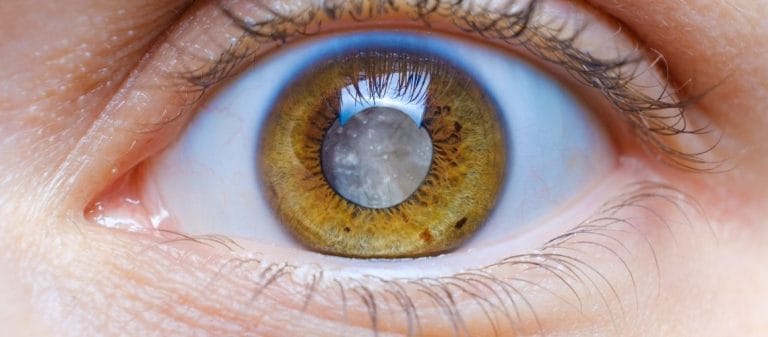
A cataract is a condition that develops when the eye's natural lens loses its transparency and can be classified into different types. Nuclear, cortical and subcapsular cataracts are among the most common types of cataracts. Each type has a different effect on visual quality and may progress at a different rate.
A nuclear cataract develops in the center of the eye's lens and particularly affects distance vision. A cortical cataract develops in the outer layer of the lens and produces white, wedge-shaped opacities. A subcapsular cataract may progress more rapidly and increase sensitivity to light.
Congenital cataracts are a specific type found in infants and may develop due to genetic factors or infections. Traumatic cataracts may occur following eye injuries. These types can be managed more effectively when diagnosed early.
Correctly identifying the type of cataract is highly important when planning treatment. An ophthalmologist's examination and detailed evaluation help determine the appropriate timing of surgery and lens selection.
There are several main types of cataracts: nuclear sclerotic, cortical and posterior subcapsular cataracts. These types differ according to the part of the eye's natural lens in which they develop. The lens is a small but extremely important structure inside the eye. Like the lens of a camera, it refracts light and helps focus it onto the retina. A loss of transparency in any part of the lens can affect vision in different ways and to varying degrees.
- A nuclear sclerotic cataract develops in the very center of the lens, in the area known as the nucleus. It causes the lens to harden and turn yellow over time.
- A cortical cataract begins at the outer edges of the lens, in the cortex, and usually creates whitish lines extending toward the center that resemble wedges or the teeth of a cogwheel.
- A posterior subcapsular cataract develops at the back of the lens, close to its capsule. It is particularly common in younger patients, people who use steroids and those with certain systemic conditions. It usually progresses rapidly.
These differences affect how quickly cataracts progress and which symptoms they cause. A nuclear sclerotic cataract, for example, may progress slowly over several years as part of the aging process, while a posterior subcapsular cataract may cause noticeable vision loss within a much shorter time. A cortical cataract generally progresses from the edges toward the center. The type of visual difficulty experienced in daily life varies because of these differences in the location of the cataract.
A nuclear sclerotic cataract can be compared to an egg yolk becoming harder and changing color. The term "nuclear" refers to the nucleus at the center of the lens. Over time, this nucleus becomes yellow and hard, making the passage and refraction of light more complex.
This type of cataract generally progresses slowly. During its early stages, mild blurred vision and sensitivity to oncoming headlights, particularly when driving at night, may be noticed. The hardening of the lens may initially make near vision clearer. This condition is commonly known as "second sight." A person may temporarily believe that their need for glasses has decreased, but this is not permanent. As the lens continues to harden and turn yellow, noticeable difficulty with both near and distance vision gradually develops. Colors may also appear faded or acquire a yellowish tone over time.
A nuclear cataract can feel like looking at faded, yellowing pictures in an old photo album. Although the texture and memories of the album may remain valuable, it becomes increasingly difficult to see fresh and vivid colors. Nuclear sclerotic cataracts are the most common type in older adults. Since they progress slowly, they can be monitored easily through regular eye examinations. Surgical treatment is considered when significant visual impairment develops.
A cortical cataract takes its name from the outer layer of the lens, known as the cortex. This part surrounds the lens nucleus like a shell. A disruption in the balance of water and proteins within the cortical layer produces white, opaque lines or wedge-shaped areas in the lens. These lines may resemble the cut sections of a slice of cake. The opaque areas initially appear small and peripheral, but extend toward the center as they progress and reduce visual quality.
In terms of its effect on daily life, a cortical cataract usually manifests through reflections and glare. Common complaints, particularly when driving at night, include rays of light, glare or light scatter around oncoming headlights. Dazzling in very bright environments is also common. Although cortical cataracts may develop as a natural part of aging, they may occur earlier in people with diabetes or those exposed to ultraviolet light for extended periods.
A cortical cataract causes cloudiness that progresses toward the center as though glue had been applied to the edges of a sheet of glass. At first, it may appear as an insignificant spot or thin line that is easy to overlook. Over time, however, it becomes more noticeable and causes significant visual impairment. The process can still be monitored closely through regular eye examinations and corrected surgically when necessary.
A posterior subcapsular cataract (PSC) develops at the back of the lens, close to the capsule. Sometimes the most critical part of a room is its back wall. For light to fall clearly onto the retina, this back wall must also be clean and smooth. In PSC, transparency is lost at precisely this point.
This type of cataract generally progresses more rapidly and can cause noticeable vision loss even during its early stages. Difficulty reading, sensitivity to light, particularly in bright sunlight, and impaired night vision are characteristic symptoms of PSC. Some patients report seeing a hazy veil in front of their eyes or halos around light sources.
The rapid progression of a posterior subcapsular cataract may interfere with daily activities within a short time. Activities such as climbing stairs, working at a computer, reading a book or driving at night may become difficult because of blurred vision and glare. Factors such as steroid use at a young age, diabetes and certain inflammatory eye diseases may trigger this type of cataract. When diagnosed early, clear vision may be restored with cataract surgery performed at the appropriate time.
As its name suggests, a congenital cataract is present at birth and may be hereditary or developmental. Although the risk of cataracts is low for most infants, some babies are unfortunately born with this problem or develop it during the early stages of life. Since vision develops rapidly during infancy and childhood, early diagnosis of a congenital cataract is critically important. If an infant cannot see clearly, the visual pathways cannot establish sufficient connections with the brain and amblyopia may develop.
A congenital cataract may affect one or both eyes. Genetic factors, infections during pregnancy and other health problems frequently play a role. If other members of the family have experienced a similar condition, for example, the likelihood of congenital cataracts in the infant increases. Routine eye examinations during the newborn period are therefore vitally important. Early diagnosis allows surgical intervention or other treatment approaches to support the development of normal vision during these critical early years.
A traumatic cataract develops following physical damage to the eye. This damage may result from a direct blow, an injury involving a sharp or penetrating object, or even exposure to chemicals or radiation. The eye's lens is surrounded by a thin and delicate capsule. If the integrity of this capsule is disrupted, the proteins inside the lens may deteriorate and the clouding process may begin.
A strong blow to the eye while playing basketball or a glass fragment entering the eye at home may cause damage severe enough to produce a traumatic cataract. A traumatic cataract does not always develop immediately. The effects of the accident may appear weeks or months later. An eye examination is therefore essential after any eye trauma. The injury may also cause the lens to move out of position, known as luxation, or lead to secondary problems that increase intraocular pressure. Rapid and appropriate intervention is highly important for protecting eye health in such cases.
Yes, metabolic disorders may also contribute to cataract development. In people with diabetes, for example, blood sugar that remains uncontrolled for an extended period may disrupt the balance of fluids and proteins within the lens, creating conditions for cortical or posterior subcapsular cataracts. In rare metabolic diseases such as galactosemia, the lens may similarly begin to become opaque because of the accumulation of excess sugar and its derivatives.
Metabolic syndrome includes factors such as obesity, hypertension, abnormal blood lipid levels and insulin resistance. These risk factors increase the likelihood of cataract formation compared with the general population. Hypertension can particularly disrupt the exchange of oxygen and nutrients by affecting circulation within the eye. An unbalanced diet and smoking may also contribute to this process, causing cataracts to develop earlier and progress more rapidly.
Cataracts are generally considered to progress through four stages, regardless of their underlying cause: early, immature, mature and hypermature.
- Early stage: This is the period when the first signs of clouding appear in the lens. Mild haziness, discomfort from headlights at night or a slight fading of colors may be noticed. These complaints may not yet have a significant effect on daily life.
- Immature stage: The cloudy areas expand, loss of clarity becomes more noticeable and night vision problems increase. The eye may begin to have a slightly dull appearance when viewed from the outside.
- Mature stage: The lens has become almost completely opaque. Vision is substantially reduced, and activities such as reading and driving become very difficult or impossible.
- Hypermature stage: At this stage, the lens may become considerably harder or, conversely, shrink as it loses fluid. If the contents of the lens leak outside the capsule, undesirable conditions such as inflammation within the eye and glaucoma may develop.
Different types of cataracts progress through these stages at different rates. Posterior subcapsular cataracts, for example, tend to progress relatively quickly, while nuclear sclerotic cataracts may progress more slowly. A cortical cataract may also remain stable for a period before beginning to progress more rapidly again. These stages can be monitored through regular eye examinations.
Cataract surgery is generally considered when visual quality has declined substantially or the condition interferes with daily activities. In some situations, however, medical evaluation may be required without delay:
- Traumatic Cataract: A serious eye injury may damage or tear the lens or cause it to move out of position, known as luxation. Such emergencies may lead to more serious problems, including increased intraocular pressure or retinal damage.
- Posterior Subcapsular Cataract: Since this type progresses rapidly, it may cause significant vision loss within a short time. PSC may dramatically reduce the quality of life of someone who drives regularly or constantly requires clear near vision for work.
- Congenital Cataract in Infants: If an opacity in an infant's lens is not treated immediately during the critical period of visual development, amblyopia may develop and cause permanent visual problems.
In some cataract cases, the lens may also affect the balance of fluid within the eye and trigger conditions such as glaucoma that require urgent intervention. If sudden vision loss occurs together with severe pain or eye redness, an ophthalmologist should be consulted without delay.










































