
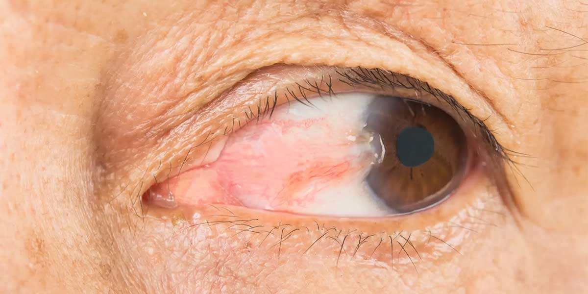
A pterygium is a usually triangular change on the ocular surface that develops when the conjunctival tissue covering the white part of the eye grows towards the cornea. Commonly known as pterygium (surfer's eye), this condition is referred to as pterygium in medical literature. It develops more frequently on the side of the eye closest to the nose, but in some people it may occur on the other side or in both eyes. Prolonged exposure to ultraviolet radiation, wind, dust, and dry eye may contribute to its development. Small, non-progressive growths may be monitored regularly, while enlarging tissue may cause redness, stinging, astigmatism, and reduced vision. Surgical treatment may involve removing the tissue and covering the exposed area with an appropriate graft. The method used is selected according to the size of the pterygium, how far it extends onto the cornea, and the condition of the ocular surface.
What Causes a Pterygium?
One of the most significant factors in the development of a pterygium is prolonged exposure to ultraviolet radiation from sunlight. The condition may occur more frequently in people who work outdoors, spend long periods in sunny environments, or do not wear protective glasses. Dust, smoke, wind, and dry air may also cause repeated irritation of the ocular surface. Dry eye and chronic inflammation may contribute to changes in the conjunctival tissue. Individual predisposition and environmental conditions may also play a combined role in the development of the condition. A pterygium is not contagious, and not every case of eye redness indicates a pterygium. Protection from sunlight and keeping the ocular surface moist may help reduce risk factors.
What Are the Symptoms of a Pterygium?
A pterygium may not cause a noticeable complaint during its early stages and may be identified during a routine eye examination. As the tissue grows, redness, prominent blood vessels, and a fleshy appearance may develop at the inner corner of the eye. Stinging, burning, itching, tearing, and a foreign-body sensation are common complaints. Discomfort may increase particularly in windy weather or after spending long periods in front of a screen. Tissue growing onto the cornea may alter the regular shape of its surface, causing astigmatism or a change in the existing glasses prescription. Blurred vision may become more pronounced in advanced cases that approach the centre of vision. The severity of the symptoms varies according to the size of the growth and the degree of inflammation on the ocular surface. Sudden pain, rapid growth, or an unusual appearance requires further assessment.
How Is a Pterygium Diagnosed?
A pterygium is diagnosed through a detailed examination of the ocular surface and assessment of how far the growth extends onto the cornea. A slit-lamp examination helps show the thickness of the tissue, its vascular structure, and how far it has progressed onto the cornea. Visual acuity and glasses prescription measurements are important for determining the effect of the growth on vision. When necessary, corneal topography may be used to assess whether the pterygium has caused astigmatism or surface irregularity. Dry eye, allergies, and other ocular surface conditions are also assessed during the examination. A pterygium must be distinguished from similar-looking growths such as a pinguecula, which does not extend onto the cornea. Other ocular surface disorders are also considered in lesions with irregular borders, rapid growth, or an appearance that differs from the usual presentation. The treatment decision is based not only on the appearance of the tissue but also on symptoms and measurement findings.
Does Every Pterygium Require Surgery?
Not every pterygium requires surgical treatment, and small, non-progressive growths may be monitored through regular examinations. Artificial tear drops may help reduce dryness and the foreign-body sensation on the ocular surface. During periods of pronounced inflammation, appropriate eye drops may be used for a limited time according to examination findings. These treatments may relieve symptoms, but they do not eliminate the existing tissue or guarantee that it will shrink. Wearing sunglasses with ultraviolet protection and protecting the eyes in dusty environments are among the supportive measures. Progression of the growth, its effect on vision, and the discomfort it causes in daily life are considered when deciding on surgery. Regular monitoring helps determine whether the growth changes over time. Avoiding unnecessary surgery is important, as is promptly assessing progression that threatens vision.
What Is Pterygium Surgery?
Pterygium surgery is an ocular surface operation intended to remove abnormal conjunctival tissue extending onto the cornea in a controlled manner. During the procedure, the pterygium tissue is carefully separated from the cornea and the white part of the eye. How the exposed area will then be covered is determined according to the characteristics of the growth and the condition of the ocular surface. A thin piece of tissue taken from healthy conjunctiva in the same eye is frequently placed over the area from which the pterygium was removed. This tissue may be secured with sutures or, in suitable cases, with a biological tissue adhesive. The aim is to restore the ocular surface, reduce the complaints caused by the pterygium, and limit the risk of recurrence. Surgery does not produce the same result in every patient and does not completely eliminate the possibility of the tissue growing again. The surgical plan is prepared by considering the individual's ocular anatomy and previous treatments.
When Is Pterygium Surgery Performed?
Pterygium surgery may be considered when the growth progresses over the cornea or begins to affect visual quality. The tissue approaching the centre of the cornea, causing significant astigmatism, or changing the glasses prescription may influence the decision to operate. Recurrent redness, persistent stinging, and ocular surface discomfort that continues despite treatment are also important criteria. In some people, the growth may make contact lens use difficult or restrict eye movements. Concerns about appearance may also be considered, but the risks and potential benefits of surgery should be assessed together. Additional assessment may be required for growths that enlarge rapidly, have unusual-looking borders, or do not have a clear diagnosis. There is no single size threshold for the timing of surgery that applies to everyone. The decision is made by jointly assessing the pattern of progression, the effect on the cornea, and the individual's complaints.
How Is Pterygium Surgery with a Graft Performed?
Pterygium surgery with a graft is based on covering the exposed area with the patient's own healthy conjunctival tissue after the abnormal tissue has been removed. This tissue, called a conjunctival autograft, is usually taken from the upper part of the same eye, from a healthy area beneath the upper eyelid. The graft is not taken from the eyelid but is prepared from the thin membrane covering the white part of the eye. It is then positioned appropriately over the area from which the pterygium was removed and secured with sutures or tissue adhesive. This approach may help restore the exposed ocular surface and reduce the risk of regrowth. The size and placement of the graft are determined according to the width of the pterygium and the condition of the surrounding tissues. Previous surgery in the same area or a limited amount of healthy conjunctiva may affect the choice of method. After the procedure, the position and healing of the graft are monitored through regular follow-up examinations.
What Is Surgery with Tissue Adhesive?
Pterygium surgery with tissue adhesive is a method in which the conjunctival graft is secured to the ocular surface with an appropriate biological adhesive. Products commonly known as fibrin adhesives may be used to reduce or, in some cases, completely eliminate the need for sutures. In suitable patients, suture-related stinging and foreign-body sensation may be more limited. However, using an adhesive does not mean that healing will be faster in every patient or that the pterygium will not recur. Problems such as displacement of the graft, separation of its edges, shrinkage, or local inflammation may still occur. In some cases, sutures may be used in addition to the adhesive to secure the graft adequately. The fixation method is selected according to the size of the pterygium, the characteristics of the ocular surface, and the surgical assessment. Not rubbing the eye and following the postoperative plan are important for protecting the graft.
The Difference Between Sutured and Adhesive Methods
Sutures and tissue adhesives used in pterygium surgery are different methods of securing the graft to the exposed area. With the sutured method, the graft is attached to the surrounding tissues using fine surgical threads. This approach may be necessary for large or specially shaped grafts, but it may cause stinging and discomfort during the first few days. With the adhesive method, suitable biological products are used to attach the graft to the ocular surface. Although the procedure time and suture-related discomfort may be reduced in some patients, risks such as separation or shrinkage of the graft must also be assessed. It is not possible to say that one method is more suitable for all patients. Previous operations, graft size, dryness of the ocular surface, and the degree of adhesion achieved during the procedure influence the choice. In some cases, both techniques may be used together to achieve more balanced graft placement.
When Is an Amniotic Membrane Used?
An amniotic membrane is appropriately prepared donor tissue that can be used to repair the ocular surface. In pterygium surgery, it may be considered when a sufficient graft cannot be taken from the patient's own conjunctiva or when a large area needs to be covered. Recurrent pterygium, previous eye surgery, and situations in which preservation of conjunctival tissue is desired may affect the choice of method. The membrane can be placed over the area left by the removed tissue and secured with sutures or tissue adhesive. This procedure is intended to support healing of the ocular surface and reduce the effects of inflammation. However, it does not need to be preferred over a conjunctival autograft in every patient. The risk of recurrence, existing ocular surface conditions, and the size of the tissue are assessed together. The appropriate method is determined according to the preoperative examination findings and the requirements identified during surgery.
Why May Mitomycin C Be Used?
Mitomycin C is a medicine that may be considered in selected pterygium operations to reduce the risk of regrowth. This substance may suppress the cellular processes that contribute to excessive tissue proliferation on the ocular surface. It may be considered as part of the surgical plan, particularly in recurrent cases or those considered to have a high risk of regrowth. However, it does not need to be used routinely in every pterygium operation. Inappropriate use of the medicine may lead to delayed wound healing, thinning of the ocular surface, or other serious problems. Whether it is used, the dose, and the duration of contact are therefore determined solely according to clinical assessment. Previous eye surgery and the condition of the corneal and scleral tissues are important in the decision-making process. A definitive choice should not be made without assessing the potential benefit together with the risks of the medicine.
How Is Pterygium Surgery Performed?
Pterygium surgery is generally performed under local anaesthesia after numbing the ocular surface. First, the tissue extending onto the cornea is separated in a controlled manner together with the surrounding conjunctiva and underlying abnormal tissues. The surface of the cornea is carefully cleaned to reduce any remaining irregularities. A conjunctival autograft, or an amniotic membrane in suitable cases, is then prepared for the exposed area. Positioning the graft in the correct orientation without creating tension is important for healing of the ocular surface. Sutures, biological tissue adhesive, or both methods may be used for fixation. A protective eye dressing or bandage contact lens may be applied at the end of the procedure in some patients. The duration of surgery varies according to the size of the tissue, previous operations, and the technique selected. If the removed tissue has an unusual appearance, the need for pathological examination is assessed separately.
Who Is Not Suitable for Surgery?
Pterygium surgery should not be planned before certain ocular and general health conditions have been brought under control. If active conjunctivitis, keratitis, or another infection of the ocular surface is present, the existing infection must be treated first. Advanced dry eye, significant ocular surface inflammation, and uncontrolled allergies may adversely affect healing. In Sjögren's syndrome or other immune system disorders, suitability is assessed according to individual risks. Whether diabetes, hypertension, and similar systemic conditions are under control is examined before surgery. The use of blood-thinning medicines or the presence of a clotting disorder should be taken into account when planning the procedure. Having undergone another eye operation recently may alter the timing of surgery. These circumstances do not always mean that surgery is definitively unsuitable. The appropriate approach is determined through reassessment after the existing risks have been managed.
How Should You Prepare Before Surgery?
Before surgery, the size of the pterygium, how far it extends onto the cornea, and the inflammation on the ocular surface are assessed in detail. Visual acuity, glasses prescription, and, when necessary, corneal topography are measured to assess existing astigmatism. If dry eye or allergies are identified, these conditions may need to be brought under control before surgery. Regularly used medicines, blood thinners, previous operations, and known medicine allergies should be reported. If contact lenses are used, the required period without them before the procedure is determined according to the individual situation. Surgery may be postponed if there is active redness, discharge, or evidence of infection in the eye. Wearing sunglasses and protecting the eye from environmental irritants are also important during the preoperative period. The anaesthetic method and postoperative care plan are explained in advance according to the examination findings. The purpose of preparation is to select the appropriate technique and reduce preventable risks.
Recovery After Surgery
Redness, stinging, tearing, and a foreign-body sensation may occur during the first few days after pterygium surgery. The severity of these complaints varies according to the surgical method used and the sensitivity of the ocular surface. While stinging may be more pronounced in patients with sutures, temporary discomfort may also occur with grafts secured using adhesive. Vision may be blurred during the early period and may change gradually as the corneal surface heals. Redness in the eye may take several weeks to resolve completely in some people and longer in others. The timing of a return to work and driving is assessed according to the nature of the person's work and their level of vision. Graft position, closure of the ocular surface, and the course of inflammation are monitored at follow-up examinations. It cannot be said that the recovery period will be the same for all patients or will be complete by a specific date.
What Should Be Considered After Surgery?
Prescribed eye drops should be used for the stated duration and at the stated frequency after surgery. Antibiotic drops help reduce the risk of infection, while the duration of medicines used to control inflammation is adjusted according to examination findings. Drops containing corticosteroids should be used under supervision because of their effects on eye pressure and other potential effects. During the first few days, the eye should not be rubbed, subjected to pressure, or brought into contact with unclean water. Recommendations given at the follow-up examination should be followed regarding the return to swimming, eye make-up, intense physical activity, and contact lens use. Avoiding dusty environments and wearing sunglasses with ultraviolet protection outdoors help protect the ocular surface. If worsening pain, marked discharge, increasing redness, or a sudden reduction in vision develops, an assessment should be performed without delay. Planned follow-up examinations should not be missed so that changes related to the sutures or graft can be identified promptly. Long-term protection from sunlight should continue after healing is complete.
Possible Side Effects and Risks
Temporary pain, redness, tearing, light sensitivity, and blurred vision may occur after pterygium surgery. Dryness of the ocular surface or a foreign-body sensation may become more pronounced during healing. Displacement of the graft, separation of its edges, shrinkage, or granuloma formation around the graft may require additional assessment. Suture-related irritation, infection, bleeding, and delayed wound healing are also among the possible complications. Corneal scarring or surface irregularity may affect visual quality in some patients. In rare cases, significant thinning of ocular surface tissues or adhesions between surrounding tissues may develop. Risks related to tissue healing should also be considered in patients treated with mitomycin C. The likelihood of complications varies according to the size of the pterygium, previous operations, and accompanying eye conditions. No surgical method can be said to eliminate all risks completely.
Can a Pterygium Recur After Surgery?
A pterygium may regrow in some patients even when it has been removed using appropriate surgical methods. The risk of recurrence depends on factors such as the patient's age, the size of the tissue, previous operations, and the degree of inflammation on the ocular surface. Intense exposure to sunlight, wind, and dust may also affect the likelihood of regrowth. The risk of recurrence may be higher with methods that leave bare sclera than with procedures in which the area is covered with an appropriate graft. A conjunctival autograft, an amniotic membrane in selected patients, and adjunctive treatments when necessary may be considered to reduce this risk. However, no technique can be said to prevent recurrence with certainty. If recurrence develops, how far the new growth extends onto the cornea and the changes it causes on the ocular surface are reassessed. Whether another operation is required is determined according to the individual's complaints and examination findings. Regular monitoring helps identify early changes.
Is It Possible to Reduce the Risk of Recurrence?
Protecting the ocular surface and following the recommended treatment plan after surgery are important for reducing the risk of recurrence. Wearing sunglasses with ultraviolet protection, using a wide-brimmed hat in sunny weather, and reducing exposure to direct sunlight may be helpful. Avoiding environmental factors such as dust, smoke, and wind contributes to controlling irritation of the ocular surface. If dry eye is present, artificial tear drops may be used for the period considered appropriate. If there is a habit of frequently rubbing the eyes because of allergies, this should be brought under control. Anti-inflammatory drops should be used after surgery only for the recommended period and within the prescribed follow-up schedule. Graft healing and the formation of new blood vessels are assessed during follow-up examinations. Although these measures may help reduce risk factors, they do not guarantee that the pterygium will not recur. Long-term monitoring is particularly important in people with a previous history of recurrence.
How Is the Success of Surgery Assessed?
The outcome of pterygium surgery is not assessed solely by looking at the change in the appearance of the eye. Removal of the tissue from the cornea, healing of the ocular surface, and a reduction in daily discomfort are among the main assessment criteria. Astigmatism or blurred vision caused by the pterygium may be measured again after the corneal surface has recovered. Although vision may improve in some patients, the expected improvement may remain limited because of pre-existing corneal scarring. The graft remaining in position, the absence of infection, and no evidence of new tissue growth are also important during follow-up. The result of surgery is affected by the size of the pterygium, the technique selected, accompanying dry eye, and the individual's healing characteristics. A continued need for glasses does not always mean that the operation has been unsuccessful. No method can be said to achieve the same visual result in everyone or to prevent recurrence with certainty.
Pterygium Surgery and Follow-Up in Izmir
When pterygium surgery is planned in Izmir, the extent of the growth onto the cornea, irritation of the ocular surface, and its effect on vision are assessed together. Dry eye, allergies, outdoor working conditions, and previous operations are considered when selecting treatment. Options such as pterygium surgery with a graft, a tissue adhesive technique, or an amniotic membrane may be assessed according to the individual's findings. Not every pterygium requires surgery, and regular monitoring with supportive drops may be sufficient in some patients. When surgery is performed, postoperative follow-up dates, the drops to be used, and measures for protection from sunlight are planned separately. When seeking pterygium treatment in Izmir and the Konak area, a detailed ocular surface examination is important for determining the appropriate method. The success of treatment is related to postoperative care and regular follow-up as well as the operation itself.
Pterygium Surgery Prices
Pterygium surgery prices may vary according to the size of the growth, how far it extends onto the cornea, and the surgical method selected. The use of a conjunctival autograft, fixation with tissue adhesive, the need for an amniotic membrane, and additional assessments may affect planning. The scope of the procedure may differ in cases that have previously been operated on or have recurred. When researching pterygium surgery prices for 2026, it is not possible to make an individual assessment based solely on the name of the procedure. Whether surgery is necessary and which technique is suitable must first be determined according to the examination findings. Individual information about fees can be provided after the treatment plan has been clarified. You can contact us through the website for information about the examination process and suitable treatment options.




































