
Contents:
Eye diseases, medically covered by the field of ophthalmology, include all health conditions that affect vision and the eye's delicate structures, such as the cornea, lens, and retina. This field covers a wide range of conditions, from common problems such as cataracts and glaucoma to strabismus and macular degeneration, as well as refractive errors such as myopia and the effects of diabetes on the eyes. Ophthalmologists are medical specialists who diagnose these conditions using modern technology, plan medical treatments such as medication or glasses, and aim to protect and improve vision through advanced surgical procedures such as laser treatment or microsurgery when necessary.
Who Is the Best Ophthalmologist in Turkey?
Many renowned and experienced ophthalmologists practising in Turkey provide diagnosis and treatment using advanced technologies in areas such as laser treatments, cataract surgery, retinal diseases, and corneal transplantation. The physician's area of expertise, techniques used, and patient satisfaction are among the main factors influencing success. The choice of the best ophthalmologist may vary depending on the patient's visual problem and treatment expectations.
Who Should We Consult About Eye Health?
When you experience an eye-related problem or need a routine check-up, you may encounter professionals with different titles. Understanding their duties and authority makes it easier to receive the right assistance at the right time. The eye care team mainly consists of:
- Ophthalmologist
- Optician
- Optometrist
An ophthalmologist is a medical doctor who completes six years of medical school, passes the Medical Specialization Examination in Turkey, known as the TUS, and then undergoes at least four more years of specialist training dedicated exclusively to eye diseases and surgery. This demanding education, which takes a total of 10-12 years, authorizes them to diagnose all types of eye conditions, prescribe medication, and perform surgery for diseases such as cataracts, glaucoma, strabismus, and retinal detachment. Laser treatments are also performed by ophthalmologists. The most fundamental feature distinguishing them from other eye care professionals is that they are medical doctors. This enables them to understand how systemic conditions such as diabetes or hypertension affect the eyes and to assess the patient as a whole.
Opticians are technicians who fulfil prescriptions issued by ophthalmologists. They graduate from two-year opticianry programs. Their role is to prepare lenses according to the prescription, fit them into the selected frame, make the necessary adjustments so that the glasses fit the face correctly, and provide information about contact lens use. Opticians do not perform eye examinations, make diagnoses, or prescribe medication.
Although the profession is not yet widespread in Turkey, some countries also have professionals known as optometrists. Optometrists are not medical school graduates, but they complete four-year university optometry programs focused on detecting visual defects and prescribing glasses or contact lenses. In Turkey, these duties are largely performed by ophthalmologists.
In Which Fields Do Ophthalmologists Specialize?
Although the eye is a small organ, it has such a complex structure that many ophthalmologists undertake additional training in a specific field after their specialist education, either through a subspecialty or fellowship. This enables them to provide more focused and advanced treatment to their patients. Some of these specialties include:
- Retinal and Vitreous Diseases
- Glaucoma
- Corneal and Anterior Segment Diseases
- Pediatric Ophthalmology and Strabismus
- Oculoplastic Surgery
- Neuro-ophthalmology
- Uveitis and Ocular Immunology
The existence of these subspecialties is clear evidence that the concept of eye disease is too broad to be grouped under a single heading. For example, a retinal specialist performs advanced procedures for conditions such as the effects of diabetes on the eyes or macular degeneration, while an oculoplastic specialist deals with conditions such as drooping eyelids or aesthetic procedures around the eyes. This structure allows every patient to consult a doctor with the knowledge and experience most appropriate for their specific condition.
Because ophthalmologists assess not only the eyes but also the effects of systemic diseases, they work in collaboration with many medical specialties:
- Neurology: For problems involving the visual pathways and optic nerve.
- Endocrinology: For diabetic retinopathy and the ocular effects of thyroid diseases.
- Rheumatology: For inflammatory conditions such as Behçet's disease and uveitis.
- Pediatrics: For the diagnosis and treatment of childhood eye diseases.
- Radiology: For advanced imaging of the eye and orbital structures.
- ENT (Ear, Nose, and Throat): For procedures involving structures around the eyes.
- Plastic Surgery: For eyelid, tear duct, and aesthetic procedures.
How Is a Detailed Eye Examination Performed?
Although many people think an eye examination consists only of reading letters, a comprehensive examination actually involves much more. Let us examine step by step what you can expect when you enter the examination room. An accurate diagnosis depends on all these steps being completed properly.
Medical History and Symptoms: First, your doctor listens to you. Information about your visual symptoms, when they started, your general health, medications, and whether anyone in your family has an eye disease provides the first clues for diagnosis.
Visual Acuity Measurement: This is the familiar letter-reading test. You are asked to read letters or shapes that gradually become smaller from a specified distance. This test measures your eye's ability to see clearly.
Refraction Examination: This examination determines whether you have a refractive error such as myopia, hyperopia, or astigmatism. Using an instrument called a phoropter, which is positioned in front of your eyes, your doctor tries different lenses and asks, "Is this clearer, or is this one clearer?" This process determines the prescription that provides the clearest vision.
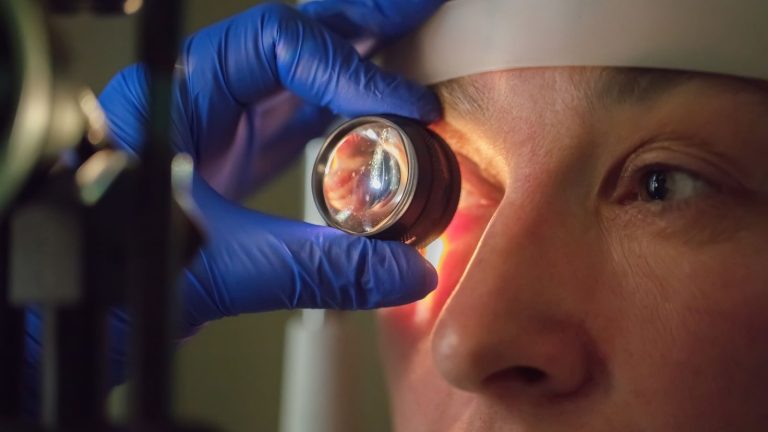
Slit-Lamp Examination: This device is a specialized microscope with a bright light source. Your doctor uses it to examine the front of your eye, including the cornea, lens, iris, and eyelids, in detail. Even a very small scratch, sign of infection, or early cataract can be detected during this examination.
Intraocular Pressure Measurement (Tonometry): This test is vital for glaucoma screening. The pressure of the fluid inside the eye is measured using instruments that either blow air onto the eye or make direct contact with it. The procedure is completely painless and takes only seconds.
Dilated Eye Examination: This is perhaps the most important step of the examination. Drops are placed in your eyes to enlarge your pupils temporarily. Instead of looking through a small keyhole, this allows the doctor to examine the back of your eye as if looking into a room with the door wide open. Only in this way can the retina, macula, and optic nerve be assessed in detail. Many serious conditions, including the effects of diabetes or hypertension on the eyes, macular degeneration, glaucoma damage, and retinal tears, can be detected through this examination. The drops remain effective for several hours, and blurred near vision and sensitivity to light during this period are normal.
More advanced examinations may sometimes be required in addition to these basic steps. For example, Optical Coherence Tomography, or OCT, analyzes the layers of the retina and optic nerve at the micron level, almost as if taking a CT or MRI scan of the eye. This technology can detect diseases such as macular degeneration or glaucoma years before the patient develops symptoms or vision loss.
What Are the Most Common Refractive Errors?
Refractive errors are not actually diseases but mismatches in the eye's optical structure. Just as the lens of a camera may fail to focus, the eye may be unable to focus light onto the nerve layer called the retina. This is the underlying cause of the most common visual problems.
Myopia (Nearsightedness): In a myopic eye, light rays from distant objects focus in front of the retina rather than directly on it. As a result, nearby objects appear clear, while distant signs, television subtitles, or the face of someone approaching from a distance appear blurred. It generally occurs because the eyeball is slightly longer than normal or because the cornea bends light too strongly. Genetic predisposition plays an important role, and myopia usually begins during school age and increases during adolescence.
Hyperopia (Farsightedness): This is the opposite of myopia. In this case, light focuses behind the retina. It occurs because the eyeball is shorter than normal. At a young age, the natural lens inside the eye is flexible and can compensate for this defect to some extent, allowing the person to see clearly at a distance. However, this continuous focusing effort can cause eye strain, discomfort, and headaches, particularly during near activities such as reading. People with high hyperopia may have blurred vision at both distance and near.
Astigmatism: The cornea, the clear front layer of a perfectly shaped eye, has the same curvature in every direction, similar to a basketball. In astigmatism, however, the cornea has a more asymmetrical shape, resembling an egg or the back of a spoon. Light entering the eye therefore cannot focus at a single point. This causes vision to appear blurred, shadowed, or doubled at both distance and near. People often report light scatter and difficulty driving at night.
Presbyopia (Age-Related Difficulty with Near Vision): This is not a refractive error but a natural consequence of ageing that occurs in everyone. When we are young, the natural lens inside the eye is flexible like the zoom lens of a camera and can easily focus on nearby objects by changing shape. However, usually after the age of 40-45, this lens gradually loses its flexibility and becomes more rigid. Focusing on nearby objects such as a phone screen, book, or menu therefore becomes difficult. Holding reading material farther away by extending the arms is the most typical sign of presbyopia.
Which Treatments Are Used for Vision Problems?
Fortunately, a wide variety of effective methods are available today for correcting refractive errors. These methods range from simple external aids to long-term solutions.
Glasses: Glasses are the easiest, safest, and most widely used method. Individually prepared lenses provide clear vision by allowing light to focus correctly on the retina. Modern technology makes it possible to produce very lightweight and aesthetically designed glasses.
Contact Lenses: These small lenses are placed directly on the surface of the eye and are an excellent alternative, particularly for people who do not want to wear glasses or who participate in sports. They provide a wider and more natural field of vision. However, contact lens use requires strict attention to hygiene and care. If the lenses are not kept clean or are worn longer than recommended, they may cause serious sight-threatening problems such as corneal infections.
Refractive Surgery (Laser Eye Surgery): These surgical methods have been developed for suitable candidates who want a long-term alternative to glasses or contact lenses. The aim is to correct the refractive error by reshaping the corneal surface with a laser. The most commonly used laser methods include:
- LASIK
- PRK
- SMILE
Each method has its own advantages, and the most appropriate option for each patient is determined following a highly detailed eye examination and diagnostic tests. Factors such as corneal thickness, prescription, and the patient's lifestyle play a role in this decision. For patients with very high prescriptions who are unsuitable for laser treatment, or for those who also have cataracts, another option is to remove the natural lens and replace it with a prescription intraocular lens.
What Is a Cataract and What Are Its Symptoms?
A cataract occurs when the natural lens inside the eye, which functions like a camera lens, gradually loses its transparency and becomes cloudy like frosted glass. This causes progressive loss of vision, faded color perception, and reduced quality of life. The main cause of cataracts is ageing. Just as our hair turns grey and our skin develops wrinkles, the natural lens of the eye also ages. However, factors such as diabetes, long-term corticosteroid use, trauma to the eye, and intense exposure to UV light may accelerate cataract development.
Cataracts usually progress slowly and may not be noticed initially. As they advance, however, certain typical symptoms appear. If you notice these symptoms, it is important to consult an ophthalmologist:
- Painless, gradually progressive blurred or hazy vision
- Colors appearing paler and less vivid
- Light scatter and glare, particularly while driving at night
- Needing brighter light for reading
- Double vision in one eye
- Frequent changes in glasses prescription
How Is Cataract Surgery Performed?
Cataracts cannot currently be treated with medication or glasses. The only definitive solution for restoring clear vision is to remove the cloudy lens surgically and replace it with an artificial lens. Fortunately, modern cataract surgery using the phacoemulsification technique is one of the most successful and safest operations in medicine.
Cataract operations are now highly comfortable procedures that usually require no stitches, are performed under topical anesthesia with eye drops, and take an average of 15-20 minutes. During surgery, the eye is entered through a very small incision of approximately 2 millimeters in the cornea. A device that emits ultrasonic waves breaks up and removes the cataractous lens inside the eye. A folded artificial intraocular lens, or IOL, is then inserted through the same small incision. It unfolds inside the eye and settles into the correct position. The patient can usually return home on the same day.
Different types of lenses can be implanted during surgery. You can discuss the options with your doctor and choose the lens best suited to your lifestyle and expectations.
Monofocal Lenses: These lenses provide clear vision at only one distance, usually distance vision. Patients generally need reading glasses for near vision after surgery.
Multifocal/Trifocal Lenses: These "smart" lenses aim to provide clear vision without glasses at both distance and near. They may greatly reduce or completely eliminate the patient's dependence on glasses.
Toric Lenses: If the patient has corneal astigmatism, these lenses correct both the cataract and astigmatism simultaneously to provide clearer vision.
What Is Glaucoma, Known as the Silent Threat?
Although glaucoma is commonly referred to as "eye pressure," it is not simply a condition involving elevated intraocular pressure. Glaucoma is a chronic disease in which the optic nerve, consisting of millions of nerve fibers that carry images from the eye to the brain, is progressively and irreversibly damaged. The most insidious aspect of the disease is that it generally causes no symptoms until its advanced stages. Damage to the optic nerve gradually destroys peripheral vision, but because central vision remains intact for a long time, the patient may not realize that there is a problem. Glaucoma is therefore called the "silent thief of sight." Because damage cannot be reversed once it occurs, early diagnosis is vital.
A fluid called aqueous humor is continuously produced inside the eye to nourish it and maintain its shape, and it leaves the eye at the same rate. When a blockage or resistance develops in the drainage channels through which this fluid exits, it begins to accumulate inside the eye like water from a tap above a blocked sink. This causes intraocular pressure, or IOP, to rise. The elevated pressure mechanically compresses the delicate optic nerve fibers and disrupts their blood flow, causing them to die gradually.
What Are the Symptoms and Risk Factors of Glaucoma?
Open-angle glaucoma, the most common type of glaucoma, causes no symptoms. It is usually diagnosed only when elevated intraocular pressure or optic nerve damage is detected during a routine eye examination.
Angle-closure glaucoma, a less common form, may present as a sudden attack. It occurs when the drainage route for the fluid inside the eye becomes completely blocked and requires emergency treatment. Its symptoms are severe:
- Severe eye pain and headache
- Nausea and vomiting
- Blurred vision
- Colored halos around lights
- Marked redness of the eye
Glaucoma can affect anyone, but some people are at greater risk:
- Being over the age of 40
- Having a family history of glaucoma
- High intraocular pressure
- Myopia or hyperopia
- Diabetes or hypertension
- Long-term corticosteroid use
- Previous eye trauma
How Is Glaucoma Treated?
The purpose of glaucoma treatment is not to restore vision that has already been lost, but to protect existing vision and stop the disease from progressing. The only way to achieve this is to lower intraocular pressure to a safe level at which the optic nerve will not be damaged. Treatment usually progresses step by step, beginning with the simplest method.
Medication (Eye Drops): This is the first and most common step in treatment. Drops with different mechanisms of action lower pressure either by reducing the production of fluid inside the eye or by facilitating its drainage. For treatment to be successful, the drops must be used regularly every day and exactly as instructed by the doctor.
Laser Treatments: These are used when eye drops are insufficient or cannot be used by the patient. In a procedure called SLT, or Selective Laser Trabeculoplasty, low-energy laser pulses are applied to the obstructed drainage channels to improve their function. It is a brief, painless outpatient procedure.
Surgical Treatments: Surgical options are considered when medication and laser treatment cannot lower intraocular pressure to the target level. In traditional trabeculectomy surgery, a new channel is created to allow fluid inside the eye to drain out. Minimally Invasive Glaucoma Surgery, or MIGS, methods, which have become increasingly popular in recent years, use microscopic implants in an effort to lower pressure more safely.
What Is Macular Degeneration, Which Threatens Central Vision?
Age-Related Macular Degeneration, or AMD, is the most common cause of permanent central vision loss in people over the age of 60. The disease affects a small but vital area called the macula, located at the center of the retina. The macula enables us to read, thread a needle, recognize someone's face, and see fine details clearly. Macular degeneration damages this central area, while peripheral vision is generally preserved. It therefore does not cause complete blindness, but it can seriously affect the patient's quality of life and abilities such as reading and driving. The most important risk factors are advanced age, genetic predisposition, and smoking.
What Are the Types and Symptoms of Macular Degeneration?
Macular degeneration has two main types, each with a different course and treatment.
Dry Type: This accounts for approximately 85-90% of all cases. Small, yellow metabolic deposits called drusen accumulate beneath the macula. Over time, this disrupts the nourishment of retinal cells and causes them to die gradually, known as atrophy. Vision loss generally progresses slowly over a period of years.
Wet Type: Although less common, this is the form primarily responsible for severe vision loss. Abnormal, leaking new blood vessels form beneath the macula. These fragile vessels leak fluid and blood under the retina, rapidly and severely damaging the visual cells. Vision loss is much more sudden and severe than in the dry type.
The most important symptoms of the disease include:
- A blurred, dark, or empty area in the center of vision
- Straight lines, such as door frames or window frames, appearing wavy, curved, or broken
- Letters or words disappearing while reading
- Difficulty recognizing people's faces
Colors appearing duller and paler
For the dry type, special vitamin formulations containing antioxidants and minerals, known as AREDS2 supplements, which have been shown to slow disease progression, are used. Treatment of the wet type has been transformed in recent years by intravitreal anti-VEGF injections. These medications stop abnormal blood vessels from growing, prevent leakage, and aim to preserve existing vision or even improve it to some extent in certain patients. Treatment usually begins with monthly injections, and the intervals are adjusted according to the patient's condition.
How Does Diabetes Affect Vision: What Is Diabetic Retinopathy?
Diabetes is not only a disease that raises blood sugar. It is a systemic condition that damages small blood vessels throughout the body. The eye is the organ in which this damage can be seen most easily and at the earliest stage. Diabetic retinopathy is a serious eye disease caused by high blood sugar damaging the structure of the retinal blood vessels. It is the most common cause of preventable blindness among the working-age population.
Damaged retinal vessels begin to leak, bleed, and become blocked over time. If this leakage accumulates in the center of the retina, or macula, it causes a condition called macular edema, which leads to blurred vision. In advanced stages, the retina, deprived of oxygen because of blocked vessels, attempts to form new blood vessels. However, these new vessels are extremely fragile and abnormal. They may bleed easily and fill the eye with blood, causing vitreous hemorrhage, or create traction on the retina and lead to retinal detachment.
The most insidious aspect of diabetic retinopathy is that it usually causes no symptoms until it affects the center of vision. It is therefore vital for everyone diagnosed with type 1 or type 2 diabetes to undergo a detailed eye examination at least once a year, even if they have no symptoms.
Which Treatments Are Used for Diabetic Retinopathy?
The cornerstone and most important step in treating diabetic retinopathy is controlling the underlying diabetes itself.
Blood Sugar Control: Keeping blood sugar, blood pressure, and cholesterol levels within the ideal ranges is the most effective way to slow the progression of the disease.
Laser Photocoagulation (Argon Laser): In this treatment, laser pulses are applied to the retina to close leaking vessels or prevent abnormal new blood vessel formation.
Intravitreal Injections: Particularly when fluid accumulates in the center of vision, causing macular edema, or in advanced disease, anti-VEGF medications are injected into the eye to reduce leakage and cause abnormal vessels to regress.
Vitrectomy Surgery: This is a surgical procedure in which the gel and blood inside the eye are removed and the retina is repaired when advanced complications such as intraocular bleeding or retinal detachment develop.
It should be remembered that these treatments aim to stop or slow the damage. The best treatment is to prevent the disease from reaching these stages through good diabetes management from the outset.
What Is Retinal Detachment, Which Requires Emergency Treatment?
Retinal detachment occurs when the retina, the nerve layer lining the back wall of the eye like wallpaper, separates from the supporting tissue beneath it. Because the retina receives nutrients and oxygen from this underlying tissue, its visual cells begin to die rapidly if the separation continues. Retinal detachment therefore requires urgent surgical treatment to prevent permanent vision loss.
The most common type results from a tear or hole in the retina. The gel-like fluid inside the eye, known as the vitreous, passes through the tear and separates the retina from the underlying wall. High myopia, previous eye surgery, and a severe blow to the eye are risk factors for retinal tears and detachment.
What Are the Warning Signs of Retinal Detachment?
Retinal detachment is usually painless, but it produces several characteristic warning signs before or when it begins. If you experience any of these symptoms, you should consult an ophthalmologist without delay.
Sudden flashes of light or a lightning-like sensation in your field of vision
The sudden appearance of numerous black dots, spots, or images resembling falling soot
Cobweb-like floaters
A dark shadow or curtain that begins at one side of your field of vision and moves toward the center
The only treatment for retinal detachment is surgery. The aim of surgery is to drain the fluid beneath the retina, seal the tear, and enable the retina to reattach. Depending on the condition of the tear, treatment may involve laser procedures, placing gas inside the eye, or more complex vitrectomy surgery. Early intervention is the most critical factor in preserving vision.
What Are the Most Common Eye Problems in Children?
A child's visual system actively develops during the first 8-10 years of life. Any eye problem present during this critical period may cause permanent visual impairment if left untreated. Monitoring eye health in children is therefore very important.
The most common problems include strabismus and amblyopia. Strabismus means that the eyes are not aligned, with one eye turning inward, outward, upward, or downward relative to the other. Amblyopia occurs when an eye has a completely normal physical structure but remains visually weak because the brain does not use it sufficiently.
These two conditions are closely related. The brain suppresses the image coming from an eye that constantly deviates to prevent double vision. If this suppression continues for a long time, the visual center associated with that eye in the brain cannot develop sufficiently, and amblyopia becomes permanent. Another common cause of amblyopia is a large prescription difference between the two eyes. The brain always prefers the eye that sees more clearly and reduces its use of the other eye.
How Are Amblyopia and Strabismus Treated?
The golden rule in treating amblyopia is early diagnosis. The earlier treatment begins, the greater the chance of success. The basic principle of treatment is to make the brain use the amblyopic eye.
Correct Use of Glasses: If the underlying cause is a refractive error, the first step is to prescribe glasses with the correct power. In some cases, wearing glasses alone may correct both strabismus and amblyopia.
Patching Treatment: This is the most effective method. The healthy, better-seeing eye is covered with a special patch for specified periods each day, forcing the brain to use the amblyopic eye.
Medicated Eye Drop Treatment: In young children who do not accept patching, atropine drops may be placed in the better-seeing eye to blur it temporarily and encourage the use of the amblyopic eye.
If the underlying strabismus cannot be corrected with glasses, the alignment of the eyes may be corrected surgically by operating on the eye muscles. Congenital conditions such as cataracts or glaucoma in children are also serious problems that require urgent treatment and may cause permanent blindness if left untreated. Every child should therefore undergo an eye examination after birth and again during the preschool period.
What Is Neuro-ophthalmology, Which Examines the Connection Between the Eyes and the Brain?
Neuro-ophthalmology is one of the most fascinating and complex subspecialties of ophthalmology. Rather than addressing problems within the eye itself, this field focuses on visual disorders caused by problems in the neural connections between the eyes and the brain. Like a detective, a neuro-ophthalmologist may use a symptom in the eye to diagnose an underlying neurological disease such as a brain tumor, stroke, or multiple sclerosis. Because vision is a complex process that uses a large part of the brain, many diseases affecting the nervous system produce their first symptoms in the eyes.
Which Conditions Fall Within the Field of Neuro-ophthalmology?
If a patient has sudden or gradually developing unexplained vision loss, double vision, drooping eyelids, or an abnormality of the pupils, assessment by a neuro-ophthalmologist may be required.
- Optic Neuritis: This condition, usually seen in young people and characterized by sudden, painful vision loss in one eye, is often the first sign of Multiple Sclerosis, or MS.
- Ischemic Optic Neuropathy: This is sudden, painless vision loss caused by blockage of the blood vessels supplying the optic nerve. It is generally seen in older patients with diabetes or hypertension.
- Tumors Compressing the Optic Nerve: Tumors arising from nearby tissues such as the brain or pituitary gland may compress the optic nerve and cause gradual visual field loss.
- Unexplained Vision Loss: If the patient continues to experience visual symptoms despite a normal eye examination, the problem may be located in the visual pathways of the brain.
- Double Vision: Paralysis of the nerves controlling eye movements, thyroid disease, or neuromuscular conditions such as myasthenia gravis may cause double vision.
- Pupil Abnormalities: A difference in size between the two pupils or abnormal reactions to light may indicate a serious underlying neurological condition.
- Neuro-ophthalmology reminds us that an eye problem may be only one small part of a much larger picture. It serves as a vital bridge between ophthalmology, neurology, and neurosurgery.
Why Does Turkey Have Some of the Best Ophthalmologists?
- Academic and Clinical Experience: Ophthalmologists in Turkey are internationally recognized for both their academic achievements and extensive clinical experience.
- Technological Infrastructure: Clinics are equipped with advanced technologies such as laser devices, OCT, and artificial intelligence-supported imaging systems.
- Wide Range of Treatments: Comprehensive services are provided in many areas, including cataract, retinal, glaucoma, and refractive surgery.
- Patient-Centered Care: Personalized treatment plans and comprehensive patient follow-up contribute to high levels of satisfaction.
Why Does Turkey Have Some of the Best Eye Hospitals?
Eye hospitals in Turkey have advanced technological infrastructure and experienced specialists in laser treatments, cataract surgery, and the management of retinal and corneal diseases. Modern methods such as excimer laser and femtosecond laser achieve high success rates, and personalized treatment approaches are used. In addition, short recovery periods, patient satisfaction, and a service structure suitable for medical tourism have made Turkey one of the regional leaders in eye care.
You can read reviews of ophthalmologists in Turkey on platforms such as DoktorTakvimi, Google Maps, Kadınlar Kulübü, or Ekşi Sözlük.










































