
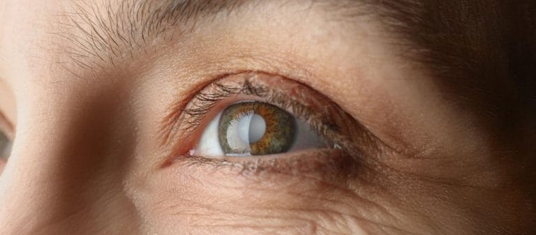
A secondary cataract is clouding that develops in the lens capsule after cataract surgery. Although it is commonly described as "the cataract returning," it is actually a different condition.
Symptoms include blurred vision, light scattering, and difficulty reading. These complaints generally develop several months or years after surgery.
The treatment method is laser capsulotomy. During this brief and painless procedure, the cloudy tissue behind the lens is opened, allowing clear vision to be restored. Patients can quickly return to their daily lives after the procedure.
Secondary cataracts cannot be completely prevented, but they can be treated effectively. Regular examinations allow early diagnosis and intervention before visual quality deteriorates.
| Definition | Formation of a membrane or layer behind the lens following cataract surgery, causing blurred vision, posterior capsule opacification |
|---|---|
| Causes | Cell proliferation and membrane formation in the lens capsule that remains behind the lens after cataract surgery |
| Symptoms | Recurrence of blurred vision, light scattering, increased glare, and reduced visual quality |
| Diagnostic Methods | Eye examination and biomicroscopic examination, slit-lamp examination |
| Treatment Methods | YAG laser capsulotomy, opening the membrane in the posterior lens capsule with a laser, generally performed in a single session |
| Complications | Rarely, a temporary increase in intraocular pressure, mild discomfort after laser treatment, retinal detachment, or vision loss |
| Prevention | Cannot be completely prevented. Early diagnosis and treatment are possible through regular examinations after cataract surgery |
| Who Is More Frequently Affected | May develop over time in any patient who has undergone cataract surgery, but is more common in younger patients |
| Recovery Time | Vision generally improves rapidly immediately after laser treatment, with full clarity achieved within a few days |
| When to Consult a Doctor | If new symptoms develop, such as recurrent blurred vision, sudden vision loss, pain, or flashes of light |
A secondary cataract is the gradual clouding of the capsule located behind the lens following cataract surgery. It is not a recurrence of a true cataract, but it causes similar visual problems. It generally develops over several months or years. Treatment involves opening the cloudy area with a laser, using YAG laser capsulotomy, and is a quick, painless procedure.
The symptoms of a secondary cataract are very familiar to people who have experienced a primary cataract or know about the condition. After becoming accustomed to clear vision following the first surgery, patients may gradually begin to experience "hazy" or "filtered" vision again. You can imagine it as a window becoming dirty over time. A view that was initially crystal clear becomes obscured by a veil.
The most common symptom is blurred vision. Patients may describe it by saying, "It feels as though there is a thin veil in front of my eye," or "It feels as though my glasses are dirty, but cleaning them does not help." This blurring is frequently accompanied by sensitivity to light. Glare, light scattering, and rings known as halos may appear around light sources, particularly at night. These halos may make car headlights or streetlights appear brighter and more diffuse than they actually are. As a result, driving at night may become difficult for many people.
Fading colors are another common symptom. Colors that once appeared vivid may look duller and less vibrant when a secondary cataract develops. Although this change may sometimes go unnoticed, people who work with art or have a high sensitivity to color may recognize it easily. For example, the colors in a favorite painting may no longer appear as striking, or it may become difficult to distinguish between shades of clothing.
Double vision, monocular diplopia, may also occur in rare cases. This means seeing two slightly displaced copies of the same image even when looking through only one eye. It may be particularly uncomfortable while reading a book or looking at a phone screen. Glare may also become intense enough to interfere with daily life. The eyes may tire quickly in summer sunlight or in brightly illuminated environments.
These symptoms generally become more pronounced over time. They may begin with mild discomfort or occasional episodes of blurred vision before eventually causing a substantial loss of comfort. Whenever you notice an obvious decline in visual quality, the healthiest approach is to consider whether a secondary cataract may be developing and arrange an eye examination.
Understanding the anatomical and physiological basis of a secondary cataract makes it easier to understand how the condition develops. When the natural lens inside the eye becomes opaque because of aging or other factors, resulting in a primary cataract, it is surgically removed. However, the entire lens structure is not removed. The posterior part of the membrane-like layers forming the anterior and posterior lens capsules is generally left in place. This posterior capsule is important for keeping the artificial lens stable.
However, some cells known as lens epithelial cells may remain around the lens. These may be cells that are difficult to remove completely during surgery or that remain concealed in the peripheral areas of the eye even after cleaning. These cells may become active and proliferate as part of the eye's natural healing processes, forming a "thin membrane" over the posterior capsule. As this membrane thickens or changes, the posterior capsule gradually loses its transparency.
Various growth factors and chemical signals play a role in the migration and proliferation of lens epithelial cells. Certain substances, such as transforming growth factor beta, TGF-β, affect the differentiation and proliferation of these cells. Although these substances are responsible for tissue healing in many parts of the body, they may contribute to the unwanted development of cataract-like opacities in the eye.
Conditions such as trauma and infection may also trigger this process. Increased inflammation in the fluids inside the eye or changes occurring after tissue damage may alter the behavior of the lens epithelial cells. This can accelerate the clouding process or make it more pronounced. Systemic diseases such as diabetes, high blood pressure, or metabolic disorders may also contribute to the same mechanism by affecting tissue healing in the eye.
When the lens is removed during the initial surgery, it may be compared to clearing all the weeds from soil, with the soil representing the inside of the eye. However, invisible seeds or roots may remain beneath the surface. Over time, these roots grow and emerge again. The mechanism through which a secondary cataract develops is similar to unwanted weeds growing back.
Although a secondary cataract can potentially develop in anyone who has undergone cataract surgery, certain groups and circumstances carry a higher risk. Patients often ask, "Does it happen to everyone?" A single answer that applies to everyone would not be accurate because every eye, body, and lifestyle has different characteristics.
- The renewal and response mechanisms of the cells in our eyes change with age. Natural lens cells may be less active in older adults, while tissues in younger people regenerate much more quickly. Secondary cataracts may therefore develop less frequently or later in some people who undergo surgery at an advanced age. In younger patients, the risk may be higher because tissue regeneration and cell proliferation occur more rapidly. Secondary cataracts are particularly common and almost unavoidable after cataract surgery during childhood.
- A family history, systemic conditions such as diabetes, high blood pressure, and certain metabolic syndromes may increase the risk by affecting healing and inflammation inside the eye. Patients with diabetes, for example, generally develop cataracts at a younger age and frequently have a higher risk of developing secondary cataracts after surgery.
- The way cataract surgery is performed, how thoroughly the area is cleaned, and the type of intraocular lens used all influence this risk. Modern surgical methods and artificial lenses with sharp, square edges may help restrict the migration of lens epithelial cells toward the posterior capsule. The surgeon's experience is also relevant to how effectively epithelial cells around the lens can be removed during surgery.
- Additional eye conditions such as uveitis, intraocular inflammation, trauma, retinitis pigmentosa, or high myopia may alter the anatomy and physiology of the eye, creating conditions that facilitate the development of a secondary cataract. Because the eye tissues are already more sensitive in these disorders, the postoperative healing process may also be more difficult.
- Some studies have identified small but statistically significant differences between women and men. However, this factor has a much weaker influence than metabolic diseases such as diabetes.
Examination and various eye tests are extremely important in diagnosing a secondary cataract. Eye care professionals begin by listening to the patient's complaints. Symptoms such as blurred vision, flashes of light, faded colors, or difficulty seeing at night may cause the doctor to consider whether a secondary cataract has developed. A comprehensive eye examination is then performed.
- Visual Acuity Test: During the first stage, vision tests determine how clearly the patient can see. Testing may be performed with or without glasses. If visual acuity has decreased significantly after it was expected to return to normal following surgery, the doctor may suspect a secondary cataract.
- Biomicroscope, Slit-Lamp Examination: This examination allows the ophthalmologist to inspect the front part of the eye under magnification. The position of the artificial lens, the amount of clouding in the anterior and posterior capsules, and structures such as the iris and cornea are evaluated. Slit-lamp examination is particularly effective for viewing opacity on the posterior capsule. By reflecting light from different angles, the doctor can easily detect clouding in areas that should be transparent.
- Fundus Examination: This method is used to examine the retina and optic nerve in the posterior segment and helps confirm the diagnosis of a secondary cataract. If the posterior capsule is highly opaque, the fundus examination may become difficult. This provides the doctor with a clue that the opacity is preventing a clear view of the retina.
- Retroillumination: Light can be directed into the eye to assess the appearance of the posterior capsule. The incoming light makes opaque areas more visible and facilitates diagnosis.
- Other Imaging Techniques, OCT and Similar Methods: In complicated cases or when another underlying eye disease is suspected, advanced imaging methods such as optical coherence tomography, OCT, may be used. This method allows the retinal layers and posterior capsule to be examined in cross-section.
After diagnosis, the doctor develops a treatment plan by considering the degree of the secondary cataract and how much it affects the patient's vision. If the patient experiences serious difficulty while driving or performing everyday tasks, treatment generally becomes unavoidable. In mild cases, however, monitoring through examinations may be preferred if the patient does not yet experience significant discomfort. Although this diagnostic process may appear simple, it requires considerable technical detail and experience.
Secondary cataracts can now be treated using clear and effective methods. Many patients may worry and ask, "Will I need another operation?" Fortunately, a secondary cataract can usually be corrected with a procedure that is much simpler than surgery.
- YAG Laser Capsulotomy: This method is considered the gold standard for treating secondary cataracts. A YAG, yttrium-aluminum-garnet, laser is used to create a small "window" in the opaque area of the posterior capsule. This allows light to focus clearly on the retina again. It is similar to creating an opening in a foggy window that is large enough to see through. The procedure is generally completed within minutes and does not require stitches, an incision, or a dressing. Vision usually improves noticeably on the same day or within a few days.
- Surgical Method: In very rare cases in which YAG laser treatment cannot be performed, or when unusual complications develop, the opaque capsule may be removed surgically. However, this method is not frequently preferred in modern medical practice because laser treatment provides a practical alternative with a low level of risk.
- Can It Be Corrected With Medication or Glasses?: The clouding caused by a secondary cataract cannot be eliminated completely with medication or glasses. During the early stages, people with mild clouding may achieve partial relief by updating their glasses prescription. However, this does not provide a permanent solution. Complaints such as "I can see a little more clearly after changing my glasses, but everything still looks somewhat blurred" may continue.
- Care After Treatment: After the laser procedure, the ophthalmologist may prescribe certain eye drops for a short period, for example, to control intraocular pressure or prevent inflammation. The recovery period is considerably more comfortable than recovery after conventional cataract surgery. Most people can comfortably return to their daily lives the day after the procedure.
YAG laser capsulotomy is the most commonly used method among these treatments and provides successful results. There is therefore no need to panic when a secondary cataract develops. For most patients, visual quality can be restored with a minor laser procedure without repeating the major cataract operation performed years earlier.
"Treatment is available, but is there a way to prevent the condition from developing at all?" is one of the most frequently asked questions about secondary cataracts. Advances in surgical techniques and artificial lens, intraocular lens, designs have substantially reduced the incidence of secondary cataracts. However, there is still no guarantee that the risk can be eliminated completely.
- Advanced Surgical Techniques: In modern cataract surgery, the lens is fragmented and removed through a very small incision using a method known as phacoemulsification. This method also removes as many epithelial cells as possible from around the lens, leaving the posterior capsule relatively clean. The surgeon's careful technique helps minimize the risk of a secondary cataract by reducing the number of cells remaining after surgery.
- Advanced Intraocular Lenses: Modern intraocular lenses have been designed with features such as sharp, square edges to restrict cell migration. This makes it more difficult for epithelial cells to move toward the posterior capsule.
- Capsule Polishing: During surgery, the surgeon may use specialized instruments or laser assistance to remove cells that may be attached to the surface of the posterior capsule. This procedure is also intended to reduce future opacity.
- Medication and Growth Factor Research: Experimental studies are investigating whether medications can be used to prevent the proliferation of lens epithelial cells. Research is continuing, particularly into blocking growth factors such as TGF-β. However, no definitive treatment option is currently available for widespread use in this area.
- Regular Examinations: Regular eye examinations after surgery allow potential clouding to be detected early. When the condition is identified and treated during its early stages, it can be managed much more easily and quickly for the patient.
Despite all these measures, secondary cataracts may not be completely preventable. The principal reason is that the biological processes in the eye cannot be controlled entirely. Healing and cell renewal occur throughout the body, and the eye is no exception. The good news is that even if a secondary cataract develops, modern laser treatment can largely resolve the problem. Preventive efforts therefore aim only to reduce the probability of a secondary cataract. Eliminating the risk completely is not always possible.
Primary and secondary cataracts are often confused, but they describe fundamentally different processes. Understanding this difference is important for providing clearer answers to questions such as, "Has the cataract in my eye returned?"
- Origin and Cause: A primary cataract is the clouding of the natural lens inside the eye as part of the aging process. Many factors play a role, including genetics, environmental influences such as ultraviolet sunlight, nutrition, and general health. A secondary cataract is clouding that develops on the posterior capsule remaining after the natural lens has been removed surgically. Instead of "deterioration of the natural lens," it involves "clouding of the membrane structure left behind after surgery."
- Appearance and Timing: A primary cataract develops gradually over several years. A secondary cataract usually appears several months or years after surgery. Both conditions may blur vision in a similar way, which is why patients perceive their symptoms as similar.
- Treatment Methods: Surgery is the only definitive and permanent treatment for a primary cataract. When the lens becomes opaque, surgery is required to replace it with an artificial lens. A secondary cataract is generally treated with YAG laser capsulotomy. The artificial lens itself is not removed during this procedure. Only the opaque area of the posterior capsule is "opened."
- Risk of Recurrence: Once a primary cataract has been removed and an artificial lens implanted, the same natural lens cannot become cloudy again because it is no longer present. However, a secondary cataract may develop after surgery. The probability of clouding recurring after YAG laser capsulotomy is very low, although it is not impossible.
- Biological Processes: A primary cataract is a natural process caused by aging or structural deterioration of the lens. A secondary cataract is related entirely to healing, cell migration, and changes developing on the posterior capsule. Chemical and structural changes in the lens are the focus in a primary cataract, while cell proliferation is involved in a secondary cataract.
Considering these differences, a person who experiences renewed blurred vision may fear that "the same cataract has returned." In reality, a cataract does not return. A different process occurs at a different anatomical location. Known in modern medical terminology as posterior capsule opacification, a secondary cataract can be treated relatively quickly and easily.
The quality of the surgery, the technique used, and the available technology closely affect the risk of developing a secondary cataract. The eye is an extremely sensitive organ, and even a minor procedure may have a noticeable long-term effect. Meticulous technique during surgery may therefore limit the development of secondary cataracts.
- Capsule-Preserving Techniques: During surgery, the anterior lens capsule is generally removed while the posterior capsule is preserved. This allows the artificial lens to remain stable and protects the internal anatomy of the eye. During this stage, however, as many lens epithelial cells as possible must be removed from the remaining posterior capsule. When this stage, known as capsule "polishing" or "cleaning," is performed carefully, the probability of developing a secondary cataract decreases.
- Phacoemulsification and the Femtosecond Laser Method: Femtosecond laser-assisted cataract surgery, which has become popular in recent years, allows capsule incisions to be made with considerable precision and regularity. This may reduce the number of cells remaining after surgery. Fragmenting and removing the lens with the phacoemulsification technique also means smaller incisions and minimal tissue trauma.
- Primary Posterior Capsulotomy: In high-risk patients, particularly children, the surgeon may perform a vitrectomy after opening the posterior capsule during surgery. This helps prevent lens epithelial cells from accumulating in the area. However, the method is not always preferred in adults because it may present certain disadvantages for the anatomical balance of the eye.
- Surgeon's Experience: No matter how advanced the technique is, the surgeon's experience remains vitally important. Minor errors during surgery may leave excessive numbers of cells on the posterior capsule, which can later develop into a secondary cataract.
- Artificial Lens Selection: The material and design of the intraocular lens play an important role in the development of secondary cataracts. Lenses manufactured from biocompatible materials with sharp-edged designs make epithelial cell migration more difficult. Certain specialized coating technologies may also reduce cell adhesion.
All these developments in surgery have provided significant progress in reducing secondary cataract rates. However, zero risk still does not exist. This opacity may occasionally develop even when the best techniques are used by the most experienced surgeons. Nevertheless, secondary cataracts may develop later and follow a milder course in patients who undergo surgery using modern methods.
YAG laser capsulotomy is the most frequently used treatment for secondary cataracts and is a relatively quick, painless, and comfortable procedure. Its recovery process is therefore much shorter and easier than recovery after conventional cataract surgery. Most patients experience considerable relief immediately after the procedure or the following day.
- First Hours After the Procedure: Patients may experience mild redness or discomfort after the laser treatment. Light sensitivity may persist for a while, similar to the sensation after looking at a bright light. Resting for a few hours is generally sufficient for the eye to recover.
- First Day and Beyond: Most patients report that their vision becomes noticeably clearer by the following day. The ophthalmologist may prescribe anti-inflammatory eye drops or drops that regulate intraocular pressure for several days. Very strenuous physical activities or situations that place the eye under pressure, such as heavy lifting or intense sports, may not be recommended during the first 24 to 48 hours. However, patients can generally return to daily life quickly.
- Long-Term Recovery: Because the YAG laser acts directly on the posterior capsule, there is a possibility of renewed opacity, although this rate is low. A single treatment session provides a long-term solution for most patients. Some people may experience mild visual fluctuations for several weeks. Patients with retinal problems or elevated intraocular pressure are monitored more closely during this period.
- Potential Complications: Although very rare, the laser may cause complications such as a temporary increase in intraocular pressure or retinal detachment. Regular examinations should therefore not be neglected after the procedure. When identified early, these complications can be treated promptly.










































