
Contents:
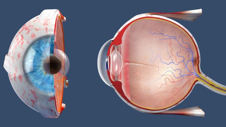
The retina is the neural layer at the back of the eyeball that initiates the visual process by converting light falling on it into electrical signals. Eye anatomy refers to all the structures that protect and nourish the retina and ensure that images are focused correctly. The cornea, lens, iris, vitreous and optic nerve are among the main components of this system. Light from the external environment passes through the transparent structures of the eye and reaches the retina. Signals generated in the retina are transmitted to the brain through the optic nerve, where they are transformed into a meaningful image. Visual quality depends on the coordinated functioning of the other anatomical structures of the eye as well as the health of the retina.
The Three Main Anatomical Layers of the Eye
The eyeball consists of three main layers with different functions. The outermost layer contains the sclera and cornea, which protect the eye from environmental factors. The middle layer is the vascular layer, which contributes to nourishing the eye and regulating the amount of light entering it. This layer, called the uvea, consists of the choroid, ciliary body and iris. The retina, which contains the nerve cells that detect light, is located in the innermost layer. These layers work together to preserve the structural integrity of the eye and maintain visual function.
The sclera is the dense connective tissue that forms the visible white part of the eye. It gives the eye its shape and protects its internal structures. It also provides the surface to which the muscles responsible for eye movements attach. The cornea, located at the front of the sclera, is transparent. Light first enters the eye through the cornea, where it is significantly refracted and directed towards the retina. The transparency and regularity of the corneal surface directly affect image clarity.
Functions of the Vascular Layer and Uvea
The uvea, which forms the middle layer of the eye, is rich in blood vessels. The choroid, located at the back of this structure, contributes particularly to nourishing the outer layers of the retina. The ciliary body, located in front of the choroid, is responsible for producing intraocular fluid. The muscles in the ciliary body also change the shape of the lens, allowing the eye to focus at different distances. The iris, which gives the eye its colour, is the frontmost part of the uvea. The pupil at the centre of the iris enlarges and contracts according to the amount of surrounding light, regulating the light entering the eye.
The retina is the neural layer lining the inner surface of the eye. It contains light-sensitive cells and neural networks that process information from these cells. The retinal pigment epithelium beneath the retina helps photoreceptor cells maintain their functions. This structure is important for nourishing the retina and removing cellular waste. The close anatomical relationship between the retina and choroid helps provide the oxygen and nutrients required by the visual cells. Damage to any of these layers can affect visual quality in different ways.
Functions of the Cornea and Lens
For a clear image to form, light must be focused on the correct point of the retina. The cornea and lens work together during this process. The cornea is a transparent, curved structure that provides a significant proportion of the eye's refractive power. The lens is located behind the iris and fine-tunes the focus of light. The flexibility of the lens contributes to clear vision of objects at different distances. The coordinated functioning of these two structures allows the image to be focused on the central part of the retina.
The lens is connected to the ciliary body by fine fibres. When the ciliary muscles contract, the tension on these fibres decreases and the lens becomes more rounded. This makes it easier to focus on objects at close distances. When the muscles relax, the fibres tighten and the lens becomes flatter, adapting for distance vision. This mechanism is called accommodation. As people age, reduced lens flexibility can cause difficulty with near vision, known as presbyopia.
Intraocular Fluids and Pressure
The structures between the cornea and lens in the front part of the eye form the anterior segment. This section contains the anterior chamber between the cornea and iris and the posterior chamber between the iris and lens. Both chambers contain a clear intraocular fluid called aqueous humour. Produced by the ciliary body, this fluid helps nourish the cornea and lens, which do not contain blood vessels. The intraocular fluid drains through structures such as the trabecular meshwork and the canal of Schlemm. The balance between fluid production and drainage plays a role in regulating intraocular pressure.
Problems with the drainage of intraocular fluid may cause increased intraocular pressure in some people. High intraocular pressure is an important risk factor for glaucoma, which can damage the optic nerve. However, glaucoma does not always occur with high pressure, and assessment is not based solely on pressure measurements. The posterior segment behind the lens forms a large part of the eyeball. This area is filled with a transparent, gel-like substance called the vitreous. The vitreous helps the eye maintain its shape and is closely associated with the retina.
Structure and Cells of the Retina
The retina is a complex neural tissue consisting of layers with different functions. Its primary role is to convert light energy into electrical signals and transmit this information to the optic nerve. The retina contains photoreceptors, bipolar cells, ganglion cells and various supporting cells. Photoreceptors are divided into two main groups called rods and cones. Rod cells contribute to vision in low-light conditions, while cone cells are responsible for colour and detailed vision. Signals from these cells are processed by other retinal cells and converted into a form that can be transmitted to the brain.
The photoreceptors that detect light are located in the part of the retina close to the choroid. Light therefore passes through the inner layers of the retina before reaching the photoreceptors. The extensions of the retinal ganglion cells join together to form the optic nerve. The optic disc, where the optic nerve exits the eye, does not contain photoreceptors. This area corresponds to the natural blind spot in the visual field. Müller cells and other supporting cells help maintain the structural integrity and cellular balance of retinal tissue.
The Role of the Macula and Fovea in Vision
The macula is the region at the centre of the retina responsible for detailed vision. It plays an important role in functions such as reading, recognising faces, driving and distinguishing fine details. The lutein and zeaxanthin pigments in the macula help filter certain wavelengths of light. These pigments also have antioxidant properties associated with protecting macular tissue. Healthy macular function is important for maintaining central vision. Conditions affecting this area can impair central vision while peripheral vision remains preserved.
At the centre of the macula is a small area called the fovea. The fovea is the area with high visual acuity and a dense concentration of cone cells. Its anatomical arrangement helps light reach the photoreceptors more directly. The absence of blood vessels at the centre of the fovea contributes to image clarity. Distinguishing letters while reading or noticing the details of an object largely depends on the function of this area. Oedema, a hole or structural changes affecting the fovea can cause significant problems with central vision.
Retinal Blood Vessels and Nutrition
The retina is one of the tissues that requires high levels of oxygen and nutrients. This requirement is met by two different circulatory systems. The inner layers of the retina are supplied by the central retinal artery and its branches. These vessels reach the eye through the optic nerve and spread to different areas of the retina. The outer retinal layers and photoreceptors are supplied mainly by the choroidal circulation. Healthy function of both systems is necessary for the visual cells to maintain their functions.
Occlusions in the retinal blood vessels can disrupt the blood supply to the affected area. This may manifest as a sudden reduction in vision or loss of part of the visual field. Disorders of the choroidal circulation may particularly affect the photoreceptors and retinal pigment epithelium. The movement of substances between the blood and retinal tissue is regulated by the blood-retinal barrier. In conditions such as diabetes, disruption of this barrier can cause vascular leakage and fluid accumulation in the retinal tissue. Assessing changes in the vascular structure plays an important role in the diagnosis and follow-up of various retinal conditions.
How Do Visual Signals Reach the Brain?
The visual process continues when electrical signals generated in the retina are transmitted to the brain. The extensions of the ganglion cells join together to form the optic nerve. The optic nerves from both eyes meet at the optic chiasm, located at the base of the brain. At this point, some of the nerve fibres cross to the opposite side, while others continue on the same side. Information relating to the left half of the visual field is therefore directed to the right side of the brain, while information relating to the right half is directed to the left side. Visual information passes through various neural pathways and reaches the visual cortex at the back of the brain.
The visual cortex allows signals from the eyes to be interpreted as a meaningful image. Vision therefore occurs through the combined function of the eyes, neural pathways and brain. Damage to the optic nerve can affect visual acuity or the visual field. Problems affecting the optic chiasm or the subsequent neural pathways can cause visual field loss involving both eyes. The distribution of these losses can provide information about the anatomical location of the problem. When necessary, visual field findings are assessed from both ophthalmological and neurological perspectives.
Age-Related Macular Degeneration and Its Symptoms
Age-related macular degeneration is a retinal condition affecting the macula, which is responsible for central vision. It is also commonly known as macular degeneration. The condition becomes more common with advancing age and is classified into two main groups, dry and wet. In the dry form, deposits called drusen and changes in the tissue over time may be observed in the macula. In the wet form, abnormal blood vessels can develop and leak fluid or blood. The course, type and effects of the condition on vision vary from person to person.
Central vision may become blurred in macular degeneration, or objects may appear different from their actual shape. Straight lines appearing wavy or distorted are among the symptoms requiring attention. A dark, empty or blurred area may be noticed in the centre of vision. Difficulty may arise in daily activities such as reading, recognising faces and seeing fine details. Some people may report that colours appear less vivid. If these symptoms begin recently or become more pronounced, an eye examination should be performed without delay.
How Does Diabetic Retinopathy Develop?
Diabetic retinopathy is an eye condition caused by damage to the retinal blood vessels due to diabetes. Blood sugar remaining high for a prolonged period can damage the structure of small blood vessels, causing leakage or occlusion. The condition may not cause symptoms during its early stages. Regular fundus examinations are therefore important for people with diabetes. Blood sugar, blood pressure and other accompanying health conditions can affect the course of the condition. The frequency of examinations is determined according to the person's general health and retinal findings.
During the early stages, microaneurysms, small retinal haemorrhages and lipid deposits leaking outside the blood vessels may be observed. Cotton-wool spots may also develop due to circulatory problems in the nerve fibre layer. Fluid accumulation in the macula is called diabetic macular oedema. This can cause blurred central vision. In advanced stages, new and abnormal blood vessels may develop in retinal tissue that does not receive an adequate blood supply. These vessels can bleed easily and, in some cases, create traction on the retina.
Retinal Detachment and Warning Signs
Retinal detachment is the separation of the neural retina from the supporting tissue beneath it. This separation can disrupt the nutrition of the photoreceptors and increase the risk of vision loss. The extent of the detachment and whether it affects the macula are among the factors determining its effects on vision. A sudden increase in floaters, flashes of light and a curtain-like shadow in the visual field are warning signs. Some people may also experience a sudden reduction in vision. These symptoms require assessment without delay.
Rhegmatogenous retinal detachment develops when fluid passes beneath the retina through a tear or opening. Age-related changes in the vitreous can create traction on the retina and cause a tear in some people. Tractional retinal detachment occurs when membranes or bands on the retinal surface pull the tissue out of position. This may be associated particularly with advanced diabetic retinopathy. In exudative detachment, fluid accumulates beneath the retina without a visible retinal tear. This fluid accumulation may result from inflammatory conditions or different problems involving the choroid and retinal pigment epithelium.
Retinal Examination and Imaging Methods
Fundus examination plays an important role in assessing retinal conditions. When necessary, drops can be used to dilate the pupil so that the retina, macula and optic nerve can be examined in detail. The structure of the retinal blood vessels, areas of haemorrhage, tears and changes in the macula are assessed during the examination. Additional imaging methods may be used when required to examine the different tissue layers. The type of examination performed is determined according to the symptoms and clinical findings. Changes detected at an early stage can help establish an appropriate follow-up and treatment plan.
Optical coherence tomography, or OCT, is an imaging method that uses light waves to produce cross-sectional images of the retina. The thickness and structural characteristics of the retinal layers can be assessed with this examination. It can be used to examine conditions such as macular oedema, fluid accumulation beneath the retina, macular holes and epiretinal membranes. It also contributes to assessing the nerve fibre layer around the optic nerve during glaucoma follow-up. Fundus photography helps document findings on the retinal surface. When necessary, ocular angiography can be performed to examine vascular structures, leakage and circulatory disorders.
Treatment Options for Retinal Conditions
The treatment of retinal conditions is planned according to the type and stage of the condition and the person's general health. Not every retinal problem is treated using the same method. Regular monitoring may be sufficient in some cases, while medication, laser treatment or surgery may be required for others. The aim of treatment is to reduce the effects of the existing problem and preserve visual function as much as possible. The response to treatment may vary from person to person. Assessment is therefore performed by considering examination findings together with imaging results.
Intravitreal anti-VEGF injections are among the treatment options that can be used for conditions such as wet age-related macular degeneration and diabetic macular oedema. VEGF is a protein that can increase abnormal blood vessel formation and vascular leakage in certain eye conditions. Anti-VEGF medications aim to reduce fluid accumulation in retinal tissue by suppressing this mechanism. The treatment plan is determined according to the characteristics of the condition and the findings obtained during follow-up examinations. Some people may require repeated injections at specific intervals. Changes resulting from treatment depend on factors such as the initial level of vision and the stage of the condition.
Laser photocoagulation is another method used for certain retinal conditions. Panretinal photocoagulation, used in advanced diabetic retinopathy, can help control the formation of abnormal blood vessels. Laser treatment around retinal tears may be preferred in suitable cases to reduce the risk of the tear progressing. The same treatment approach is not used for every retinal tear or vascular problem. Suitability for laser treatment is assessed according to the retinal findings. When necessary, laser treatment may be planned together with other methods.
Vitrectomy is a surgical method in which the vitreous gel inside the eye is removed and certain structural problems affecting the retina are addressed. It may be considered in cases such as retinal detachment, persistent intraocular haemorrhage and advanced diabetic retinopathy. It can also be used to treat epiretinal membranes on the macular surface or tissues causing traction on the retina. Macular holes are among the other conditions that may be treated with vitrectomy in suitable cases. The decision to perform surgery is made by considering the characteristics of the condition, expected benefits and potential risks. You can contact me through the website to arrange an examination and assessment of your retinal health.










































