Central serous retinopathy is an important retinal disease caused by fluid accumulation beneath the center of vision, known as the macula, and it directly affects visual quality. It may cause blurred central vision, straight lines appearing curved or wavy, faded color perception, and objects appearing different in size. Also known in the medical literature as central serous chorioretinopathy, the disease is commonly abbreviated as CSR or CSCR in clinical practice.
When planning treatment for central serous retinopathy, I evaluate how long the symptoms have been present, the extent to which vision has been affected, the patient's occupational visual requirements, and high-resolution retinal imaging findings as a whole. While close and regular monitoring may be sufficient for some patients, active and targeted treatment may be required in cases that are persistent, frequently recurrent, or carry a risk of tissue damage.
My primary treatment goals are to promote the absorption of fluid beneath the retina, restore the relationship between the retinal pigment epithelium and visual cells, and minimize the risk of permanent tissue loss, known as atrophy, in the center of vision. I particularly explain the following critical point to my patients: A partial improvement in vision does not necessarily mean that the fluid beneath the retina has disappeared completely. Therefore, I base follow-up and treatment decisions not only on the patient's current symptoms but also on microscopic findings obtained through OCT (Optical Coherence Tomography).
What Is Central Serous Retinopathy and How Does It Develop?
The retina is a sensitive layer of nerve tissue lining the inner back wall of the eye. It converts light stimuli into electrical signals and transmits them to the brain. The macula, located at the exact center of the retina, is responsible for sharp visual functions such as reading, recognizing faces, driving, and distinguishing fine details.
The primary mechanism in central serous retinopathy is increased permeability in the vessels of the choroid, the layer beneath the retina that supplies it with nutrients. Fluid crosses the retinal pigment epithelium (RPE) barrier and leaks beneath the macula. This leakage causes the retina to lift, with serous fluid accumulating beneath it. The accumulated fluid separates the light-sensing photoreceptor cells from their nourishing layer, resulting in impaired function.
Although the disease usually causes noticeable symptoms in one eye, detailed retinal and choroidal examinations may reveal silent vascular changes in the other eye as well.
What Causes Central Serous Retinopathy? What Are the Risk Factors?
It is not appropriate to attribute the disease to a single cause. It generally develops through a combination of structural susceptibility and environmental or hormonal triggers:
- Corticosteroid Use: This is one of the strongest and most established risk factors. Corticosteroids are not only administered as systemic tablets or injections. Nasal sprays, asthma inhalers, skin creams, intra-articular injections, and even certain herbal products or dietary supplements may increase cortisol levels in the body.
- Stress and Type A Personality: An intense workload, excessive stress, and increased sympathetic nervous system activity may raise choroidal vascular pressure and trigger leakage.
- Sleep Disorders and Shift Work: Irregular sleep patterns and obstructive sleep apnea syndrome are also among the risk factors.
- Hormonal Changes: Pregnancy, high endogenous cortisol levels such as those seen in Cushing syndrome, and hypertension may contribute to the condition.
⚠️ Critical Warning: If you are found to be using medication containing corticosteroids, you must not stop it suddenly on your own. Gradual dose reduction or transition to an alternative treatment should be planned in coordination with the physician who prescribed the medication.
What Are the Symptoms of Central Serous Retinopathy?
Patients generally present with the following symptoms, which may begin suddenly in one eye or become noticeable over several days:
- Blurred Central Vision: A dark, gray, or shadowed area in the center of the viewed point, known as a positive scotoma.
- Metamorphopsia (Distortion of Lines): Straight lines, door frames, or lines of text appear broken, curved, or wavy.
- Micropsia (Objects Appearing Smaller): Objects appear smaller or farther away than when viewed with the other eye.
- Impaired Color and Contrast Perception: Colors appear faded or dull, and light-dark contrast is reduced.
- Difficulty Focusing at Near Distances: Difficulty distinguishing letters while reading or adjusting the reading distance.
These symptoms are generally painless. Similar complaints may also occur in other serious retinal diseases, such as age-related macular degeneration or macular edema. A detailed retinal examination should therefore be performed without delay when symptoms begin.
How Is Central Serous Retinopathy Diagnosed?
I use advanced retinal imaging technologies during the diagnostic process:
- Detailed Fundus Examination: After the pupil has been dilated with eye drops, the macula and surrounding retinal tissue are evaluated with a biomicroscope.
- Optical Coherence Tomography (OCT): This is the gold standard for diagnosis and follow-up. It displays cross-sectional images of the retinal layers at micron-level resolution, clearly revealing the amount of fluid, the height of the leakage, and the condition of the photoreceptor layer.
- Fluorescein Angiography (FFA): A special dye administered intravenously is used to identify leakage points in the retinal vascular network, including mushroom-shaped or smokestack leakage patterns.
- Indocyanine Green Angiography (ICGA): This evaluates the structure of the choroidal vessels and deeper areas of excessive permeability, allowing the treatment area to be identified precisely.
- OCT Angiography (OCTA): This examines the retinal and choroidal vascular networks in three dimensions without using dye. It is particularly important for excluding choroidal neovascularization, which is abnormal new blood vessel formation, that may mimic or accompany the disease.
- Fundus Autofluorescence (FAF): This reveals the pattern of chronic retinal pigment epithelium damage and traces of previous episodes.
How Is Central Serous Retinopathy Treated?
When creating a treatment plan, the duration of the disease, whether it is acute or chronic, previous episodes, the condition of the other eye, the patient's occupational requirements, and OCT and angiography findings are evaluated together.
1. Monitoring and Lifestyle Changes in Acute Central Serous Retinopathy
In acute cases occurring for the first time without signs of permanent retinal damage, the fluid may resolve spontaneously within the first 1-3 months in many patients. During this period:
- Corticosteroid use is discontinued whenever possible after consultation with the relevant physician.
- Stress management and a regular sleep schedule are encouraged.
- OCT and visual acuity monitoring continue at monthly intervals.
However, the rule that "every patient should wait for 3-4 months" is not valid. Active treatment may be started earlier in patients with severe vision loss, those who drive regularly or perform visually demanding work, those with vision loss in the other eye, or those with recurrent episodes.
2. Active Treatment Methods for Chronic and Recurrent Disease
Active treatments are considered when fluid persists for more than 3 months, recurrent episodes develop, or a risk of photoreceptor loss becomes apparent:
- Photodynamic Therapy (PDT): This is one of the strongest treatment options for chronic and treatment-resistant CSR, with effectiveness demonstrated in international clinical studies. The light-sensitive substance verteporfin is administered intravenously and accumulates in choroidal vessels with increased permeability. A special cold laser is then applied to the targeted area to activate the medication. The aim is to close the excessively permeable choroidal vessels, stop the leakage, and promote fluid absorption. To reduce side effects, "half-dose" or "half-energy" protocols are now frequently and safely preferred.
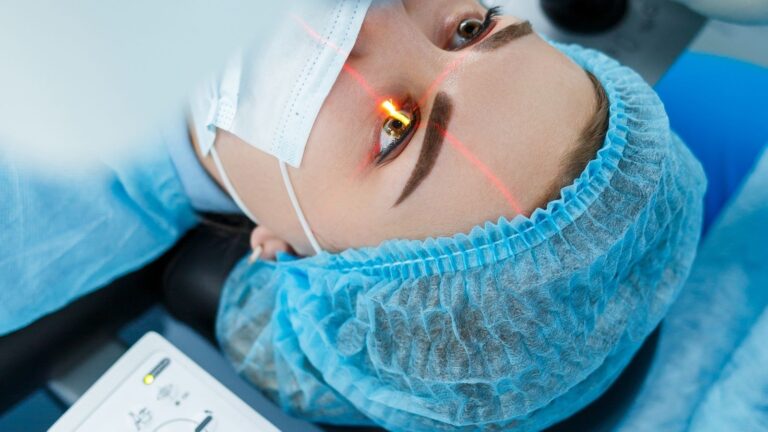
- Subthreshold Micropulse Laser: This tissue-sparing laser method stimulates the retinal pigment epithelium to pump fluid without creating a visible burn or scar. It can be safely used for leakage close to the center of vision or beneath the fovea.
- Conventional Focal Laser: This may be used when the leakage point is sufficiently far from the center of vision, known as the fovea. A focal laser is applied to close the leakage point. Because of the risk of scarring, it is not preferred for areas close to the center.
- Intravitreal Anti-VEGF Injections: Intravitreal injections are not routinely administered for CSR. However, if choroidal neovascularization, which is an abnormal new blood vessel network, develops secondary to the disease, anti-VEGF injections become necessary. This distinction should be clearly established with OCTA.
Recovery and Recurrence Risk in Central Serous Retinopathy
Fluid absorption does not always mean that vision will immediately return completely to its previous level. Even after the fluid has resolved, recovery of the photoreceptor cells and improvement in image quality may take several months. In chronic cases where the fluid has remained for a long time, mild color fading, reduced contrast, or slight visual distortion may become permanent.
Central serous retinopathy has a tendency to recur. Approximately 30-50% of patients may experience another episode in the same eye or the other eye after the first year. Therefore, periodic retinal examinations should not be neglected even if all symptoms have resolved.
Central Serous Retinopathy Treatment Prices
Treatment costs vary according to whether the disease is acute or chronic, the advanced imaging tests required, including OCT, angiography, and OCTA, the specific treatment method used, including monitoring, photodynamic therapy, micropulse laser, or focal laser, and the scope of follow-up. It is not medically appropriate to provide a standard treatment price without a detailed retinal examination and imaging. After the examination, a personalized treatment plan is clarified and detailed information is provided.