Türkçe
 Deutsch

Dry eye is a common but quality-of-life-impacting condition resulting from complex disorders of the tear film and ocular surface. It generally occurs due to inadequate tear production or excessive tear evaporation, which can lead to eye surface discomfort, vision disturbances, and, over time, tissue damage. Factors like meibomian gland dysfunction (MGD) may increase evaporation, while inflammation underpins the disease. The treatment process involves the use of artificial tears and anti-inflammatory medications, tear preservation, and MGD therapy. With early diagnosis and appropriate treatment, symptoms can largely be brought under control.
| Possible Causes | – Reduced tear production – Increased tear evaporation – Meibomian gland dysfunction (impaired oil glands in the eyelids) – Aging – Prolonged screen use – Contact lens use – Environmental factors (wind, dry air, air conditioning) – Autoimmune diseases (e.g., rheumatoid arthritis, Sjögren’s syndrome) – Certain medications (antihistamines, antidepressants, blood pressure drugs) – Post-eye surgery (e.g., LASIK) |
| Symptoms | – Sensation of dryness in the eyes – Burning or stinging – Eye redness – Blurred vision (temporary) – Foreign body sensation in the eye – Tearing (reflex tearing due to dryness) – Light sensitivity |
| Risk Factors | – Aging – Hormonal changes in women (menopause) – Long periods of screen use – Extended or incorrect use of contact lenses – Prolonged exposure to windy or dry air – Smoking – Chronic illnesses (e.g., diabetes, autoimmune diseases) |
| Diagnostic Methods | – Eye examination – Schirmer test (measures tear quantity) – Tear film layer tests (e.g., BUT – Tear Break-Up Time) – Meibography (imaging of the meibomian glands) |
| Treatment Methods | – Artificial tear drops or gels – Medications that reduce eye dryness (e.g., cyclosporine drops, lifitegrast) – Lipid-based tear drops (for oil gland dysfunction) – Warm compress and eyelid massage – Environmental control (using a humidifier, protecting against air conditioning and wind) – Punctal plug insertion or surgical intervention in advanced cases |
| Prevention | – Regularly moisturizing the eyes – Frequent blinking and following the 20-20-20 rule when using screens – Wearing protective eyewear in windy and dry weather – Adhering to proper contact lens guidelines – Drinking plenty of water and maintaining a balanced diet (foods rich in omega-3 fatty acids and vitamin A) – Avoiding smoke and pollutants |
What Is Dry Eye?
The historical development of dry eye disease is tied to the deepening scientific understanding of ocular surface disorders and the tear film. Initial observations date back to 1892 with Jan Mikulicz-Radecki’s description of “Mikulicz syndrome,” involving swelling of the parotid and lacrimal glands. However, because this term encompassed various clinical situations, it led to diagnostic ambiguities. In 1933, Swedish ophthalmologist Henrik Sjögren introduced the term “keratoconjunctivitis sicca,” distinguishing dry eye from other causes of dryness. Sjögren’s work laid the foundation for recognizing dry eye as a distinct clinical entity.
In the 1960s and 1970s, research explored the detailed structure and function of the tear film. This period highlighted how the lacrimal glands, meibomian glands, and ocular surface epithelia interact harmoniously to maintain tear film integrity. The concept of the “lacrimal functional unit” described this integrated nature. Additionally, it was during this era that the mucin-containing glycocalyx—crucial for preserving the corneal epithelium—was discovered.
Significant advances in defining and classifying dry eye came in 1995, when the National Eye Institute (NEI) defined the condition based on discomfort and damage caused by tear film abnormalities, distinguishing “aqueous-deficient” from “evaporative” forms. In 2007, the International Dry Eye Workshop (DEWS) expanded the definition, emphasizing tear film instability, hyperosmolarity, and inflammation as core components. The 2017 DEWS II report further refined these definitions, adding neurosensory abnormalities and loss of tear film homeostasis as contributors to disease etiology.
What Causes Dry Eye?
Dry eye results from a complex interplay of factors disrupting the balance of the tear film and ocular surface. Causes are multifactorial, ranging from lifestyle and environmental influences to systemic diseases.
- Meibomian Gland Dysfunction (MGD):
The meibomian glands secrete lipids for the tear film’s outer layer, preventing tear evaporation. When these glands fail, the lipid layer is disrupted, accelerating tear evaporation. Aging, hormonal shifts, and certain systemic diseases can raise MGD risk. - Insufficient Tear Production:
Inadequate lacrimal gland function can lead to insufficient tear quantity. Causes include autoimmune disorders (e.g., Sjögren’s syndrome), aging, or the effects of certain medications that reduce tear output. - Inflammation:
Chronic inflammation impairs tear production and damages epithelial cells, exacerbating dryness. Environmental triggers or systemic autoimmune responses can spark this inflammatory cycle. - Environmental Factors:
Low humidity, wind, smoke, or extensive screen use increase tear evaporation—a primary concern in modern lifestyles. - Hormonal Changes:
Postmenopausal women in particular may experience changes that adversely affect tear production and lipid balance. - Systemic Illnesses and Medications:
Conditions like rheumatoid arthritis or diabetes raise dry eye risk, as do certain drugs like antihistamines and antidepressants. - Neurological and Anatomical Factors:
Nerve damage or eyelid dysfunction can disrupt tear distribution, leading to dryness.
How Common Is Dry Eye?
Dry eye is broadly recognized as a public health concern, presenting varying prevalence worldwide depending on factors such as age, gender, region, and diagnostic criteria. Global studies indicate prevalence rates between 5% and 50%. This wide range arises from differences in methodology and definitions. For instance, the TFOS DEWS II report highlights the challenge of consistent prevalence data.
Age and gender are key predictors. The incidence escalates notably with age, particularly in individuals over 50. Data from the United States indicate that 8.4% of those under age 60 report symptoms, rising to 19% among those aged 80 and up. Women are generally more susceptible than men, with hormonal changes—especially after menopause—playing a significant role in tear production.
Geographical variances also matter. While Brazil’s prevalence is around 13%, it can reach 41% in Mexico. Among office workers and students, rates can exceed 70%, underscoring the importance of lifestyle and environment.
How Does Dry Eye Develop?
Dry eye disease arises from tear film instability and inflammation involving multiple interrelated mechanisms. The tear film comprises three layers—lipid, aqueous, and mucin. Dysfunction in any one layer accelerates evaporation, driving the tear film into a hyperosmolar state. Hyperosmolarity triggers inflammatory responses on the ocular surface, resulting in epithelial damage and goblet cell loss, further destabilizing the tear film.
Hyperosmolarity-induced inflammation is mediated by cytokines and matrix metalloproteinases (MMPs). These mediators cause apoptosis of epithelial and goblet cells, disrupting the mucin layer responsible for proper tear film adherence to the ocular surface. Without mucin, the tear film degrades more quickly.
Meibomian gland dysfunction (MGD) is a significant contributor to evaporative dry eye, reducing lipid secretion and undermining tear film stability. Additionally, corneal nerve damage can reduce reflex tear production and blink rate, hindering tear film renewal.
What Are the Symptoms of Dry Eye?
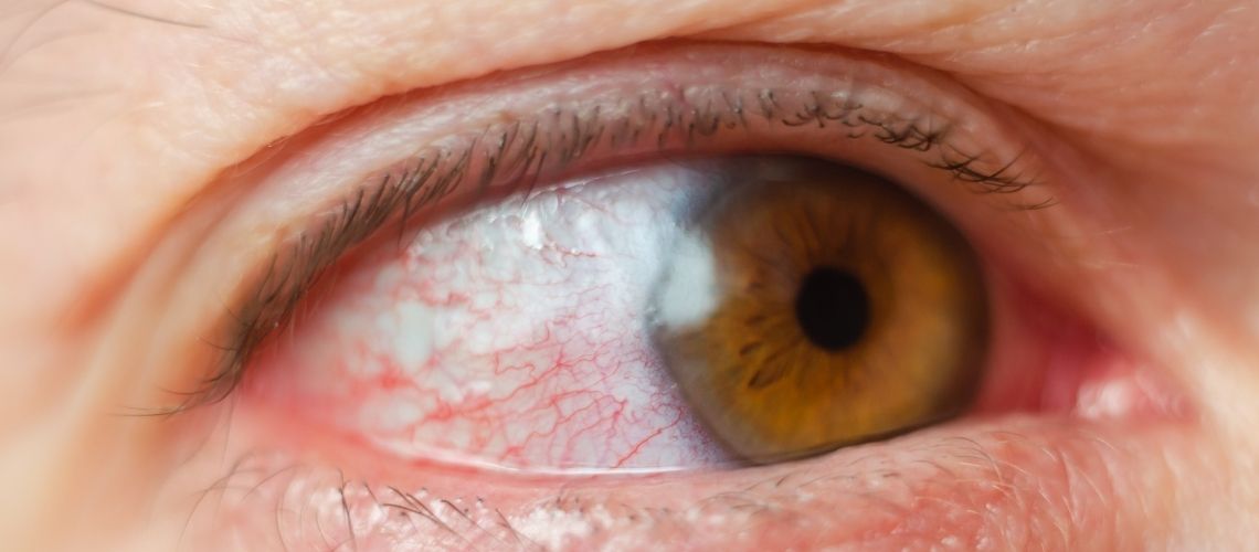
Dry eye symptoms are varied but typically include discomfort, burning, stinging, or the sensation of a foreign body in the eye. Some patients report a constant gritty feeling, while reflex tearing may paradoxically arise from dryness. Blurred or fluctuating vision often becomes noticeable during tasks like reading, computer use, or driving, impacting daily life. Light sensitivity (photophobia) is also common, and redness and fatigue of the eyes may intensify after prolonged visual activities. In severe cases, chronic inflammation can cause lasting damage to the ocular surface.
How Is Dry Eye Diagnosed?
Dry eye disease affects quality of life and requires accurate diagnosis. The diagnostic process involves a thorough patient history, clinical examination, and specialized tests.
An initial step is evaluating the patient’s complaints using standardized questionnaires like the Ocular Surface Disease Index (OSDI), which covers symptom frequency, impact on daily life, and environmental triggers over the past week. An OSDI score above 15 suggests possible dry eye and the need for further assessment.
A comprehensive eye examination then checks for eyelid abnormalities, meibomian gland function, tear film stability, and ocular surface condition. Eye blinking frequency and completeness are also assessed for clues about excessive evaporation.
Various objective tests further clarify the diagnosis:
- Tear Film Break-Up Time (TBUT): Measures tear film stability. Values under 10 seconds indicate instability.
- Schirmer Test: Uses a strip of paper in the lower eyelid to gauge tear production; results under 10 mm suggest insufficient tear flow.
- Tear Osmolarity: Levels above 308 mOsm/L signify dry eye, providing high diagnostic accuracy.
- Ocular Surface Staining: Dyes like fluorescein or lissamine green detect epithelial damage.
- Meibomian Gland Evaluation: Reviews the function and structure of these glands, which can be critical in evaporative dry eye.
How Is Dry Eye Treated?
Dry eye management demands an individualized approach focusing on increasing tear production or reducing tear evaporation to relieve ocular surface discomfort. Treatment is chosen based on disease severity and underlying causes:
- Artificial Tears and Drop Therapy:
Initial treatment often involves artificial tear solutions that lubricate the ocular surface, offering temporary relief. Mild cases can use low-viscosity drops, while more severe cases may benefit from thicker gels or night-time ointments. Preservative-free formulas help prevent irritation in frequent users. - Punctal Plugs:
If artificial tears alone are insufficient, punctal plugs may be used to block tear drainage channels. By retaining tears on the ocular surface, they provide symptomatic relief, especially for patients with autoimmune-related reduced tear production. This is a minimally invasive office procedure, but follow-up is required to watch for rare complications like foreign-body sensation or infection. - Laser Treatments:
In cases where corneal surface irregularities exacerbate dryness, procedures like phototherapeutic keratectomy (PTK) can smooth the cornea, potentially stabilizing the tear film. Though not a routine therapy, it can improve visual acuity and lessen symptoms if standard treatments fail. - Botulinum Toxin Injections:
Botox (BTX-A) injections may assist dry eye by reducing muscle spasms that affect tear dynamics. Primarily used for conditions such as benign essential blepharospasm or hemifacial spasm, it can raise the tear meniscus height and ease dryness. However, injections must be repeated periodically as the effect is temporary. - Supportive Therapies:
Managing meibomian gland dysfunction (MGD) is pivotal. Techniques such as lid hygiene, warm compresses, and lid massage can restore gland function. Inflammation can be controlled with topical corticosteroids or immunomodulators like cyclosporine A, reducing ocular surface inflammation and enhancing tear production.
What Are the Complications of Dry Eye?
Although it often appears mild, untreated dry eye can lead to serious complications, substantially compromising patients’ quality of life. Understanding these complications underscores the importance of proper management and early intervention:
- Ocular Surface Damage:
Chronic dry eye can result in corneal abrasions, epithelial defects, and in severe cases, corneal ulcers. The cornea is integral to clear vision, so damage can lead to vision loss. - Heightened Infection Risk:
The tear film barrier is essential for protecting the ocular surface from pathogens. When tears are insufficient, bacteria and viruses can penetrate more easily, raising the likelihood of conjunctivitis and keratitis and complicating disease management. - Chronic Inflammation:
Persistent inflammation can lead to conjunctival scarring and goblet cell loss. These cells produce mucus for tear film stability, and their depletion further disrupts the tear film, exacerbating dryness. - Reduced Quality of Life:
Symptoms like irritation, photophobia, and blurred vision can hamper daily tasks such as reading, driving, or computer use. Over time, this may cause psychological distress and social withdrawal. - Corneal Scarring and Neovascularization:
In advanced cases, prolonged dryness leads to corneal scarring and abnormal blood vessel growth, threatening corneal clarity and permanent vision loss.
When Is Treatment for Dry Eye Indicated?
The timing of dry eye treatment depends on disease severity, how symptoms affect daily life, and any underlying causes. Patients with mild to moderate symptoms often start with artificial tears or lubricating drops. These therapies are especially beneficial for discomfort from prolonged screen use, environmental dryness, or contact lens wear, and can be used upon symptom onset or prophylactically.
Moderate to severe cases unresponsive to artificial tears might benefit from punctal plug insertion, which helps preserve the tear film, particularly beneficial in patients with autoimmune-related tear deficiency. Punctal plugs are frequently employed for long-term control.
More resistant or complicated cases may warrant advanced therapies like laser procedures or botulinum toxin injections if standard measures fail and ocular surface abnormalities heavily contribute to symptoms. Corneal irregularities can be corrected with laser treatments to improve tear film distribution.
When Is Treatment for Dry Eye Contraindicated?
Dry eye treatments should be planned according to each patient’s ocular and general health status. Certain circumstances may restrict treatment options. A thorough evaluation is crucial before initiating therapy:
- Preservative Sensitivity:
Artificial tears are a cornerstone therapy. However, patients sensitive to preservatives risk irritation or allergic reactions, necessitating preservative-free products. - Active Ocular Infections:
Interventions like punctal plugs that aim to conserve tears must not be done during active infections, which could spread. Treatment should proceed once infection is controlled. - Severe Dry Eye Syndrome:
Laser procedures that refine the corneal surface can raise complication risks and impair healing in severely dry eyes. Bringing dryness under better control is often recommended prior to laser treatment. - Autoimmune Diseases:
Conditions like Sjögren’s syndrome may hamper healing. Surgical or invasive options can be risky, prompting consideration of alternative methods. - Neuromuscular Disorders:
While botulinum toxin injections can help certain ocular issues, they may exacerbate muscle weakness in conditions like myasthenia gravis, making them contraindicated. - Pregnancy and Lactation:
The safety of botulinum toxin during pregnancy and breastfeeding is not fully established. Avoiding such therapies is advised for this period.
What Is the Recovery Process Like for Dry Eye?
The healing process for dry eye is individualized, contingent on disease severity, underlying causes, and treatment strategies. Typically, therapy aims to address specific deficits in tear production or evaporation and to resolve any ocular surface pathology.
Artificial Tears and Eye Drops:
For initial therapy, patients often use artificial tears or lubricants, relieving dryness by maintaining ocular surface moisture. Preservative-free formulas are crucial for frequent use to reduce irritation.
Punctal Plugs:
In advanced cases, punctal plugs help retain tears. After this minimally invasive procedure, patients often experience improvement within a few days, enhanced through routine follow-ups.
Ocular Surface Interventions:
Laser procedures or botulinum toxin injections address specific corneal and eyelid issues. For corneal irregularities exacerbating dryness, laser therapy can improve tear film distribution, provided instructions are followed rigorously for optimal healing.
How to Prevent Dry Eye
Controlling environmental factors is essential for preventing dry eye. Humidifiers can counteract dry indoor air, particularly beneficial in winter when heating systems lower humidity. Avoid direct airflow from hairdryers, fans, or air conditioners. In windy or polluted settings, wraparound sunglasses shield the eyes from environmental strain.
Digital screens play a major role in modern dry eye cases. Limiting screen time and taking regular breaks helps preserve ocular surface moisture. The 20-20-20 rule—looking at an object 20 feet away for 20 seconds every 20 minutes—effectively supports eye health. Avoiding smoke (both active and secondhand) is likewise important, as irritants can worsen dryness.
Omega-3 fatty acids help support tear production and reduce inflammation. Foods like salmon, walnuts, and chia seeds are recommended. Vitamin A–rich foods (e.g., carrots, dark leafy greens) play a vital role in maintaining a healthy ocular surface.
Artificial tears may suffice for mild symptoms, providing short-term relief. If discomfort persists, consulting an ophthalmologist and exploring personalized treatment options is essential.
Frequently Asked Questions
Is there a link between dry eye and diet?
Yes. Diet can influence dry eye. Foods rich in omega-3 fatty acids can enhance tear quality and mitigate dryness symptoms. Fatty fish like salmon, tuna, and sardines are good sources of omega-3.
Does dry eye worsen seasonally?
Yes. In colder months, dryness often intensifies as indoor heating depletes humidity and disrupts tear stability.
Which medications can cause dry eye?
Certain drugs—including antihistamines, antidepressants, birth control pills, and blood pressure medications—may trigger dryness by reducing tear production.
Can contact lens use be affected by dry eye?
Yes. Dry eye can make contact lens wear uncomfortable and irritating, as dryness prevents the lens from staying well-lubricated.
Do children also experience dry eye?
Yes, although more common in older adults, children can also develop dry eye due to environmental factors or extensive screen use.
What happens if dry eye is left untreated?
Failure to manage dry eye can result in corneal damage, higher infection risk, and possible vision loss, all of which degrade overall ocular health and quality of life.
How is dry eye diagnosed?
An ophthalmologist diagnoses dry eye through thorough examination and tests evaluating tear production. These findings guide treatment planning.
Which habits help prevent dry eye?
Setting limits on screen usage, taking regular breaks, using humidifiers, and avoiding smoke all help. Incorporating omega-3–rich foods into one’s diet is also beneficial.