Türkçe
 Deutsch

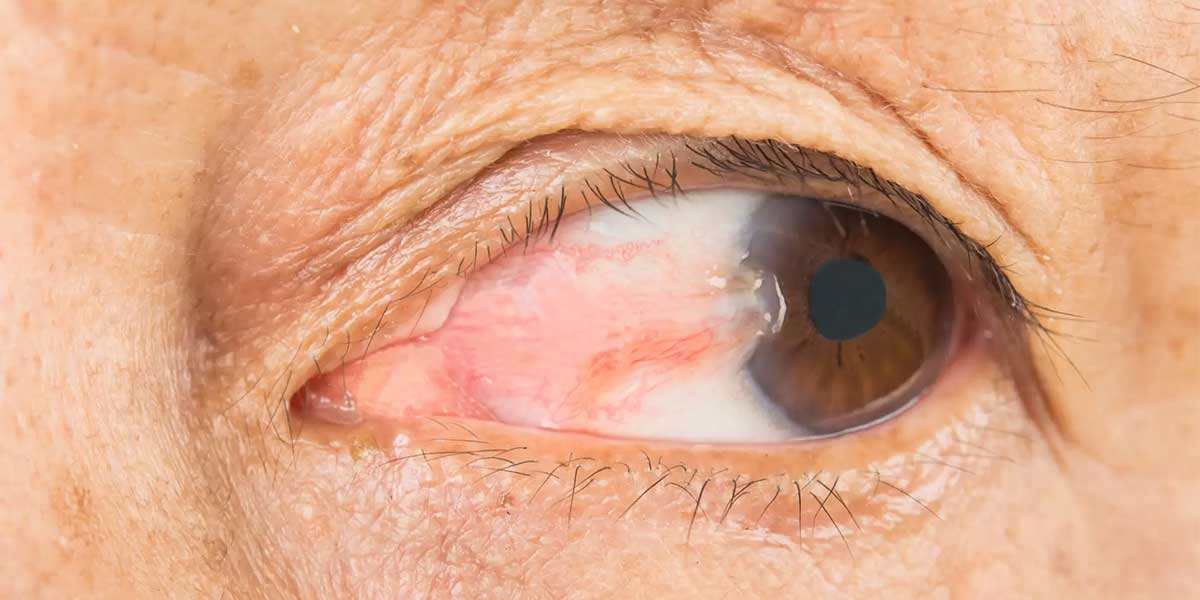
Pterygium, commonly known as “Bird’s Wing” among the public, is a disease medically referred to as pterygium but also known as “eye flesh.” It is the name given to a piece of tissue located on the transparent layer called the cornea, which is the frontmost part of the eye. It is generally more common in men. Pterygium disease is more frequently encountered in people who work outdoors and are exposed to sunlight and wind.
Turkey is one of the best country for Pterygium Surgery at a afforable price. If you are looking for a place for Pterygium Surgery abroad with best clinics and doctors, you are definitly on the right spot. Check Dr. Berkay Akmaz's Pterygium Surgery prices, reviews and packages.
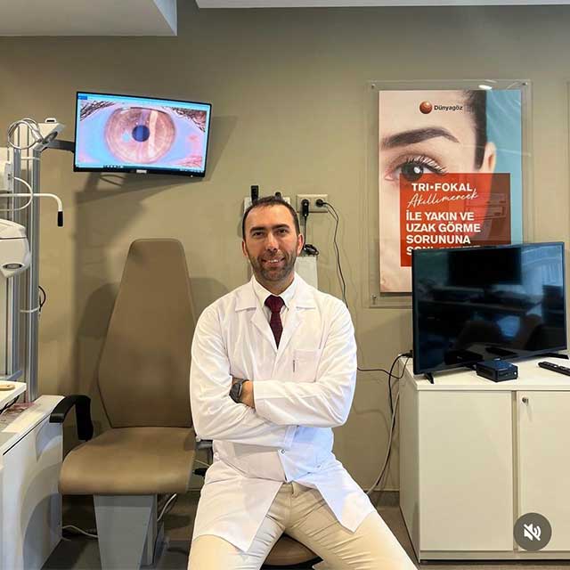
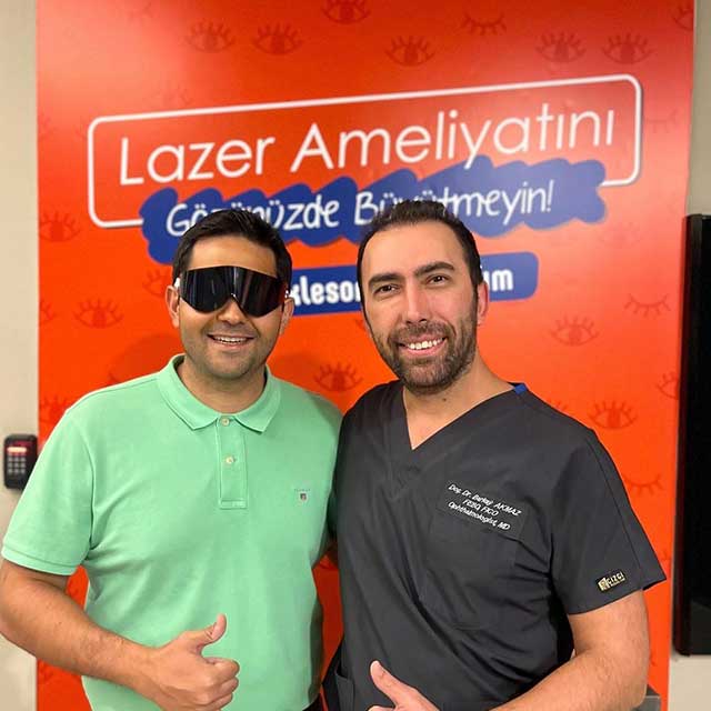
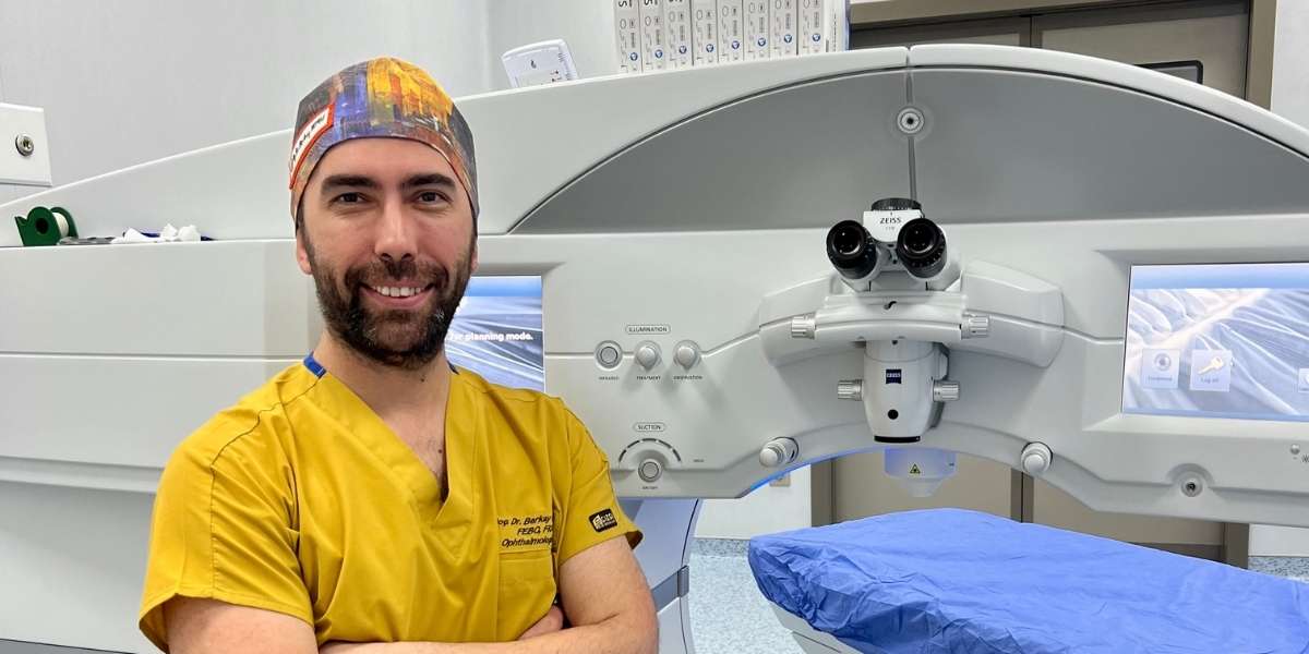
Ophthalmology Specialist – Assoc. Prof. Dr. Berkay Akmaz was born in İzmir in 1985. Due to his father's profession, he spent his primary school years in different cities. He began his education at Gülhane Military Medical Academy in 2003 and graduated with the title of Medical Doctor from Ege University Faculty of Medicine in 2010. In 2010, Assoc. Prof. Dr. Berkay Akmaz ranked 25th in the Medical Specialization Exam (TUS) in Turkey and started his specialization in the Department of Ophthalmology and Surgery at Dr. Lütfi Kırdar Kartal Education and Research Hospital. He completed his specialization training in 2014.






| Definition | Pterygium is a triangular-shaped tissue growth that extends from the conjunctiva of the eye towards the cornea; commonly known as “eye flesh”. |
| Symptoms | Redness in the eye, irritation, burning, itching, sensation of a foreign body, vision impairment in advanced cases. |
| Causes | Prolonged exposure to ultraviolet (UV) rays, working in dusty or windy environments, dry eye syndrome. |
| Risk Factors | Living near the equator, working outdoors, male gender, genetic predisposition. |
| Diagnostic Methods | Eye examination, slit lamp biomicroscopy, visual acuity test. |
| Treatment Options | Lubricating (moisturizing) eye drops, anti-inflammatory drops, surgical intervention. |
| Surgical Indications | Pterygium that obstructs the visual axis, cosmetic discomfort, frequent inflammation, interference with contact lens use. |
| Surgical Methods | Excision and Autograft Technique: Removal of the pterygium and placement of a graft taken from the patient’s own conjunctiva; Amniotic Membrane Transplantation; Mitomycin-C Application. |
| Operation Duration | Approximately 30-45 minutes and performed under local anesthesia. |
| Recovery Process | May experience mild pain and discomfort in the first few days; full recovery typically takes 2-4 weeks. |
| Risks and Complications | Risk of recurrence, infection, scar formation, corneal clouding, astigmatism. |
| Recurrence Rate | Varies depending on the surgical technique; recurrence rate is between 5-10% with the autograft method, higher with simple excision. |
| Postoperative Care | Use of antibiotic and steroid eye drops, avoiding rubbing the eyes, protection from sunlight, regular doctor check-ups. |
| Prevention | Wearing UV-protected sunglasses, protecting the eyes in dusty and windy environments, keeping the eye surface moist with artificial tears. |
| Alternative Treatments | Medication can alleviate symptoms but does not reduce pterygium; surgery is generally the most effective treatment method. |
| Social and Functional Impact | Can reduce visual quality, cause aesthetic discomfort, affect daily activities. |
| Success Rate | Has a high success rate and low recurrence rate when performed by experienced surgeons. |
| Technology and Developments | Sutureless surgical techniques and fibrin glues accelerate the healing process and enhance patient comfort. |
| Insurance Coverage | Generally covered by insurance in cases of medical necessity. |
| Precautions | Follow the doctor’s recommendations post-surgery, use prescribed drops regularly, do not miss follow-up appointments. |
| Lifestyle Recommendations | Limit sun exposure, use artificial tears, avoid smoking. |
| Affected Age Group | Generally more common in adults aged between 20-50 years. |
What Is Pterygium Surgery?
Pterygium surgery aims to carefully remove the abnormal tissue formed on the cornea. First, the surgeon begins the procedure by making an incision at the limbus. Then, the pterygium is cleaned with special dissection techniques, and the cornea is polished. Afterwards, the conjunctiva and Tenon’s capsule are carefully dissected to remove scar tissue.
Surgeons often apply a conjunctival autograft or amniotic membrane graft to reduce the risk of recurrence. Because autografts have lower recurrence rates compared to the bare sclera technique. Additionally, medications like mitomycin-C can be used to support the prevention of regrowth, and postoperative care is important.
Initially, the bare sclera technique was preferred, but today more advanced methods are used. Especially, conjunctival autograft applications stand out due to their potential to reduce recurrence rates. Although the FDA has not approved a specific drug for pterygium treatment, medications like mitomycin-C are preferred off-label.
Current studies in the field of pterygium surgery focus on reducing recurrence rates and speeding up the healing process. With the widespread adoption of this surgical procedure, new techniques that improve treatment outcomes are continuously being developed. Thus, postoperative complications have decreased, and patient satisfaction has increased.
How Does Pterygium Surgery Work?
Pterygium surgery involves several different techniques aiming to reduce recurrence rates. Among these, the simplest method, the Bare Sclera Technique (BSR), involves the removal of the pterygium and leaving the sclera to heal naturally. However, due to its high recurrence rate, it is less preferred today.
Conjunctival Autograft (CAU) is performed by transplanting a graft taken from the patient’s own conjunctiva to the scleral area after pterygium excision. This method covers the excised area with healthy conjunctival tissue, suppressing fibroblastic proliferation and reducing the risk of recurrence. It has a high success rate because it creates a normal conjunctival surface.
Another technique, Amniotic Membrane Graft (AMG), uses the amniotic membrane to cover the sclera and reduce inflammation. It is especially useful in cases where a conjunctival autograft is not suitable. AMG supports wound healing and prevents scar formation.
In some cases, surgeons use an antimetabolite called Mitomycin-C (MMC) to inhibit fibroblast activation. MMC is applied intraoperatively to reduce recurrence. By suppressing fibrovascular growth, it lowers the risk of pterygium reformation. It is usually combined with CAU or AMG.
Finally, one of the modern techniques, Contact Inhibition, involves the proper suturing of conjunctival tissue. This technique ensures there are no gaps, preventing cell proliferation and reducing the likelihood of recurrence.
When Should Pterygium Surgery Be Performed?
Pterygium surgery is a treatment method preferred when the pterygium causes problems in certain situations. It is especially important for patients with visual impairment because as the growth progresses towards the cornea, it can obstruct vision and lead to visual defects like astigmatism. Cosmetic concerns also cause some patients to request surgery since pterygium can affect the appearance of the eye and cause discomfort.
Moreover, if the pterygium shows progressive growth, surgical intervention may be necessary, as this condition can lead to more serious complications over time. Persistent eye irritation, redness, foreign body sensation, or dryness are also significant indications for surgery.
Surgical indications include:
- Corneal Involvement: If the pterygium has spread more than 3 mm onto the cornea or is causing significant corneal changes, intervention is required.
- Restriction of Ocular Motility: Although rare, growth can restrict eye movements and may need to be corrected surgically.
- Suspicion of Malignancy: Pterygia that carry the risk of malignancy with signs of abnormal growth or color change are removed surgically.
Surgery is recommended when these symptoms affect daily life or when the risk of ocular complications increases if the growth is left untreated. Additionally, adjuvant therapies like conjunctival autograft and mitomycin-C are used to reduce the risk of recurrence. These methods can be effective in decreasing the likelihood of regrowth after surgery.
Who Is Not Suitable for Pterygium Surgery?
Pterygium surgery may not be suitable for some patients. Such contraindications include factors that may increase the risks of surgery or jeopardize the healing process. Here are the conditions where pterygium surgery is not recommended:
- Active Ocular Infections: Infections like conjunctivitis or keratitis present on the eye surface can make pterygium surgery risky because infection can negatively affect postoperative healing.
- Severe Dry Eye Syndrome: Patients experiencing severe dry eye problems may face increased dryness, discomfort, and graft adhesion issues after surgery. Therefore, surgery should be approached cautiously in such cases.
- Autoimmune Diseases: Autoimmune diseases like rheumatoid arthritis or Sjögren’s syndrome can complicate wound healing. Surgical intervention is generally not recommended due to the high risk of postoperative complications in such situations.
- Uncontrolled Systemic Diseases: Individuals with uncontrolled systemic diseases like diabetes or hypertension may increase the risk of infection and graft failure during pterygium surgery. Therefore, careful evaluation is necessary for patients with such conditions.
- Recently Underwent Ocular Surgery: In individuals who have recently undergone eye surgery, a new surgical intervention may increase the risk of scar formation or graft failure.
- Bleeding Disorders: Patients with bleeding disorders like coagulopathy may carry a risk of excessive bleeding during surgery. Therefore, careful risk assessment is important in such cases.
How Is Pterygium Surgery Performed?
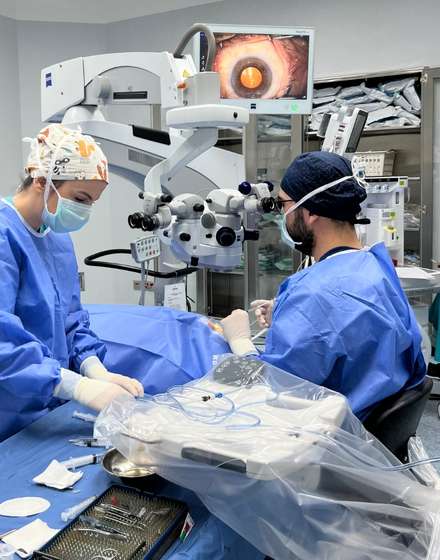
Pterygium surgery begins by preparing according to the size and location of the pterygium. After applying local anesthesia, the surgeon uses special surgical instruments to remove the pterygium. During these procedures, the pterygium tissue is carefully separated from the cornea and conjunctiva surfaces. Thus, the underlying Tenon’s capsule is removed to minimize the risk of recurrence.
After completely removing the pterygium, the corneal surface is smoothed. The surgeon can choose one of two different grafting techniques to further reduce the risk of recurrence:
- Conjunctival Autograft: Conjunctival tissue taken from under the upper eyelid is placed in the excised area. The graft is secured with stitches or fibrin glue.
- Amniotic Membrane Transplantation: In large pterygium cases, the amniotic membrane obtained from human placenta is placed over the defect to support the healing process.
After the closure process is completed, the graft is secured with stitches or tissue adhesive. Using tissue adhesives in this procedure significantly increases postoperative patient comfort. Therefore, although using tissue adhesive increases the cost, it enhances patient comfort and success rate. Afterwards, a shield is placed over the eye for protection.
What Are the Side Effects of Pterygium Surgery?
After pterygium surgery, patients may encounter various side effects and complications. These commonly include redness, irritation, tearing, and a feeling of discomfort in the eye. Additionally, blurred vision and light sensitivity may occur after surgery. These effects are usually temporary and decrease with appropriate care. Dry eye is a common condition due to the impact on the tear film during the procedure. Patients can use artificial tear drops to alleviate this discomfort.
Some patients may experience a foreign body sensation in the eye during the healing process. This feeling usually decreases over time but can be persistent in some cases. Long-term risks include astigmatism resulting from changes in the shape of the cornea. If astigmatism develops, correction can be achieved with glasses, contact lenses, or additional surgical procedures if necessary.
Common Side Effects:
- Redness and irritation
- Tearing and discomfort
- Blurred vision and light sensitivity
- Dry eye
- Foreign body sensation
Another significant long-term risk is the regrowth of the pterygium. Although new techniques like conjunctival autograft are effective in reducing this risk, they cannot eliminate it entirely.
The risk of infection is also present and manifests with symptoms like redness, pain, and discharge. In this case, prompt antibiotic treatment is necessary. Additionally, postoperative scar tissue formation can affect the cornea. In rarer cases, corneal thinning or perforation may occur, and a complication known as symblepharon can lead to adhesion of the conjunctiva to the cornea.
How Successful Is Pterygium Surgery?
The success rates of pterygium surgery vary depending on the techniques used, as well as patient characteristics and postoperative care. Success is generally evaluated based on three main factors: recurrence rates, cosmetic results, and long-term effects.
- Recurrence Rates: Recurrence is a significant challenge in pterygium surgery. Recurrence rates vary between 5% and 88% depending on the surgical technique and methods used. Techniques like conjunctival autograft (CAG) provide effective results by limiting recurrence between 5% and 20%. Combining this method with mitomycin-C (MMC) can reduce recurrence rates to as low as 5-10%. An advanced technique called P.E.R.F.E.C.T. stands out as one of the most successful methods with a recurrence rate below 1% in studies.
- Cosmetic Results: Cosmetically satisfactory results play a significant role in the success of the surgery. The combination of femtosecond laser-assisted surgery and conjunctival autograft offers good to excellent cosmetic results in 93% of patients. Poor cosmetic results are observed only in about 3% of cases, usually due to incomplete removal of the pterygium.
- Long-Term Results: Long-term observations show that most recurrence cases occur within the first 6 months after surgery. The conjunctival autograft technique is widely accepted as a standard method in many cases by providing long-term protection against recurrence.
How to Prepare for Pterygium Surgery?
When preparing for pterygium surgery, it is essential to evaluate the patient’s risk factors and overall health condition. It is known that excessive exposure to sunlight is associated with recurrence in recurrent pterygium cases. Therefore, the patient is advised to avoid excessive sunlight and use protective eyewear before surgery. Additionally, determining the size, thickness, and location of the pterygium with a comprehensive ophthalmological examination is important.
The following steps are taken to gather information about the condition and vascularity of the pterygium:
- Evaluating the thickness and vascularity of the pterygium.
- Performing imaging tests like slit-lamp examination.
Preoperative medication therapy is important in inflamed pterygium cases. Anti-inflammatory eye drops (e.g., corticosteroids) may be recommended to reduce inflammation. Additionally, topical mitomycin-C (MMC) application may be considered in some cases to reduce the risk of recurrence. These applications are important for providing faster healing after surgery and reducing complications.
The patient’s lifestyle should also be considered during the preparation process for pterygium surgery. It is recommended to limit outdoor activities related to eye health and use UV-protective sunglasses and wide-brimmed hats. Preoperative antibiotic prophylaxis may be considered against the risk of infection, determined by the surgeon based on the patient’s risk assessment.
How Should Postoperative Care Be Managed After Pterygium Surgery?
Proper care after pterygium surgery supports the healing process and prevents complications. First, medication use should be carefully followed. Postoperative antibiotic and anti-inflammatory eye drops prescribed by the doctor reduce the risk of infection and control inflammation. Steroid eye drops are prescribed in a gradual dose to suppress inflammation. Pain and discomfort are considered normal, and some measures should be taken to manage them.
- Pain Management: Pain relievers recommended by the doctor can be used to alleviate mild pain. Cold compresses and artificial tears also relieve discomfort.
- Protective Measures: Protecting the eyes from sunlight is essential. Sunglasses that block UV rays should be preferred. Environmental irritants like dust, smoke, and wind should be avoided.
Additionally, attention should be paid to eye hygiene. Especially in the first days, direct contact should be avoided, and the eye area should be gently cleaned as specified by the doctor. Wearing an eye shield during sleep helps protect the eye.
- Follow-Up Appointments: Attending scheduled follow-up appointments with the ophthalmologist is important. This allows monitoring of the healing process and early intervention in case of any complications.
Finally, to prevent recurrence, moisturizing eye drops are used, and prolonged sun exposure is avoided. Some patients may continue long-term anti-inflammatory therapy depending on their risk factors.
How Does Resistance Develop Against Pterygium Surgery?
Resistance mechanisms developing after pterygium surgery are characterized by complex processes leading to high recurrence rates. The reasons for pterygium recurrence are based on many biological and molecular factors. First, fibrovascular proliferation and inflammation processes may continue following surgery, increasing the risk of recurrence.
Growth factors like VEGF and TGF-β play an important role in tissue regeneration and angiogenesis processes. Overexpression of these molecules in pterygium tissue contributes to the recurrence of pterygium. At the same time, chronic inflammatory conditions strengthen the pro-inflammatory environment in corneal and conjunctival tissues.
Another resistance mechanism is related to the dysregulation of the p53 gene. This tumor suppressor gene normally triggers cell cycle regulation and apoptosis. However, in pterygium tissue, the function of p53 may be impaired, creating an environment that prevents cell death. Decreased p53 activity leads to abnormal cell proliferation. Additionally, the MDM2 protein further suppresses p53. This situation increases the cells’ ability to survive and proliferate after surgery.
Growth factors and cyclins also play a significant role in the recurrence process. Disrupted expression of cell cycle proteins like cyclin D1 and p27 causes uncontrolled cell proliferation. Additionally, growth factors like VEGF and FGF support neovascularization, facilitating the regrowth of pterygium.
Exposure to UV radiation and oxidative stress are among other important causes of pterygium recurrence. UV rays cause DNA damage and inflammation in cells, increasing the risk of recurrence after surgery. Oxidative stress creates permanent damage in pterygium tissue.
The impact of surgical techniques is also important. Especially the bare sclera excision method has higher recurrence rates compared to other surgical techniques. Techniques like conjunctival autografting or amniotic membrane grafting have a higher likelihood of reducing recurrence rates. Additionally, the use of antimetabolites can reduce the risk of recurrence, but these treatments may carry some side effects.
On the other hand, patient-specific factors also affect the likelihood of pterygium recurrence. Recurrence probability is higher especially in younger patients or in larger pterygium cases.
Recent Posts

Dermatochalasis (Eyelid Sagging)
Anasayfa » Eyelid » Dermatochalasis (Eyelid Sagging)Dermatochalasis (eyelid sagging) is a condition caused by excess [...]
Read MoreMar
Congenital Cataract
Anasayfa » Cataract » Congenital CataractCongenital cataract is an eye disease that occurs when the [...]
Read MoreMar

Causes of Blurred Vision in One Eye
Anasayfa » Eye Health » Causes of Blurred Vision in One EyeThe causes of blurred [...]
Read MoreDec

Comments from Patients with Pseudotumor Cerebri
Anasayfa » Uncategorized » Comments from Patients with Pseudotumor CerebriComments from patients with pseudotumor cerebri [...]
Read MoreDec

Reviews of People Who Have Undergone Corneal Surgery
Anasayfa » Eye Health » Reviews of People Who Have Undergone Corneal SurgeryReviews from people [...]
Read MoreDec
Keratoconus Patient Reviews
Anasayfa » Eye Health » Keratoconus Patient ReviewsKeratoconus patient reviews provide important insights into the [...]
Read MoreDec

How Long Does Cataract Surgery Take?
Anasayfa » Cataract » How Long Does Cataract Surgery Take?The answer to the question of [...]
Read MoreDec
How Do Under-Eye Dark Circles Go Away?
Anasayfa » Uncategorized » How Do Under-Eye Dark Circles Go Away?The question of how under-eye [...]
Read MoreDec
Gallery