Türkçe
 Deutsch

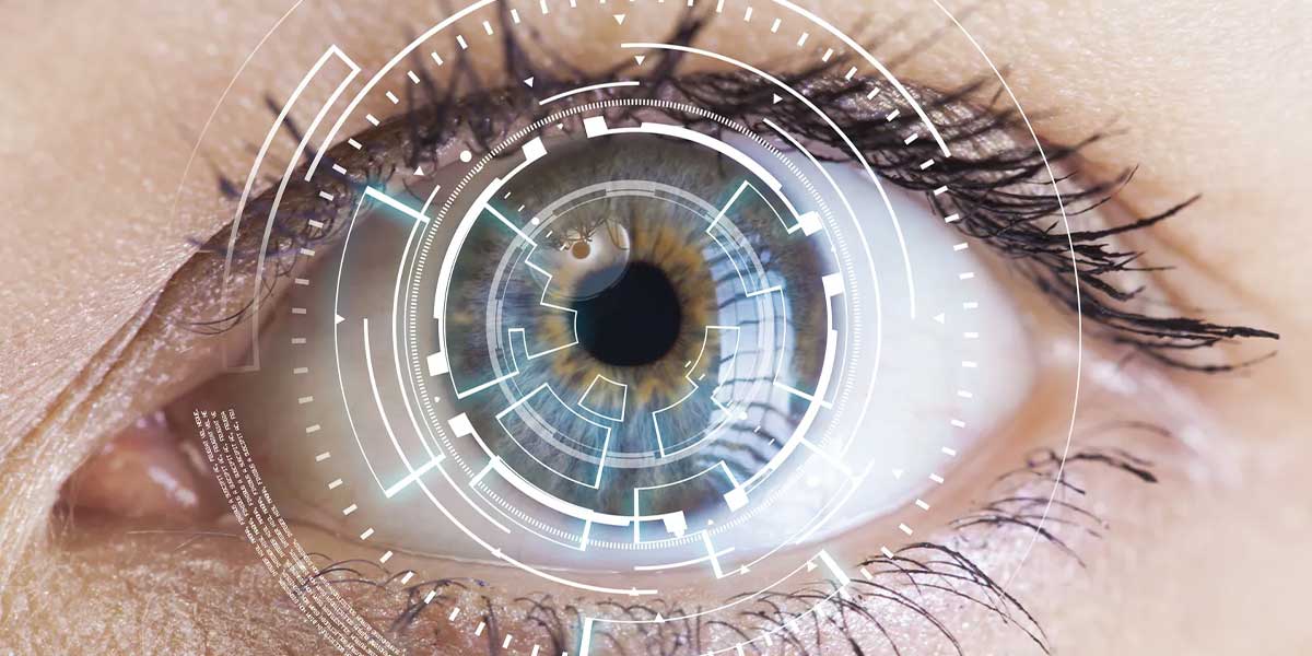
Macular degeneration is a progressive eye disorder that particularly affects elderly individuals. In this disease, yellow deposits called drusen accumulate in the macula, the central part of the retina. There are two different types: dry and wet.
In the dry type, the macula gradually thins over time, while in the wet type, abnormal blood vessels form. However, both types increase the risk of vision loss. Genetic factors, aging, and oxidative stress are the main causes of this disease. However, early diagnosis is possible with regular eye examinations. In this way, the progression of the disease can be slowed down, and vision loss can be prevented.
Turkey is one of the best country for Macular Degeneration and Retinal Diseases at a afforable price. If you are looking for a place for Macular Degeneration and Retinal Diseases abroad with best clinics and doctors, you are definitly on the right spot. Check Dr. Berkay Akmaz's Macular Degeneration and Retinal Diseases prices, reviews and packages.
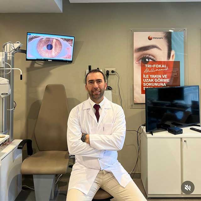
Ophthalmology Specialist – Assoc. Prof. Dr. Berkay Akmaz was born in İzmir in 1985. Due to his father's profession, he spent his primary school years in different cities. He began his education at Gülhane Military Medical Academy in 2003 and graduated with the title of Medical Doctor from Ege University Faculty of Medicine in 2010. In 2010, Assoc. Prof. Dr. Berkay Akmaz ranked 25th in the Medical Specialization Exam (TUS) in Turkey and started his specialization in the Department of Ophthalmology and Surgery at Dr. Lütfi Kırdar Kartal Education and Research Hospital. He completed his specialization training in 2014.






| Definition | Macular Disease is an eye condition that occurs due to damage to the macula, the central part of the retina. |
| Types | There are two types: Dry Type (atrophic) and Wet Type (neovascular). |
| Symptoms | Blurred central vision, wavy appearance of straight lines, dark or empty spots in the visual field. |
| Risk Factors | Advanced age, family history of the disease, smoking, high blood pressure, obesity, and poor nutrition. |
| Diagnostic Methods | Fundus examination, Amsler grid test, Optical Coherence Tomography (OCT), Fluorescein Angiography. |
| Treatment Options | For the dry type: vitamin and mineral supplements; for the wet type: anti-VEGF injections, laser therapy, photodynamic therapy. |
| Prevention | Healthy and balanced diet, quitting smoking, regular eye check-ups, protection from UV rays. |
| Nutrition Recommendations | Consumption of green leafy vegetables, omega-3 fatty acids, foods rich in zinc and antioxidants. |
| Affected Age Group | Generally seen in individuals aged 50 and above. |
| Vision Loss | As the disease progresses, it can lead to central vision loss, but peripheral vision is usually preserved. |
What Is Macular Degeneration?
Macular degeneration is an eye disease that affects the macula, the central region of the retina. Especially age-related macular degeneration leads to the loss of central vision. This disease can make daily tasks such as reading or recognizing faces difficult.
There are two types: known as dry and wet. In the dry type, gradual thinning occurs in the macular area. On the other hand, in the wet type, abnormal blood vessels form and grow under the retina. Macular degeneration is usually seen in elderly individuals and has a progressive nature. Thus, as the disease progresses from early to advanced stages, vision loss becomes more pronounced.
What Are the Causes of Macular Degeneration?
Macular degeneration has various causes, and these factors are effective in the onset and progression of the disease. The main causes of AMD include aging, oxidative stress, drusen accumulation, genetic predisposition, chronic inflammation, environmental and lifestyle factors, and lipid imbalances.
Aging is one of the most important risk factors for AMD. As age progresses, oxidative stress and chronic inflammation accumulate in the retinal tissue, leading to deterioration in the macula. This process is associated with the loss of function of the retinal pigment epithelium (RPE) and photoreceptor cells.
Oxidative damage in the retina disrupts cellular functions and damages retinal cells. Reactive oxygen species (ROS) accumulate in the retinal tissue as a result of aging, exposure to sunlight, and metabolic processes. This accumulation strains the retinal protective mechanisms, increasing cell damage.
Drusen are lipid and protein residues that accumulate between the RPE and Bruch’s membrane. These deposits are seen in the early stages of AMD and hinder the transport of nutrients. This situation leads to inflammation and degeneration in retinal cells.
Genetic predisposition is one of the factors that increase the risk of AMD. Genetic variants involved in immune response, especially mutations in the CFH gene, contribute to the progression of the disease by increasing inflammation in the retina.
Chronic inflammation plays a significant role in the development of AMD. As a result of insufficient clearance of drusen accumulation and disruption of the immune system, RPE cells are damaged. Matrix metalloproteinases (MMP2 and MMP9) are effective in this process by supporting inflammation.
Smoking reduces blood flow to the retinal tissue and increases oxidative stress. Poor nutrition, prolonged exposure to UV light, and antioxidant deficiency can also negatively affect retinal health.
The retina is rich in polyunsaturated fatty acids. However, irregular metabolism of these fatty acids leads to the accumulation of toxic byproducts in retinal cells, causing cellular dysfunction.
How Common Is Macular Degeneration?
Age-related macular degeneration (AMD) is one of the leading causes of severe vision loss worldwide. The prevalence of this disease is rapidly increasing globally, especially with the aging population. Currently, approximately 196 million people are affected by AMD, and this number is expected to reach 288 million by 2040. Studies conducted in Europe indicate that 25.3% of individuals aged 60 and over have early or intermediate AMD. Additionally, 2.4% are in the advanced stage of the disease.
- Global demographic changes and population aging play a significant role in the increase of AMD cases.
- Ethnicity and socioeconomic status are other important factors affecting the prevalence of AMD in different regions.
Research shows that AMD is more common in white populations. This rate is higher compared to Asian, African, and Latin groups. The burden of AMD is also higher in Africa and the Eastern Mediterranean regions compared to other regions. These areas feel the effects of the disease more than regions like America and Southeast Asia.
In the European Union, the number of new late-stage AMD cases is expected to increase from 400,000 to 700,000 annually by 2050. Considering aging and other demographic factors, the impact of AMD on global health is expected to increase further. Therefore, AMD is considered a major threat to vision health worldwide in the future.
How Does Macular Degeneration Develop?
The process of age-related macular degeneration arises from the interaction of genetic and environmental factors. In this disease, lipid and protein deposits accumulating under the retina disrupt the function of the retinal pigment epithelium. Over time, these accumulations lead to the degeneration of photoreceptor cells and vision loss. As the disease progresses, geographic atrophy in the dry type or choroidal neovascularization in the wet type occurs. This leads to scarring in the retinal tissue. Additionally, oxidative stress, inflammation, and disruption of the complement system accelerate the process further. All these factors come together to contribute to the death of retinal cells and lead to the advanced stages of the disease.
What Are the Symptoms of Macular Degeneration?
The symptoms of macular degeneration may vary depending on whether the disease is in its dry or wet form. Initially, patients may notice blurriness in their central vision, which over time can make tasks requiring detail, such as reading, more difficult. In addition, straight lines appearing wavy or bent is a very common symptom. Also, dark or empty areas may form in the central vision, causing blind spots in the patient’s field of view. On the other hand, difficulty adapting to low light conditions is a frequently experienced problem. Additionally, colors may appear faded and lose their vibrancy, which is among the symptoms.
How Is Macular Degeneration Diagnosed?
Macular degeneration is diagnosed through a detailed series of tests and evaluations. In this process, the doctor evaluates the patient’s retina and analyzes structural and functional changes occurring in the eye. First, a fundus examination is performed. This step helps in the early detection of yellow deposits called drusen under the retina. In advanced cases, conditions such as geographic atrophy and choroidal neovascularization can be identified. Additionally, using optical coherence tomography (OCT) technology, cross-sections of the retina are examined in detail. OCT plays a critical role in determining retinal thickness and structural changes.
On the other hand, fluorescein angiography is preferred especially in wet AMD cases. In this test, a fluorescent dye is injected to visualize abnormal leaking blood vessels under the retina. Also, the Amsler Grid test is a simple yet effective tool used to detect visual distortions. The patient reports whether there is any bending or loss in the lines on the grid. However, the sensitivity of this test is limited.
Moreover, preferential hyperacuity perimetry (PHP) offers a more sensitive method. This test detects subtle shifts in the visual field and monitors the likelihood of progression to CNV. Finally, visual acuity tests help in detecting the disease at an early stage. In these tests, the patient indicates whether they experience a decrease in near visual acuity or difficulty in reading. When all these tests are evaluated together, a clear diagnosis of macular degeneration can be made.
How Is Macular Degeneration Treated?
Various methods are applied in the treatment of macular degeneration depending on the type of the disease. In wet AMD treatment, the commonly used anti-VEGF therapy is administered via intravitreal injections to prevent the growth of abnormal blood vessels in the retina. This treatment includes drugs like ranibizumab (Lucentis), aflibercept (Eylea), and bevacizumab (Avastin) and is usually repeated every 4-8 weeks. However, due to the frequent injection requirement, it can be burdensome for patients in the long term.
Another wet AMD treatment option is photodynamic therapy (PDT). PDT targets abnormal vessels in the eye and destroys them with laser light. However, due to the success of anti-VEGF therapies, it is used less today. Nevertheless, it is considered a suitable option for some patients.
There is currently no definitive treatment for dry AMD. However, AREDS formulas containing antioxidants and mineral supplements have the potential to slow the progression of the disease. Nutritional supplements, especially vitamin supports containing minerals like zinc and copper, are recommended.
On the other hand, developing gene therapy studies appear promising. Treatments like RGX-314 in clinical trials aim to reduce the need for anti-VEGF injections. Additionally, new technologies like nanotechnology and 3D bioprinting may offer more effective treatment options in the future.
In addition, lifestyle changes can also slow the progression of AMD:
- Quitting smoking
- Maintaining a diet rich in omega-3 fatty acids
- Protecting cardiovascular health
What Are the Risk Factors for Macular Degeneration?
Age-related macular degeneration (AMD) is a complex disease that develops under the influence of many factors. Particularly, the combination of genetic and environmental factors contributes to the onset of the disease. One of the biggest risk factors is advancing age. Damage occurring in cells with age can accelerate the development of AMD. In addition, genetic predisposition is an important factor. Individuals with a family history of AMD have a higher likelihood of developing this disease.
Other prominent risk factors include:
- Smoking: Tobacco use causes increased oxidative stress and reduced blood flow to the retina. This significantly increases the risk of AMD in smokers.
- Obesity and Cardiovascular Health: Excess fat accumulation, especially in the abdominal area, raises the risk of AMD. Cardiovascular factors like hypertension and high cholesterol levels also increase this risk.
- Nutrition: A diet poor in antioxidants like lutein, zeaxanthin, and omega-3 fatty acids can increase the risk of AMD. Conversely, a diet rich in green leafy vegetables and fish can reduce the risk.
- Exposure to Sunlight: Prolonged exposure to UV rays can lead to accumulated damage in the retina, triggering the development of AMD.
- Race and Gender: The disease incidence is higher in the white race, and women are generally at higher risk.
- Environmental Factors: Occupations requiring constant exposure to toxins or intense light make the development of AMD more likely.
When Can Macular Degeneration Treatment Be Performed?
Treatment of age-related macular degeneration (AMD) is based on specific criteria to slow the progression of the disease and manage symptoms. In wet AMD, treatment is necessary when new blood vessels form under the retina, i.e., when there is active neovascularization. In this case, anti-VEGF therapies are applied to the eye, focusing on preventing further vision loss.
In dry AMD, treatment is necessary when significant drusen formation or geographic atrophy occurs. In intermediate stages, antioxidant vitamins, especially the AREDS formula, help slow the progression of the disease. In advanced stages, low vision rehabilitation can be considered, providing significant support in coping with vision loss.
When Can’t Macular Degeneration Treatment Be Performed?
Treatment for macular degeneration cannot be applied in some cases, and these situations may affect the patient’s health. First, in individuals who have had an eye infection or have an active eye infection, this treatment can worsen the infection. Additionally, those with hypersensitivity to any component of the drug should avoid this treatment. Systemic health problems also pose an obstacle to treatment. Especially in individuals with uncontrolled hypertension or who have recently had a cardiovascular event, treatment may worsen the condition. The safety of anti-VEGF agents during pregnancy and breastfeeding has not been proven.
What Is the Recovery Process After Macular Degeneration Treatment?
The recovery process after macular detachment treatment varies depending on the treatment method applied, the patient’s general health condition, and the preoperative eye structure. During the recovery process, an increase in visual acuity is possible. However, this improvement often takes several months, and it may take as long as 12 to 24 months to achieve full results. Especially if the retinal layers are intact before surgery, visual improvement may be more pronounced.
The duration and degree of improvement in visual acuity vary depending on the following factors:
- Preoperative retinal integrity
- The duration and size of the detachment
- The response to the applied treatment
Also, recovery after macular detachment treatment is generally dependent on the timing of the surgery. Repairs performed within the first week show better results compared to longer-standing detachments. However, the complete recovery of visual functions occurs over time and gradually. Various complications may be experienced in the postoperative period. These include increased intraocular pressure, accumulation of subretinal fluid, or flashes. However, these complications usually resolve within a few months and do not lead to permanent vision loss.
During the rehabilitation process, visual training is important for the patient to adapt to visual changes. Additionally, magnifiers or other visual aid devices may be recommended for patients with vision loss. These supports are particularly beneficial for those experiencing permanent vision loss and help improve the quality of daily life.
How to Prevent Macular Degeneration?
Prevention of age-related macular degeneration is possible with healthy lifestyle changes and dietary adjustments. First, appropriate nutritional supplements can slow the progression of macular degeneration. Particularly, vitamins and minerals recommended in the AREDS and AREDS2 studies are found to be effective in this regard. The recommended nutritional supplements are:
- Vitamin C (500 mg), Vitamin E (400 IU), and Zinc (80 mg): They support eye health thanks to their antioxidant properties.
- Lutein (10 mg) and Zeaxanthin (2 mg): Powerful carotenoids that protect the retina by absorbing harmful light.
- Copper (2 mg): Prevents copper deficiency when taken with zinc.
- Omega-3 fatty acids: These fatty acids found in fish can reduce the disease risk by decreasing inflammation in the retina.
The diet should also be rich in antioxidants. For this, especially dark green leafy vegetables, fruits, and vegetables rich in carotenoids and anthocyanins are recommended:
- Dark green leafy vegetables: Kale and spinach contain lutein and zeaxanthin.
- Fruits and vegetables rich in carotenoids and anthocyanins: Examples include strawberries, oranges, and peppers.
- Fish: Fatty fish like salmon and mackerel are rich in Omega-3.
Additionally, lifestyle changes have a significant impact on eye health. Measures such as quitting smoking, controlling cholesterol and blood pressure, protecting from UV light, regular exercise, and weight control reduce the risk. Environmental factors should also be considered. Controlling situations like air pollution and sleep apnea helps reduce oxidative stress and protect retinal health.
Recent Posts

Dermatochalasis (Eyelid Sagging)
Anasayfa » Eyelid » Dermatochalasis (Eyelid Sagging)Dermatochalasis (eyelid sagging) is a condition caused by excess [...]
Read MoreMar
Congenital Cataract
Anasayfa » Cataract » Congenital CataractCongenital cataract is an eye disease that occurs when the [...]
Read MoreMar

Causes of Blurred Vision in One Eye
Anasayfa » Eye Health » Causes of Blurred Vision in One EyeThe causes of blurred [...]
Read MoreDec

Comments from Patients with Pseudotumor Cerebri
Anasayfa » Uncategorized » Comments from Patients with Pseudotumor CerebriComments from patients with pseudotumor cerebri [...]
Read MoreDec

Reviews of People Who Have Undergone Corneal Surgery
Anasayfa » Eye Health » Reviews of People Who Have Undergone Corneal SurgeryReviews from people [...]
Read MoreDec
Keratoconus Patient Reviews
Anasayfa » Eye Health » Keratoconus Patient ReviewsKeratoconus patient reviews provide important insights into the [...]
Read MoreDec

How Long Does Cataract Surgery Take?
Anasayfa » Cataract » How Long Does Cataract Surgery Take?The answer to the question of [...]
Read MoreDec
How Do Under-Eye Dark Circles Go Away?
Anasayfa » Uncategorized » How Do Under-Eye Dark Circles Go Away?The question of how under-eye [...]
Read MoreDec
Gallery