Türkçe
 Deutsch

Diabetic retinopathy is known as one of the leading causes of vision loss worldwide. This condition, which arises due to chronic hyperglycemia, damages the retinal blood vessels and can lead to vision loss in the advanced stages. Furthermore, it triggers processes such as inflammation, oxidative stress, and neovascularization by causing capillary occlusion and retinal ischemia.
Advanced stages like proliferative diabetic retinopathy (PDR) are characterized by abnormal blood vessel growth. Diabetic macular edema (DME) can also lead to vision loss, and mitochondrial dysfunction and inflammation play significant roles in the development of DR.
Turkey is one of the best country for Diabetic Retinopathy Treatment at a afforable price. If you are looking for a place for Diabetic Retinopathy Treatment abroad with best clinics and doctors, you are definitly on the right spot. Check Dr. Berkay Akmaz's Diabetic Retinopathy Treatment prices, reviews and packages.
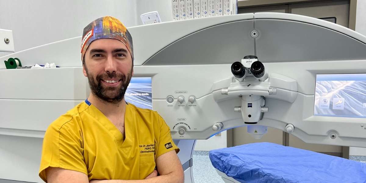
Ophthalmology Specialist – Assoc. Prof. Dr. Berkay Akmaz was born in İzmir in 1985. Due to his father's profession, he spent his primary school years in different cities. He began his education at Gülhane Military Medical Academy in 2003 and graduated with the title of Medical Doctor from Ege University Faculty of Medicine in 2010. In 2010, Assoc. Prof. Dr. Berkay Akmaz ranked 25th in the Medical Specialization Exam (TUS) in Turkey and started his specialization in the Department of Ophthalmology and Surgery at Dr. Lütfi Kırdar Kartal Education and Research Hospital. He completed his specialization training in 2014.






| Definition | Diabetic Retinopathy is an eye disease that develops as a long-term complication of diabetes, resulting from damage to the blood vessels in the retina. It can lead to vision loss and even blindness. |
| Symptoms | Initially, it may not present any symptoms; in advanced stages, blurred vision, dark or empty areas in the visual field, difficulty seeing at night, faded colors, sudden and complete vision loss. |
| Stages | Non-Proliferative Diabetic Retinopathy: Presence of microaneurysms, hemorrhages, and macular edema. Proliferative Diabetic Retinopathy: Formation of new and abnormal blood vessels (neovascularization) and fibrosis. |
| Risk Factors | Long-term presence of diabetes, poor blood sugar control, hypertension, high cholesterol, pregnancy, smoking, kidney disease. |
| Diagnostic Methods | Eye examination, Schirmer test measuring tear production, Break-Up Time (BUT) test measuring tear evaporation, staining tests (fluorescein, lissamine green). |
| Treatment Options | Control of blood sugar, blood pressure, and cholesterol; laser photocoagulation therapy; anti-VEGF injections (bevacizumab, ranibizumab); corticosteroid injections; vitrectomy surgery. |
| Prevention | Good management of diabetes, regular eye examinations, healthy and balanced diet, quitting smoking, regular exercise. |
| Regular Check-ups | Comprehensive eye examinations at least once a year after a diabetes diagnosis; more frequent checks may be necessary during pregnancy. |
| Complications | Macular edema, retinal detachment, neovascular glaucoma, vitreous hemorrhage, blindness. |
| Impact on Quality of Life | Discomfort during activities such as reading, driving, using a computer; decreased sleep quality; negative effects on social and professional life. |
| Affected Age Group | Individuals with Type 1 and Type 2 diabetes of any age; the duration and control of diabetes significantly affect the risk. |
| Related Diseases | Autoimmune diseases such as Sjögren’s Syndrome, rheumatoid arthritis, lupus can cause dry eye. |
| Nutrition Recommendations | Foods rich in Omega-3 fatty acids (fish, walnuts, flaxseeds), foods containing vitamin A (carrots, spinach). |
| Alternative Treatments | Lipiflow Therapy: Improves the oily tear layer by cleansing Meibomian glands. Intense Pulsed Light (IPL) Therapy: May enhance the function of tear glands. |
| Precautions | Avoiding or taking breaks from contact lens use or changing lens types, minimizing contact of makeup and cleaners with the eyes, not using drops or medications without doctor’s advice. |
| When to See a Doctor | If symptoms affect daily life, do not improve with artificial tear drops, or if there is vision impairment or severe pain. |
| Technologies Used in Diagnosis and Treatment | Meibography: Imaging of Meibomian glands. Osmolarity Test: Assessing tear quality. |
| Success Rate | When adhering to treatment and addressing underlying causes, symptoms are generally controllable. |
| Cost and Insurance Coverage | The cost of treatment varies depending on the medications used, treatment methods, and duration of therapy; some treatments may be covered by insurance. |
| Lifestyle Recommendations | 20-20-20 Rule: Every 20 minutes, look at something 20 seconds away at 20 feet (6 meters); drink plenty of water; quit smoking; maintain regular sleep. |
| Workplace Considerations | Position computer screens below eye level, blink frequently, adjust screen brightness and contrast, use humidifying devices. |
What Is Diabetic Retinopathy?
Diabetic retinopathy damages the retinal blood vessels as a complication of diabetes, leading to severe vision loss. There are two main stages: non-proliferative diabetic retinopathy (NPDR) and proliferative diabetic retinopathy (PDR). In the NPDR stage, small blood vessels weaken and leak blood or fluid, adversely affecting retinal functions.
PDR is characterized by the growth of abnormal blood vessels, which can lead to serious complications such as bleeding or retinal detachment. Diabetic macular edema (DME) occurs due to fluid accumulation in the central region of the retina and is a common cause of vision loss. Blood sugar control and appropriate treatments play a critical role in managing DR.
What Are the Causes of Diabetic Retinopathy?
Diabetic retinopathy is caused by various factors that damage the retinal blood vessels. Firstly, chronic hyperglycemia leads to the blockage of retinal blood vessels and a decrease in oxygen supply, causing damage. High blood sugar levels increase vascular permeability and disrupt the function of retinal blood vessels. Poor glycemic control is a significant risk factor for the onset and progression of diabetic retinopathy in both Type 1 and Type 2 diabetes patients.
Additionally, oxidative stress and chronic inflammation play critical roles in the development of DR. High glucose levels lead to increased oxidative stress and the formation of reactive oxygen species (ROS), causing damage to retinal cells. This process increases vascular permeability and neovascularization, leading to further dysfunction of retinal blood vessels.
Abnormal blood vessel growth is another significant factor in DR. In response to oxygen deprivation, the body attempts to form new blood vessels; however, these new vessels are fragile and prone to leakage. As a result, fluid or blood accumulates in the retina, leading to visual disturbances.
Furthermore, factors such as hypertension and dyslipidemia also damage the retina. Poorly controlled high blood pressure and cholesterol levels put pressure on retinal vessels, causing further damage and fluid leakage.
Additionally, rapidly lowering blood sugar levels with insulin therapy can temporarily worsen DR in some patients. This is associated with sudden changes in osmotic balance, leading to temporary retinal edema and vessel fragility.
The duration of diabetes is also important in the development of DR. Individuals with diabetes for 20 years or longer have a significantly increased risk of retinopathy. Nearly all patients with Type 1 diabetes develop retinopathy after this period.
Finally, it is believed that certain metabolites circulating in the blood may contribute to the development of DR:
- High levels of certain lipids can increase oxidative stress.
- Some amino acids can have protective effects.
How Common Is Diabetic Retinopathy?
Diabetic retinopathy (DR) stands out as a common complication affecting approximately 25% to 34.6% of diabetes patients worldwide. This condition is a leading cause of vision loss, especially among working-age adults, and its prevalence increases with age and the duration of diabetes. Predictions indicate that global DR cases will reach 161 million by 2045.
- Global Prevalence: DR affects around 103 million people worldwide, and increasing diabetes rates are driving this number higher.
- Vision-Threatening DR: Approximately 6% of diabetic patients have severe forms of DR known as vision-threatening DR.
In the United States, it is estimated that over 9.6 million people have DR, with 1.84 million individuals affected by vision-threatening forms of DR. Prevalence rates vary by age group, peaking in the 65-79 age range. Additionally, severe forms of DR, such as diabetic macular edema, are more commonly observed in males.
DR rates vary among ethnic groups, with Hispanic and Black populations experiencing higher rates of DR. These differences are attributed to socioeconomic factors, challenges in accessing healthcare services, and inadequate diabetes management strategies. With the global rise in diabetes prevalence, targeted healthcare services are needed to mitigate the impacts of DR.
How Does Diabetic Retinopathy Develop?
Diabetic retinopathy is a severe eye disease resulting from chronic hyperglycemia caused by diabetes mellitus. The development process involves several complex pathophysiological mechanisms, including retinal microvascular damage, inflammation, oxidative stress, and neurodegeneration.
High blood sugar levels lead to oxidative stress and the accumulation of advanced glycation end-products (AGEs). This accumulation disrupts cellular functions, particularly causing mitochondrial dysfunction in retinal pigment epithelial (RPE) and photoreceptor cells.
DR is known as a microvascular disease and results from the breakdown of the blood-retinal barrier. This breakdown damages retinal capillaries, increasing vascular permeability. Initially, microaneurysms—small swelling of capillaries—form, causing blood to leak into the retina. In advanced stages, capillary occlusions occur, leading to oxygen deprivation in retinal tissue. Ischemia makes it difficult for retinal cells to survive and triggers the release of vascular endothelial growth factor (VEGF).
The neurodegeneration process also plays a significant role in the progression of the disease. Hyperglycemia leads to the loss of retinal neurons and affects glial cells, contributing to the rapid progression of the disease in early DR stages. Chronic inflammation further exacerbates this damage.
The release of pro-inflammatory cytokines (e.g., TNF-α) contributes to vascular and nerve damage. The adhesion of white blood cells to vessel walls further blocks capillaries, worsening ischemic damage.
The loss of pericytes, which support retinal blood vessel structure, weakens capillary stability, leading to increased DR over time. Avascular capillaries form, resulting in retinal ischemia and neovascularization. As the disease progresses, it advances from non-proliferative diabetic retinopathy (NPDR) to proliferative diabetic retinopathy (PDR).
What Are the Symptoms of Diabetic Retinopathy?
Diabetic retinopathy often progresses silently in its early stages, making diagnosis challenging. However, as the disease advances, various symptoms emerge, potentially leading to severe vision loss. The symptoms of diabetic retinopathy vary depending on the stage of the disease, beginning in the early stage known as Non-Proliferative Diabetic Retinopathy (NPDR).
Microaneurysms: Small red dots caused by swelling of blood vessels in the retina, representing one of the earliest signs of DR.
- Bleeding: Dark floating spots or blotches can be observed due to blood leakage from damaged retinal vessels.
- Hard Exudates: Yellowish lipid deposits formed from leaking fluid accumulate in the retina.
- Cotton Wool Spots: Soft white spots indicating microinfarcts and retinal ischemia.
As the disease progresses, symptoms become more severe in the Proliferative Diabetic Retinopathy (PDR) stage.
- Neovascularization: Abnormal blood vessels grow on the surface of the retina, potentially leading to retinal detachment and vision loss.
- Vitreous Hemorrhage: Bleeding from fragile new vessels can leak into the vitreous fluid, causing sudden vision loss.
- Macular Edema: Fluid accumulation in the central retina (macula) leads to widespread vision loss.
- Reduced Color Vision: Colors may appear faded or dull.
Other symptoms include night blindness, waviness in vision, and permanent vision loss, all of which increase the severity of the disease.
How Is Diabetic Retinopathy Diagnosed?
The diagnosis of diabetic retinopathy relies on both clinical examinations and advanced imaging techniques. During this process, the presence of microaneurysms, bleeding, and other abnormalities in the retina is detected through eye examinations, determining whether DR is in the non-proliferative or proliferative stages. Common diagnostic methods include:
- Fundus Photography: This method captures high-resolution photographs of the retina’s surface to identify early-stage DR signs such as microaneurysms and bleeding. Fundus photography is one of the most widely used tools for DR screening.
- Optical Coherence Tomography (OCT): This non-invasive test creates cross-sectional images of retinal tissue and is used to detect macular edema, a common complication in DR patients. Additionally, OCT Angiography allows detailed examination of retinal microvasculature, facilitating early diagnosis of ischemia and neovascularization.
- Fluorescein Angiography (FA): In this test, a fluorescent dye is injected to capture images of retinal blood vessels. This technique aids in detecting vascular leakage, ischemia, and abnormal vessel growth, especially in advanced DR stages.
- Artificial Intelligence (AI) and Deep Learning: AI-powered systems can analyze retinal images to automatically diagnose DR with high accuracy. These systems offer significant advantages in large-scale screenings, particularly in regions with limited access to eye health specialists.
How Is Diabetic Retinopathy Treated?
Significant advancements have been made in the treatment of diabetic retinopathy in recent years. Treatment methods primarily focus on controlling retinal damage and slowing the progression of DR. These treatment options vary across different stages and involve various strategies:
- Anti-VEGF Therapy: Anti-VEGF drugs are fundamental treatment options for diabetic macular edema (DME) and proliferative diabetic retinopathy (PDR). Agents like ranibizumab, aflibercept, and faricimab effectively prevent the growth and leakage of abnormal blood vessels, yielding positive outcomes.
- Steroid Therapy: Intravitreal corticosteroids are administered to patients who do not respond to anti-VEGF therapy. Steroids such as dexamethasone implants (Ozurdex) reduce retinal inflammation, thereby helping to slow vision loss.
- Laser Photocoagulation: Laser therapy remains an option for patients unresponsive to anti-VEGF treatments. Laser photocoagulation seals abnormal blood vessels, slowing the progression of damage. However, this method generally does not significantly improve visual acuity.
- Emerging Treatments: New treatment options targeting different aspects of DR development are under investigation. For example, fenofibrate laser therapy may help reduce the need for other interventions, while tonabersat could be effective in reducing macular edema. Additionally, gene therapy and nanoparticle-based approaches are being explored as more permanent treatment solutions.
What Are the Risk Factors for Diabetic Retinopathy?
Risk factors for diabetic retinopathy encompass both modifiable and non-modifiable elements. Firstly, the duration of diabetes increases the risk of developing DR. Specifically, individuals with diabetes for over 10 years have a more pronounced risk, and those with diabetes for 20 years or longer are approximately three times more likely to develop DR compared to those with a shorter diabetes history.
Poor glycemic control plays a critical role in the progression of DR. Individuals with high blood sugar levels and HbA1c levels above 7% have a significantly increased risk of DR. Conversely, patients who maintain tight glycemic control experience lower rates of DR progression.
Another important risk factor is hypertension. Individuals with blood pressure below 140/90 mmHg have a lower risk of developing DR. Unmanaged high blood pressure can increase the risk of DR and trigger disease progression.
Lastly, cholesterol and lipid levels significantly impact the development of DR. High LDL cholesterol levels can lead to severe damage in the retina.
When Can Diabetic Retinopathy Treatment Be Performed?
Treatment for diabetic retinopathy is determined based on the progression of the disease, with treatment options varying according to stages such as non-proliferative diabetic retinopathy (NPDR), proliferative diabetic retinopathy (PDR), and diabetic macular edema (DME).
- Non-Proliferative Diabetic Retinopathy (NPDR)
In the non-proliferative stage, if there is no significant diabetic macular edema (DME), immediate treatment may not be necessary. However, if DME is present, anti-vascular endothelial growth factor (anti-VEGF) therapy is recommended. This treatment controls fluid accumulation in the retina and reduces the risk of vision loss. Specifically, anti-VEGF agents like ranibizumab and aflibercept are used.
- Proliferative Diabetic Retinopathy (PDR)
In advanced stages, PDR is characterized by neovascularization, posing a high risk of severe vision loss. In such cases, laser therapy known as panretinal photocoagulation (PRP) is performed. PRP targets ischemic retinal tissue, significantly reducing the risk of blindness and proving effective in high-risk PDR patients.
- Diabetic Macular Edema (DME)
DME can occur at any stage of DR and leads to central vision loss. In addition to anti-VEGF therapy, focal or grid laser photocoagulation is recommended. In cases of clinically significant macular edema (CSME), laser treatment can halve the risk of vision loss.
- Situations Requiring Vitrectomy
- Vitreous hemorrhage
- Tractional retinal detachment
In these cases, vitrectomy surgery may be necessary and is used to treat persistent vitreous hemorrhage.
When Can’t Diabetic Retinopathy Treatment Be Performed?
The contraindications for diabetic retinopathy treatment encompass various factors that need to be considered during the treatment process. When selecting a treatment method, the patient’s overall condition, existing medical conditions, and individual risk factors must be taken into account. Specifically, different contraindications exist for common treatment options such as anti-VEGF injections, corticosteroid applications, laser photocoagulation, and vitrectomy. These contraindications play a crucial role in the decision-making process and help determine whether a patient is suitable for a particular treatment.
- Anti-VEGF Therapy: Anti-VEGF injections may become risky in certain situations. Patients with cardiovascular diseases, especially those who have recently experienced a stroke or heart attack, may not be suitable candidates for this treatment. The potential teratogenic effects of anti-VEGF therapy during pregnancy must be considered, and safety data for this population are limited. Additionally, the requirement for regular injections may render this treatment ineffective for patients who are non-compliant.
- Corticosteroid Therapy: Intravitreal corticosteroid injections are recommended for patients who do not respond to anti-VEGF therapy; however, they may be contraindicated in certain situations. Glaucoma patients may not be suitable for this treatment due to the potential increase in intraocular pressure caused by corticosteroids. Furthermore, steroid injections carry the risk of cataract development or progression, making them a significant contraindication for patients who wish to avoid cataracts or are younger in age.
- Laser Photocoagulation: While laser therapy is used in treating proliferative diabetic retinopathy, it may be less effective in patients with macular edema. Additionally, it is contraindicated in patients with severe cataracts or vitreous hemorrhage, as successful laser treatment requires clear visualization of the retina.
- Vitrectomy: This surgical intervention may not be suitable for some patients. Particularly, those with uncontrolled diabetes or cardiovascular issues may face higher surgical risks. Moreover, vitrectomy should not be performed in the presence of active eye infections or significant ocular inflammation.
What Is the Recovery Process After Diabetic Retinopathy Treatment?
The recovery process following diabetic retinopathy treatment varies based on the treatment method applied, the patient’s overall health, and the preoperative eye structure. During the recovery period, noticeable improvements in visual acuity may occur within the first three months post-surgery; however, full recovery typically takes several months. Specifically, a reduction in central retinal thickness (CRT) contributes to positive developments in visual acuity. On the other hand, complications such as persistent macular edema can slow the recovery process and affect visual outcomes. Additionally, the duration of recovery observed post-surgery can vary depending on the extent of macular damage.
Recurring vitreous hemorrhage (RVH) is a prominent postoperative complication. Although sutureless vitrectomy performed with 23 and 25 gauge systems effectively reduces RVH rates, some patients may still experience this risk. Critical points in managing complications include:
- Additional interventions may be required to treat RVH
- Younger patients and those with aggressive disease progression have a higher risk of RVH
Managing inflammation is also important during the recovery process. Anti-inflammatory treatments such as dexamethasone eye drops can help control inflammation. Additionally, the use of modern surgical techniques can reduce the inflammatory response and support healing.
The use of anti-VEGF therapies in managing macular edema can provide some benefit in reducing macular thickness. However, these treatments may not always yield successful results. Especially in patients with initially thicker CRT, post-surgical visual improvement may be more pronounced.
How Can Diabetic Retinopathy Be Prevented?
Preventing diabetic retinopathy focuses on effective diabetes management, including both blood sugar control and lifestyle modifications. Key preventive measures include maintaining healthy blood sugar levels, managing blood pressure, and engaging in regular physical activity, all of which play critical roles in reducing the risk of DR. Additionally, improving diet, ensuring adequate vitamin intake, and quitting smoking are supplementary measures that help mitigate risk factors.
- Optimal Blood Sugar Control: Maintaining glycemic control is the fundamental strategy to reduce the risk of diabetic retinopathy. Lowering HbA1c levels prevents microvascular damage in the retina and slows the progression of DR.
- Blood Pressure Management: High blood pressure can accelerate the development of retinopathy. Therefore, especially in diabetic individuals, keeping blood pressure below 140/90 mmHg is recommended.
- Lipid Control: Managing cholesterol levels, particularly controlling LDL cholesterol, provides a protective effect against retinopathy. High LDL and triglyceride levels are associated with more severe forms of DR, such as macular edema.
- Diet and Nutrient Intake: Adhering to a Mediterranean diet rich in fruits, vegetables, and whole grains can help reduce the risk of diabetic retinopathy. Additionally, consuming foods rich in Omega-3 fatty acids and vitamin D supports retinal health.
Regular physical activity is another avenue to reduce DR risk. Exercise is crucial for diabetic individuals as it increases insulin sensitivity and lowers blood pressure. However, in advanced DR cases, high-intensity exercises should be avoided.
Quitting smoking is also part of diabetic retinopathy prevention strategies. Smoking can exacerbate microvascular complications and damage the retina. Therefore, quitting smoking supports overall vascular health and reduces DR risk.
Lastly, prolonged exposure to sunlight can damage retinal cells. Hence, wearing protective sunglasses that block ultraviolet (UV) rays is essential.
Recent Posts

Dermatochalasis (Eyelid Sagging)
Anasayfa » Eyelid » Dermatochalasis (Eyelid Sagging)Dermatochalasis (eyelid sagging) is a condition caused by excess [...]
Read MoreMar
Congenital Cataract
Anasayfa » Cataract » Congenital CataractCongenital cataract is an eye disease that occurs when the [...]
Read MoreMar

Causes of Blurred Vision in One Eye
Anasayfa » Eye Health » Causes of Blurred Vision in One EyeThe causes of blurred [...]
Read MoreDec

Comments from Patients with Pseudotumor Cerebri
Anasayfa » Uncategorized » Comments from Patients with Pseudotumor CerebriComments from patients with pseudotumor cerebri [...]
Read MoreDec

Reviews of People Who Have Undergone Corneal Surgery
Anasayfa » Eye Health » Reviews of People Who Have Undergone Corneal SurgeryReviews from people [...]
Read MoreDec
Keratoconus Patient Reviews
Anasayfa » Eye Health » Keratoconus Patient ReviewsKeratoconus patient reviews provide important insights into the [...]
Read MoreDec

How Long Does Cataract Surgery Take?
Anasayfa » Cataract » How Long Does Cataract Surgery Take?The answer to the question of [...]
Read MoreDec

How Do Under-Eye Dark Circles Go Away?
Anasayfa » Uncategorized » How Do Under-Eye Dark Circles Go Away?The question of how under-eye [...]
Read MoreDec
Gallery