
A retinal tear is a full-thickness break in the retinal layer that lines the inner surface of the eye and generates visual signals. It most commonly develops due to traction as the gel-like vitreous inside the eye separates from the retina with age. Early detection of a tear is vital because fluid can pass through the opening and collect beneath the retina, causing a retinal detachment that may result in vision loss.
It is important to assess the shape and location of the tear, vitreous traction, surrounding fluid, other weak areas of the retina, the condition of the macula and risks in the other eye as a whole. The treatment decision is made according to this clinical assessment.
Do Not Wait If You Experience These Symptoms
If new flashes of light, a sudden increase in black floaters or cobweb-like shadows, a curtain or shadow over part of the visual field or reduced vision develops in one eye, a dilated retinal examination is required on the same day. A curtain or visual field loss may be a sign of retinal detachment. The absence of pain does not mean that the condition is insignificant.
Differences Between Posterior Vitreous Detachment, Retinal Tear and Retinal Detachment
- Posterior Vitreous Detachment (PVD): This is the separation of the vitreous gel inside the eye from the retina. It is common, but a tear may develop if the retina is pulled during the separation.
- Retinal Tear: This is a full-thickness break in the retina. Symptomatic horseshoe-shaped tears with persistent traction are usually treated because of the risk of retinal detachment.
- Retinal Hole: This is generally a rounder opening without traction. Not every hole is treated. The decision depends on the presence of fluid, its location and individual risk factors.
- Lattice Degeneration: These are areas where the peripheral retina has become thinner. Its presence alone does not usually require preventive laser treatment.
- Retinal Detachment: This is the separation of the retina from the tissue beneath it. Surrounding the area with laser treatment may no longer be sufficient, and surgery may be required.
What Causes a Retinal Tear and Who Is at Risk?
The most common cause is acute posterior vitreous detachment (PVD). A tear may develop if the vitreous pulls on an area where it is strongly attached to the retina during separation.
- Acute PVD and age-related vitreous changes
- High myopia and a thin peripheral retina
- Blows to the eye or trauma
- Previous cataract surgery or other intraocular operations
- A personal history of retinal detachment in the other eye or a family history of retinal detachment
Symptoms of a Retinal Tear
- Lightning-like flashes that may also be noticed when the lights are off
- New floaters or a sudden increase in floaters, a shower of black specks or cobweb-like images
- A curtain, shadow or dark area progressing from the edge of the visual field
Some retinal tears may cause no symptoms and may be detected during a routine examination. Although flashes and floaters do not always indicate a tear, a retinal examination must be performed when these symptoms begin suddenly.
Diagnostic Methods
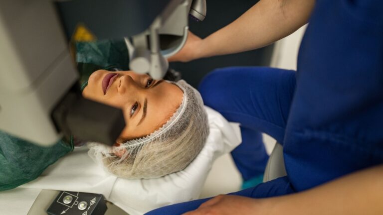
The main diagnostic method is a detailed examination of the peripheral retina after the pupil has been dilated with eye drops. When necessary, the far peripheral areas of the retina are examined using scleral indentation. B-scan ultrasonography may be used if the retina cannot be visualized because of a dense cataract or bleeding inside the eye. OCT is valuable for assessing the macula, but it is not sufficient on its own to rule out a peripheral retinal tear.
Is Laser Treatment Performed for Every Retinal Tear?
No. Symptomatic horseshoe-shaped tears with persistent vitreous traction are usually treated. In contrast, asymptomatic atrophic holes, certain operculated tears and lattice degeneration can be closely monitored in most patients. The decision to perform preventive laser treatment is made individually according to the examination findings.
Treatment Methods for Retinal Tears
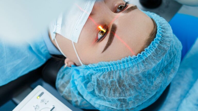
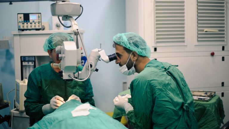
- Retinal Laser Treatment (Laser Photocoagulation): Rows of laser spots are applied around the tear to create a strong adhesion, or barrier, between the retina and the underlying tissues. The aim is to limit the progression of fluid through the tear. Laser treatment does not remove existing floaters or reattach the vitreous.
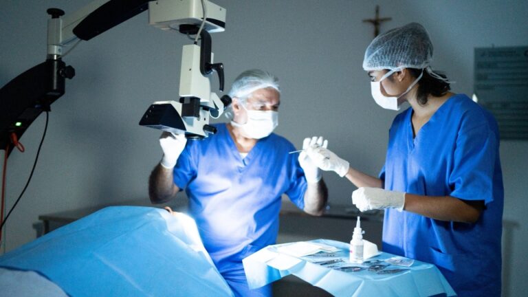
- Cryotherapy (Freezing Treatment): If the tear is located very far forward or media opacity makes laser visualization difficult, adhesion is created using a cold probe applied from outside the eye.
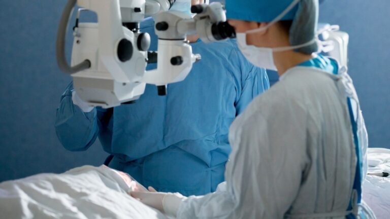
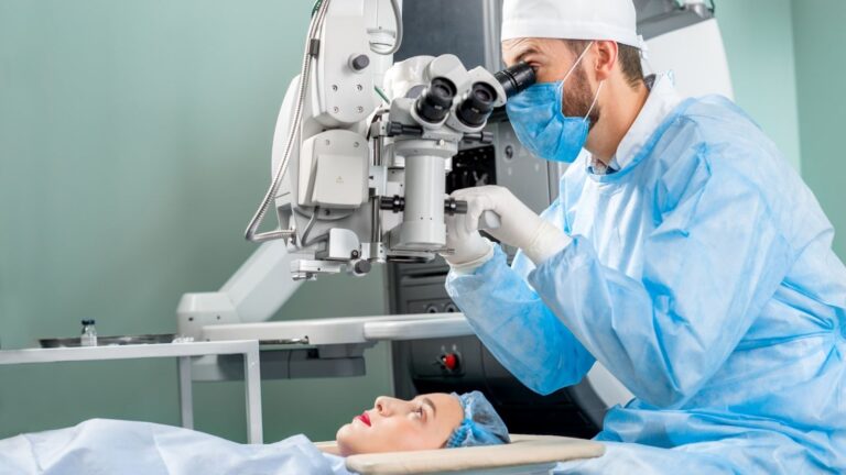
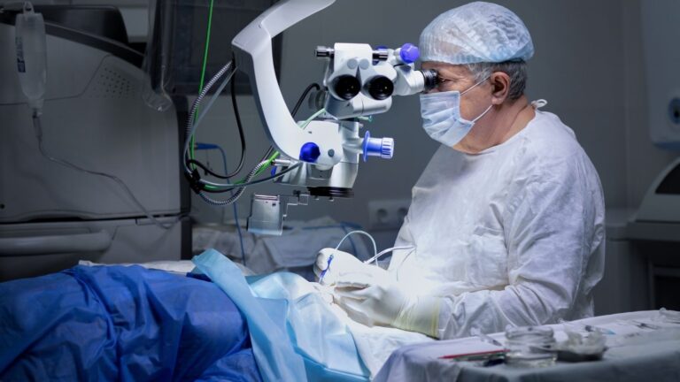
- If Retinal Detachment Has Developed: If fluid passing through the tear has caused a retinal detachment, pneumatic retinopexy, scleral buckling, pars plana vitrectomy or a combination of these methods may be planned. Gas or silicone oil may be placed inside the eye during surgery.
Retinal Tear Treatment Prices
Treatment costs vary according to the examination findings, the extent of laser treatment or cryotherapy, the need for additional imaging and, if retinal detachment is present, the surgical method used. Providing a fixed total price without an examination would not be appropriate. The condition of the retina is assessed first, and current pricing information is then provided.