Macular edema is the accumulation of fluid and thickening of retinal tissue in the macula, the area responsible for detailed central vision. It is not a disease on its own. It is a common finding associated with different conditions such as diabetic retinopathy, retinal vein occlusion, uveitis, an inflammatory response after cataract surgery, or traction on the retinal surface. Therefore, treatment is determined not simply by stating that edema is present, but by identifying its cause and the structural changes in the macula.
The treatment plan is created by evaluating visual acuity together with OCT findings, the duration of the edema, previous treatments, intraocular pressure, lens status, and the patient's systemic diseases. The aim is not only to reduce the fluid, but also to protect retinal cells and avoid unnecessary or ineffective repeated treatments. Close monitoring may be sufficient for some patients, while others may require intravitreal injections, laser treatment, steroid therapy, or vitrectomy.
What Are the Macula and Macular Edema?
Macula: The specialized area in the center of the retina responsible for reading, recognizing faces, driving, distinguishing colors, and seeing fine details. The fovea, located at the very center of the macula, provides the highest visual acuity.
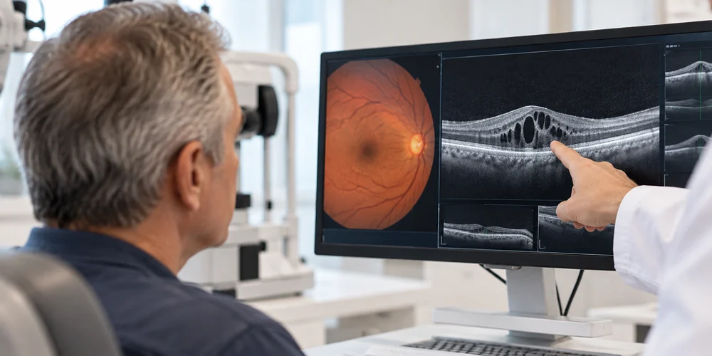
Macular Edema: When the permeability of retinal blood vessels increases, fluid and proteins inside the vessels may leak into the retinal tissue. OCT may show cystic spaces within the retina, retinal thickening, or fluid beneath the retina.
What Are the Symptoms of Macular Edema?
Macular edema is usually painless. When only one eye is affected, early changes may go unnoticed because the other eye continues to see well. The most common symptoms include:
- Blurred or hazy central vision
- Straight lines appearing curved, broken, or wavy, known as metamorphopsia
- Reduced reading speed and difficulty distinguishing small print
- Colors appearing faded or different
- A difference in image size between the two eyes, known as micropsia or macropsia
- A gray, blurred, or dark area in the center of the visual field
What Causes Macular Edema?
Diabetic Macular Edema: Diabetes-related damage to blood vessel walls causes leakage in the macula. Controlling blood glucose, blood pressure, kidney function, and blood lipid levels supports the treatment.
Edema Due to Retinal Vein Occlusion: An occlusion in the main retinal vein or one of its branches increases vascular pressure and permeability.
Macular Edema Due to Uveitis: Intraocular inflammation increases the permeability of retinal blood vessels. Identifying whether the uveitis is caused by an infection or an autoimmune condition is essential.
Macular Edema After Cataract Surgery: Pseudophakic cystoid macular edema, also known as Irvine-Gass syndrome, may develop after cataract surgery and is often controlled with anti-inflammatory eye drops.
Vitreomacular Traction and Epiretinal Membrane: Traction caused by the vitreous adhering to the macula or the development of a membrane on the retinal surface may mechanically cause fluid accumulation.
Diagnostic Methods
Optical Coherence Tomography (OCT): Provides cross-sectional images of the retinal layers and shows the amount and location of the fluid at micron-level resolution.
OCT Angiography: Provides information about retinal blood flow and nonperfused areas without using dye.
Fundus Fluorescein Angiography (FFA): Used to map vascular leakage, bleeding sites, and areas of ischemia.
How Is Macular Edema Treated?
Treatment is planned in stages according to the underlying cause, the degree to which central vision is affected, the presence of ischemia, and the patient's general condition.
1. Intravitreal Anti-VEGF Injections
Medications that suppress the VEGF protein, which plays a central role in vascular permeability, are the first treatment option for center-involving diabetic macular edema and retinal vein occlusion. The frequency of treatment is personalized according to the patient's response.
2. Steroid Treatments
Slow-release intravitreal implants or injections may be used for inflammatory macular edema, persistent cases after cataract surgery, or eyes with an inadequate response to anti-VEGF treatment. Intraocular pressure must be monitored.
3. Laser Treatment
Focal or grid laser treatment is currently used mainly for leakage outside the center of the macula or as a complementary treatment to injections in selected resistant cases.
4. Vitrectomy Surgery
When edema is caused by vitreomacular traction, an epiretinal membrane, or another mechanical membrane, the vitreous gel and membranes are surgically removed.
Intravitreal Injection Procedure and Aftercare
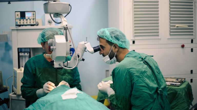
The injection is performed under sterile operating room conditions after the ocular surface is numbed with anesthetic eye drops. The procedure lasts only a few seconds and is painless. The patient feels only mild pressure.
Macular Edema Treatment Prices in Izmir
The cost of treatment varies according to the method used, including anti-VEGF injections, slow-release steroid implants, laser treatment, or vitrectomy surgery, the type of medication, the number of injection sessions, and hospital conditions. It is not medically appropriate to provide a standard price before the examination and OCT tests have been completed.