Xanthelasma refers to yellow or yellowish-white cholesterol deposits that appear on the eyelids, particularly near the nose. They do not cause pain or vision loss, but they may enlarge over time, become prominent on both eyelids, and create an aesthetically disturbing appearance.
I evaluate the depth of the xanthelasma, the area it covers, its location on the upper or lower eyelid, previous procedures, and the effect of the tissue to be removed on eyelid function. The aim is to reduce the lesion as much as possible while preserving the natural appearance and closure function of the eyelid.
What Is Xanthelasma and Why Does It Occur?
Xanthelasma palpebrarum consists of benign plaques formed by the accumulation of cells containing fat and cholesterol in the skin. It most commonly begins at the inner corner of the upper eyelid but may also occur on the lower eyelid or multiple eyelids. It is generally flat, soft, and painless. Typical xanthelasma is not a cancerous lesion. However, rapidly growing, hardening, bleeding, or ulcerating lesions should be evaluated to exclude other eyelid conditions.
The exact cause of xanthelasma is not the same in every patient. It may develop in people with normal blood lipid and cholesterol levels. In some patients, it may occur together with high cholesterol, triglyceride disorders, diabetes, or thyroid or liver diseases.
Systemic Evaluation: Particularly in cases that begin at a young age, are extensive or recurrent, or are associated with a family history, the lipid profile should be evaluated by an internal medicine specialist or cardiologist. Controlling cholesterol is valuable for general health, but it does not mean that an existing plaque will disappear spontaneously.
How Is Xanthelasma Diagnosed?
The diagnosis is usually made through examination. The color, surface structure, depth, borders, and location of the lesion on the eyelid are assessed. Syringoma, milia, sebaceous gland enlargement, cysts, xanthogranulomatous diseases, and some tumors may have a similar appearance. If the appearance is not typical or the lesion recurs and behaves differently, a biopsy or pathological examination may be required.
Xanthelasma Treatment Methods
There is no single fixed treatment for every xanthelasma. The treatment plan for small and superficial plaques differs from that for large, deep lesions or lesions approaching the eyelid margin. Skin excess, symmetry between the two sides, previous scars, skin color, and the patient's expectations are evaluated when making the decision.
1. Surgical Treatment of Xanthelasma (Excision)
Surgical excision is an effective option for well-defined lesions that can be removed without causing excessive tension in the eyelid tissue. The procedure is usually performed under local anesthesia. The plaque is removed in a controlled manner. The incision is planned to align with the natural eyelid creases and is closed with fine sutures.
Combination with Eyelid Surgery: When significant excess skin is also present with large upper eyelid lesions, treatment may be combined with an upper eyelid surgery plan known as blepharoplasty in suitable patients. Staged surgery may be required for lower eyelid or extensive lesions because removing excessive tissue may create a risk of impaired eyelid closure.
2. Laser Treatment of Xanthelasma
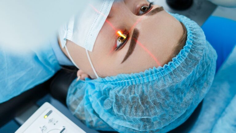
Ablative lasers such as carbon dioxide (CO2) and Er:YAG are used to reduce the tissue layer by layer in selected superficial or moderately deep xanthelasma lesions. Laser treatment eliminates the need for sutures. However, if the treatment depth is not properly controlled, depression, scarring, or changes in skin color may develop.
3. Chemical and Energy-Based Methods
Chemical applications such as trichloroacetic acid (TCA), radiofrequency, electrocautery, and cryotherapy are other methods described in the medical literature. They may be considered for small and selected lesions.
⚠️ Home Treatment Warning: Treatment with acids, wart medications, garlic, or similar products must never be attempted at home. Uncontrolled use around the eyes may cause chemical burns, pigmentation changes, permanent scarring, and eyelid damage.
Which Treatment Method Is More Suitable?
It is not appropriate to reduce the choice of treatment to the question of "laser or surgery" alone:
- For superficial and small plaques: Laser or controlled ablation methods
- For deeper and well-defined lesions: Surgical excision
- For extensive plaques or plaques involving multiple eyelids: Combined or staged approaches may be preferred
The most appropriate plan can be made after the actual borders of the lesion and the tissue defect that removal would leave in the eyelid are assessed during the examination.
Recovery and Important Considerations
The recovery process varies according to the method used and the extent of the procedure. Swelling, bruising, and tenderness along the incision line may occur after surgery. Crusting, redness, and temporary skin color changes may occur after laser treatment.
- Scabs must not be removed while the treatment area is healing.
- Prescribed products should be used as recommended, and the area should be protected from the sun.
- The timing of suture removal and wound care is determined individually.
Treatment Risks and the Possibility of Xanthelasma Recurrence
Temporary swelling, bruising, redness, infection, bleeding, scarring, lightening or darkening of the skin, and recurrence of the lesion are possible risks. Extensive tissue removal may cause functional problems such as eyelid retraction or incomplete eye closure.
Can Xanthelasma Recur? Yes. Regardless of the method used, xanthelasma may recur in the same area or another part of the eyelid. Recurrence does not mean that the treatment was performed incorrectly. The person's tissue characteristics and metabolic predisposition may persist. Safe tissue limits are prioritized to preserve eyelid function.
Xanthelasma Treatment Prices in Izmir
The fee varies according to the number of lesions, whether one or multiple eyelids are involved, the treatment method to be used, whether laser, surgery, or a combined approach, whether treatment is planned in a single session or stages, and whether pathological examination is required. It is not medically appropriate to determine an exact price without performing an examination and assessing eyelid function.