Türkçe
 Deutsch

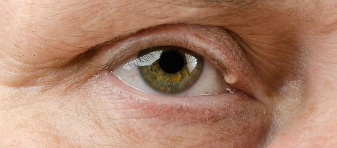
The eye is a complex and vital sensory organ that forms the sense of sight by detecting light. Eye anatomy primarily consists of optical structures that focus light from the external environment and a neural layer that converts this light into electrical signals. This magnificent system begins with transparent layers such as the cornea and lens refracting light. The image is projected onto the innermost layer, the retina, where it is processed and transmitted to the brain via the optic nerve. Thanks to the perfect harmony of each component, we gain the ability to perceive the world around us in three dimensions and color. This structure is our most important window to the world.
| Things You Should Know | Information |
| What is the eye? | A sensory organ with a complex structure that enables vision; it detects light and transmits signals to the brain. |
| Main function of the eye | To collect and refract light from the external environment, focus it on the retina, and transmit visual information to the brain through the neural conduction system. |
| Outer layer of the eye | Consists of the sclera (white of the eye) and cornea; provides structural support and protects the eye from external influences. |
| Sclera | The white part of the eye; it is a tough and fibrous structure and is a continuation of the cornea. |
| Cornea | With its transparent structure, it refracts light and transmits it to the inner structures of the eye; it is part of the optical system. |
| Middle layer (Uvea) | Composed of the iris, ciliary body, and choroid; it is rich in blood vessels. |
| Iris | The structure that determines eye color; regulates the amount of light via its central opening (pupil). |
| Ciliary body | Enables focusing of the lens; plays a role in the production of aqueous humor (intraocular fluid). |
| Choroid | Located between the retina and sclera; contains the blood vessels that nourish the retina. |
| Inner layer | The retina; the neural layer where vision begins. |
| Lens | Located behind the pupil; focuses light onto the retina with its flexible structure. |
| Eye chambers | Divided into three: anterior chamber (between cornea and iris), posterior chamber (between iris and lens), and vitreous chamber (between lens and retina). |
| Intraocular fluids | Aqueous humor (in the anterior and posterior chambers) and vitreous gel (in the posterior chamber); involved in eye nourishment and maintaining its shape. |
| Visual process | Light passes through the cornea, anterior chamber, lens, and vitreous, then focuses on the retina; here it is converted into nerve signals and transmitted to the brain via the optic nerve. |
Which Layers Protect the Outside of the Eyeball?
Our eyeball has a strong outer shell that protects it from potential dangers coming from the outside world. This shell consists of two main parts and both preserves the shape of the eye and forms the first step of the optical system.
The first is the sclera, which is the white part of our eye. This structure, composed of very durable fibrous connective tissue, covers most of the eyeball and acts like a shield. The muscles that move the eye attach to this strong layer. At the back, it also encases and protects the optic nerve that comes from the brain.
The second is the cornea. It is the transparent, dome-shaped front layer of the eye, resembling a watch glass. It is the first window through which light enters the eye and actually provides the majority of the refractive power needed for vision. The secret to the cornea’s clarity is that it contains no blood vessels. This unique feature allows it to be a perfect optical surface, but also makes it vulnerable to injury.
Which Structures Give the Eye Its Color and Provide Nourishment?
Just beneath the eye’s protective outer layer lies the vascular layer, also known in medical terms as the uvea. As the name suggests, this layer is very rich in blood vessels and is responsible for nourishing the internal tissues of the eye. Composed of three sections, this layer forms the colorful and functional center of the eye.
The parts of this layer are as follows:
- Iris: The part that gives the eye its color (blue, green, brown). The central opening is called the “pupil.” The iris functions like a camera diaphragm; in bright light, it contracts to shrink the pupil and limit light entry. In darkness, it relaxes to dilate the pupil, allowing more light in.
- Ciliary Body: Located just behind the iris, this structure has two major functions. First, it produces the intraocular fluid (aqueous humor). Second, the tiny muscles it contains help change the shape of the lens, allowing us to focus on near or distant objects.
- Choroid: The dense network of blood vessels located between the sclera and retina, covering the back wall of the eye. Its main function is to nourish the retina, where visual cells are located.
Where Does the Image Form Inside the Eye?
At the innermost layer of the eye lies the retina, made up of nerve cells, where vision takes place. The retina lines the back wall of the eyeball like wallpaper and functions similarly to a camera’s film or digital sensor. After passing through the cornea and lens, light is focused onto the retina.
The retina contains millions of specialized cells that detect light. These cells are of two types:
Cone cells: Allow us to see colors and perceive fine details. They are concentrated in the center of the retina, in a region known as the macula or yellow spot. We owe our sharp vision used for reading, recognizing faces, or driving to these cone cells.
Rod cells: Enable us to see in low light and at night. They cannot distinguish colors but are highly sensitive to light. They are located mostly around the periphery of the retina and are responsible for peripheral vision.
These retinal cells convert light energy into electrical signals. These signals are then processed by other nerve cells within the retina and transmitted to the brain via the optic nerve. The final interpretation of these signals—what we call “seeing”—takes place in the brain’s visual center.
What Are the Functions of the Fluids That Fill the Eyeball?
Our eyes are not hollow; on the contrary, they are filled with two different types of special fluids that give the eye its shape and help maintain the health of its internal, delicate tissues.
The first of these is aqueous humor. It is a clear, water-like fluid that fills the spaces in the front part of the eye, between the cornea and the lens. It is continuously produced by the ciliary body and drained through special channels (trabecular meshwork) back into the bloodstream. This constant production and drainage cycle helps maintain a steady intraocular pressure (eye pressure). It also provides nourishment to the avascular cornea and lens. In short, it is the lifeblood of the eye’s anterior segment.
The second fluid is vitreous humor. It is a transparent, gel-like substance with the consistency of egg white that fills the largest cavity at the back of the eyeball. Unlike aqueous humor, it is not continuously renewed. Its primary function is to maintain the spherical shape of the eyeball and support the retina by keeping it in place. With age, the structure of this gel can deteriorate and develop small clumps. The small dots or threads often described as “floaters” are usually the result of these changes.
What Are the Accessory Structures That Protect and Move the Eyeball?
The eyeball does not function in isolation. It is surrounded by a number of accessory structures that protect it, support it, and ensure it functions at its best.
These structures are called the “ocular adnexa” or simply “adnexa.”
The orbit, or eye socket, is a bony cavity that houses the eyeball, the muscles that move it, the nerves, blood vessels, and the fatty tissue that cushions it against impacts. This strong bony structure protects the eye from serious trauma.
There are six extraocular muscles that allow us to move our eyes in all directions. These muscles work in incredible coordination under the command of the brain to ensure that both eyes focus on the same point at the same time. Their primary movements include:
- Turning inward
- Turning outward
- Looking upward
- Looking downward
- Rotating inward and outward
Additionally, the eyelids serve as our most important barrier, protecting the eyes from external elements, foreign bodies, and excessive light. With each blink, they spread the tear film across the eye surface to prevent drying. The eyelashes act as a filter against small particles like dust.
The conjunctiva is a thin, transparent membrane that covers the eye surface and the inner part of the eyelids, ensuring the eye remains lubricated.
Finally, the tear system is responsible for keeping the eye moist and clean at all times. Tears are secreted by the lacrimal glands, wash over the eye surface, cleanse it of microbes, and then drain through small canals at the inner corner of the eye into the nasal cavity. That’s why it is completely normal for your nose to run when you cry.
What Is the Microscopic Structure of the Tissues That Maintain the Eye’s Transparency?
For the eye to see clearly, all the media through which light passes before reaching the retina must be completely transparent. The two main structures responsible for this are the cornea and the lens. Their microscopic architecture is specially designed to maintain transparency, but this design also makes them delicate and limits their ability to regenerate.
The microscopic structure of the cornea consists of five layers. The outermost epithelial layer is a self-renewing protective surface. Beneath it, the stromal layer forms the bulk of the corneal thickness and is composed of perfectly organized collagen fibers. This flawless arrangement ensures that light passes through without scattering. The innermost endothelial layer, made of a single row of cells, acts like tiny pumps, constantly removing excess water from the stroma and preventing the cornea from swelling and becoming cloudy. Endothelial cells cannot regenerate, so their number decreases with age or disease, leading to loss of corneal clarity.
The lens of the eye resembles an onion in structure. It grows throughout life as new fibers form on top of older ones. During maturation, these fibers lose their nuclei and all organelles to maximize transparency. Their interiors are filled with special proteins called crystallins. Over time, and especially due to factors like UV light, the structure of these proteins may deteriorate, causing the lens to lose its transparency and resulting in cataract, or clouding of the lens.
How Does the Eye Focus Light and Form an Image?
Vision is a magical process where physics and biochemistry intertwine. It all begins when light rays from the outside world enter our eyes. The eye is a sophisticated optical system that captures these rays and transforms them into a clear image on the retina.
Light first strikes the cornea, where most of the refraction occurs. It then passes through the pupil and reaches the lens. The role of the lens is to fine-tune this refraction. When we look into the distance, the lens flattens. However, when we want to focus on a nearby object, like a book, a reflex called “accommodation” is triggered. The ciliary muscles inside the eye contract, the fibers attached to the lens relax, and the lens becomes rounder, increasing its refractive power. This allows light rays from near objects to focus sharply on the retina. At the same time, the pupils slightly constrict and the eyes turn inward. This trio of responses is known as the “near response.”
When light hits the retina, the biochemical part of the process begins. Inside the retina’s millions of photoreceptor (light-sensing) cells are pigments sensitive to light. When a photon strikes one of these pigments, it initiates a domino effect. The impact changes the shape of the pigment, which triggers a cascade of chemical reactions within the cell. As a result, light energy is converted into an electrical signal—a language the brain can understand. This initial signal is transmitted to other nerve cells within the retina, processed, and finally sent to the brain via the optic nerve. This entire complex process takes place in less than a millisecond.
What Is Intraocular Pressure (Glaucoma) and Why Does It Occur?
Glaucoma, commonly known as “eye pressure” or “black water” among the public, is a progressive and insidious disease of the optic nerve. Because it usually progresses without symptoms, it is also referred to as the “silent thief of sight.”At the root of the disease lies an elevation in intraocular pressure beyond what the optic nerve can tolerate.
The pressure inside the eye is maintained by a delicate balance between the production and drainage of aqueous humor, the fluid inside the eye. This can be likened to a sink with a constantly running tap and a drain. As long as the water flowing from the tap (fluid production) and the water draining out (fluid outflow) are in balance, the sink doesn’t overflow. In glaucoma, the problem is not an overactive tap but a clogged drain.
This “drain” in the eye is a spongy tissue called the trabecular meshwork. With age or for various reasons, this tissue’s filtering function deteriorates. The fluid can’t drain fast enough, begins to accumulate inside the eye, and slowly raises intraocular pressure. This high pressure places mechanical stress on the most sensitive structure—the optic nerve—and disrupts blood flow, leading to the gradual death of nerve fibers. This damage is irreversible and initially causes loss of peripheral vision. The patient often only becomes aware of the condition when it’s very advanced—when it feels like they’re looking through a narrow tube. That’s why early diagnosis is critically important.
What Are the Treatment Options for Glaucoma?
The main goal in treating glaucoma is to lower intraocular pressure to stop or slow down damage to the optic nerve. While existing damage cannot be reversed, remaining vision can be preserved with proper treatment. Treatment is personalized and typically involves multiple steps.
The main types of eye drops used in treatment include:
- Prostaglandin analogs
- Beta-blockers
- Alpha-adrenergic agonists
- Carbonic anhydrase inhibitors
- Miotics
These drops work by either reducing the production of aqueous humor or improving its outflow. For treatment to be successful, it is critical that the drops are used daily, consistently, and exactly as prescribed.
If medication is insufficient or cannot be used by the patient, laser treatments are considered. Selective Laser Trabeculoplasty (SLT) is the most commonly used method. In this procedure, low-energy laser is applied to the blocked drainage channels to rejuvenate the area and improve fluid filtration. It is a painless, quick outpatient procedure and an effective treatment option.
If pressure remains uncontrolled despite medications and laser treatment, surgical methods are employed. In a trabeculectomy, a new channel is created to bypass the eye’s blocked natural drainage, allowing fluid to flow out. Glaucoma drainage implants (tube surgeries) are used in more complex cases, where a fine tube is inserted into the eye to direct the fluid to the back of the eye for absorption.
What Are the Most Common Age-Related Eye Problems?
Aging is a natural process that affects our eyes just like the rest of our body. As we age, the incidence of certain eye conditions increases. The most common age-related eye problems include cataracts, presbyopia (difficulty seeing up close), and macular degeneration.
Cataracts occur when the eye’s natural lens loses its clarity and becomes cloudy like frosted glass. This leads to gradually reduced vision, colors appearing dull or yellowish, glare while driving at night, and frequent changes in eyeglass prescriptions. Cataracts cannot be treated with medication; the only solution is surgery, where the cloudy lens is removed and replaced with a clear intraocular lens. Today, this procedure is successfully performed using sutureless and quick phacoemulsification techniques.
Presbyopia is age-related difficulty in near vision, usually starting around ages 40–45. The once-flexible eye lens hardens with age and loses its ability to focus on nearby objects (accommodation). This makes people hold phones, books, or menus farther away to see them clearly. Presbyopia can be corrected with reading glasses, contact lenses, or surgical options.
Age-related macular degeneration (AMD) is a disease affecting the macula, the central part of the retina responsible for sharp and detailed vision. There are two main types:
- Dry Type
- Wet Type
How Is Macular Degeneration (AMD) Treated?
Treatment of macular degeneration varies depending on the type (dry or wet) and stage of the disease. The goal is to slow the progression and minimize vision loss.
Currently, there is no definitive treatment for dry AMD. However, large scientific studies have shown that special nutritional supplements containing certain vitamins and minerals (AREDS2 formulation) can reduce the risk of progression to advanced stages. These supplements may be recommended for patients with moderate to advanced dry AMD. In addition, a healthy diet, avoiding smoking, and using UV-protected sunglasses are important lifestyle changes that can slow disease progression.
Wet AMD requires more urgent and active treatment. The core issue in this type is the development of abnormal, leaky blood vessels under the macula. Treatment involves anti-VEGF medications, which block the substance called Vascular Endothelial Growth Factor (VEGF) that triggers these vessels’ growth. These drugs are injected directly into the eye using a very fine needle. Contrary to popular belief, the procedure is painless and takes just a few seconds. These injections help shrink the abnormal vessels and stop leakage, stabilizing vision or even improving it in some patients. Treatment typically begins with monthly injections, and the frequency is adjusted based on the patient’s response. Early diagnosis and regular treatment are key to preserving vision in wet AMD.
What Are the Effects of Diabetes on the Eye?
Diabetes is a chronic condition that causes consistently high blood sugar levels, damaging small blood vessels throughout the body. The eye is among the most affected organs. The damage diabetes causes to the retinal blood vessels is called diabetic retinopathy, and it is the leading cause of preventable blindness in working-age individuals.
The disease generally progresses in two main stages:
- Non-proliferative diabetic retinopathy (NPDR): This is the early stage. The walls of small blood vessels in the retina weaken, allowing fluid and blood to leak out. Small balloon-like swellings (microaneurysms) form. If this leakage occurs in the macula—the area responsible for sharp vision—it leads to diabetic macular edema, causing central vision to blur.
- Proliferative diabetic retinopathy (PDR): This is the advanced stage of the disease. Due to widespread damage, the retina receives insufficient oxygen. The body attempts to compensate by creating new blood vessels. However, these new vessels (neovascularization) are highly abnormal, fragile, and prone to bleeding. They can easily hemorrhage into the eye (vitreous hemorrhage), causing sudden vision loss. Scar tissue that forms around these vessels can contract and pull on the retina, leading to tractional retinal detachment.
How Are the Damages Caused by Diabetes in the Eye Managed?
The most basic and important step in managing diabetic retinopathy is controlling the underlying condition—diabetes itself. Maintaining ideal levels of blood sugar, blood pressure, and blood lipids (cholesterol, triglycerides) can delay the onset of retinopathy and slow its progression. However, if retinopathy develops or progresses despite these precautions, various eye treatments come into play.
Treatment options are determined based on the stage of the disease and the area it affects. The main treatment methods include:
- Intraocular injections (Anti-VEGF): These are the first choice, especially for diabetic macular edema. These medications are injected into the eye to reduce fluid leakage from vessels, decrease swelling in the macula, and potentially improve vision.
- Laser photocoagulation: This treatment has two main applications. “Focal/Grid Laser” targets specific leaking vessels in macular edema, while “Panretinal Photocoagulation (PRP)” is used in advanced proliferative retinopathy. PRP involves making thousands of small laser burns in the peripheral retina to deactivate areas with low oxygen demand. This helps reduce abnormal blood vessel growth and decreases the risk of bleeding.
- Vitrectomy surgery: In advanced cases—particularly if there is persistent vitreous hemorrhage or tractional retinal detachment—vitrectomy is required. In this surgery, the blood-filled or traction-causing vitreous gel inside the eye is removed, and the retina is repositioned.
Frequently Asked Questions
What are the main anatomical structures of the eye and how do they work together to create vision?
The eye consists of the cornea, iris, pupil, lens, retina, optic nerve, and supporting tissues. These structures work together to focus light, convert it into electrical signals, and transmit visual information to the brain for interpretation.
How does the cornea contribute to the eye’s ability to focus incoming light?
The cornea is the transparent front surface of the eye and provides most of its focusing power. By bending incoming light rays toward the retina, it helps create a clear image and plays a critical role in visual sharpness.
Why are the iris and pupil important for regulating vision in different lighting conditions?
The iris controls the size of the pupil, which regulates the amount of light entering the eye. In bright environments the pupil constricts, while in dim conditions it enlarges to allow more light to reach the retina.
How does the lens change shape to help the eye focus on near and distant objects?
The lens adjusts its shape through a process called accommodation. It becomes thicker for close-up tasks and flatter for distant viewing, allowing light to focus accurately on the retina and maintain clear vision.
What role does the retina play in converting light into visual information?
The retina contains specialized photoreceptor cells called rods and cones. These cells detect light and color, converting them into electrical impulses that travel through the optic nerve to the brain for processing.
How does the optic nerve connect the eye to the brain and support vision?
The optic nerve carries electrical signals generated by the retina directly to the brain. These signals are interpreted in visual processing centers, allowing people to recognize shapes, colors, movement, and depth.
What are the functions of the vitreous and aqueous fluids within the eye?
The aqueous and vitreous fluids help maintain eye shape, support internal structures, provide nutrients, and allow light to pass through transparent tissues. Proper fluid balance is essential for healthy eye function.
How do rods and cones differ in supporting vision under various conditions?
Rods are responsible for vision in low-light environments and detecting movement, while cones provide detailed central vision and color perception. Together they allow the eye to function effectively across different lighting conditions.
What common age-related changes can affect the structure and function of the eye?
Aging may lead to cataracts, reduced lens flexibility, vitreous changes, and retinal disorders. These structural changes can affect visual clarity, focusing ability, and overall eye health, often requiring regular monitoring.
Why is understanding the structure of the eye important for preventing vision problems?
Knowledge of eye anatomy helps individuals recognize symptoms early and appreciate the importance of routine examinations. Early detection of structural abnormalities can improve treatment outcomes and help preserve long-term vision.