Türkçe
 Deutsch

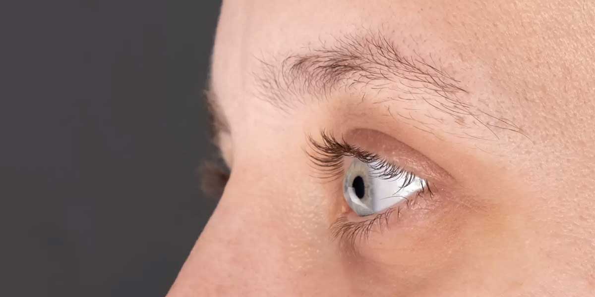
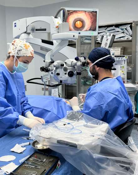
Corneal transplant is a surgical procedure that involves replacing damaged or diseased corneal tissue with a healthy cornea from a donor. Nowadays, there is increasing interest in less invasive techniques such as Descemet’s Membrane Endothelial Keratoplasty (DMEK). Since this method replaces only the inner layer of the cornea, it accelerates the healing process and improves visual outcomes.
It also significantly reduces the risk of tissue rejection. DMEK, which is particularly effective in diseases like Fuchs’ dystrophy, reduces complications and increases success rates thanks to advanced tissue preparation methods, offering an important innovation in the field of corneal transplantation.
Turkey is one of the best country for Corneal Transplant at a afforable price. If you are looking for a place for Corneal Transplant abroad with best clinics and doctors, you are definitly on the right spot. Check Dr. Berkay Akmaz's Corneal Transplant prices, reviews and packages.
Ophthalmology Specialist – Assoc. Prof. Dr. Berkay Akmaz was born in İzmir in 1985. Due to his father's profession, he spent his primary school years in different cities. He began his education at Gülhane Military Medical Academy in 2003 and graduated with the title of Medical Doctor from Ege University Faculty of Medicine in 2010. In 2010, Assoc. Prof. Dr. Berkay Akmaz ranked 25th in the Medical Specialization Exam (TUS) in Turkey and started his specialization in the Department of Ophthalmology and Surgery at Dr. Lütfi Kırdar Kartal Education and Research Hospital. He completed his specialization training in 2014.
6 Reasons to Get Corneal Transplant in Izmir






| Definition | Corneal Transplant (Keratoplasty) is the procedure of replacing a damaged or diseased cornea with healthy donor corneal tissue. The cornea is the transparent layer located at the front of the eye that allows light to be clearly focused onto the retina. |
| Purpose | To enhance visual acuity, alleviate pain or damage in the cornea, and restore the structural integrity and transparency of the cornea. |
| Applications | Keratoconus, corneal scarring, infections (e.g., herpes simplex keratitis), corneal dystrophies, traumatic corneal damage, failure of previous corneal surgeries. |
| Types of Transplant | Penetrating Keratoplasty: Full-thickness corneal transplant. Lamellar Keratoplasty: Replacement of only the damaged layers (e.g., DALK, DSAEK, DMEK). |
| Donor Tissue Source | Corneal tissues obtained from deceased individuals; screened for suitability and infection in cornea banks. |
| Operation Duration | Approximately 1-2 hours and performed under local or general anesthesia. |
| Recovery Process | Complete visual recovery may take several months to a year; sutures are usually removed after 6-12 months. |
| Risks and Complications | Rejection of the transplant, infection, astigmatism, development of glaucoma, cataract formation, retinal detachment. |
| Signs of Rejection | Redness in the eye, pain, light sensitivity, decreased vision; can be treated with early intervention. |
| Postoperative Care | Regular use of antibiotic and steroid eye drops, protecting the eye from impacts, avoiding strenuous physical activities, and attending regular eye check-ups. |
| Success Rate | Varies depending on the type of disease and the patient’s overall health; generally has a high success rate. |
| Alternative Treatments | Corneal Cross-Linking therapy, specialized contact lenses, other refractive surgical methods. |
| Prevention | While some corneal diseases are genetic or unavoidable, maintaining eye hygiene and protecting the eyes from UV rays is important. |
| Impact on Quality of Life | Regaining visual function increases independence in daily activities; improves quality of work and social life. |
| Donor Compatibility | Although blood type compatibility is not required, donor corneas are carefully examined for tissue compatibility and infection risks. |
| Affected Age Group | Applicable at any age; requires special attention and monitoring in children and the elderly. |
| Cost and Insurance Coverage | The cost of treatment varies depending on the center where the procedure is performed and the technology used; many insurance plans cover corneal transplants in cases of medical necessity. |
| Technological Developments | Femtosecond Laser Technology: Provides more precise cuts and better tissue alignment. Artificial Cornea (Keratoprosthesis): Used as an alternative when natural corneal transplant is not possible. |
| Long-term Follow-up | Regular eye examinations, vision tests, and intraocular pressure measurements to detect and treat possible complications early. |
| Precautions | Avoid rubbing the eyes, stay away from dusty and dirty environments, use prescribed drops regularly, and stay alert to any symptoms. |
| Social and Psychological Support | Psychological support and patient education are important for coping with vision loss and the surgical process. |
What is Corneal Transplant?
Corneal transplantation became widespread in the mid-20th century with surgical advancements and has since made significant progress. Especially from the 1970s onwards, new techniques have allowed for successful outcomes. In the United States, the FDA approved an artificial cornea called the Boston Keratoprosthesis in 1992, making it the only device approved for artificial corneal transplantation in the U.S.
Today, techniques such as full-thickness keratoplasty and endothelial keratoplasty are used, each aiming to accelerate the healing process. Additionally, these techniques make it possible to replace only selected corneal layers, significantly reducing the risk of rejection. These developments have opened new horizons in corneal transplantation practices.
What are the Types of Corneal Transplant?
The mechanism of action of corneal transplantation is based on improving vision and treating corneal disorders. The main goal of this procedure is to restore the structural integrity and transparency of the cornea. Different approaches are used in corneal transplantation performed with various techniques, depending on the type of tissue transplanted:
- Penetrating Keratoplasty (PK): In this method, the entire thickness of the cornea is replaced with donor tissue. Full-thickness transplantation renews all layers of the cornea with new tissue.
- Lamellar Keratoplasty (LK): Only specific corneal layers are replaced. This technique is divided into two sub-methods:
- Endothelial Keratoplasty (EK): Only the innermost layer of the cornea, the endothelial layer, is transplanted.
- Deep Anterior Lamellar Keratoplasty (DALK): In this method, the middle layers of the cornea, known as the stroma, are replaced.
Since these techniques involve the transplantation of different layers, each is suitable for specific conditions. During the procedure, the damaged corneal tissue is carefully removed and replaced with donor cornea. After the transplant, the new cornea ensures proper refraction of light, correcting visual problems.
Moreover, the avascular nature of the cornea provides limited exposure to the immune system. Nevertheless, there is still a possibility of graft rejection. Post-transplant immune responses, such as corneal neovascularization, can threaten the long-term success of the graft.
When is Corneal Transplant Performed?
Corneal transplant is a treatment option applied in cases where the cornea is permanently damaged and causes severe vision loss. The most common indications include keratoconus, Fuchs’ endothelial dystrophy, and bullous keratopathy. These conditions affect the structural or functional integrity of the cornea, significantly reducing the person’s quality of life. The most frequent situations where corneal transplant is performed are:
- Keratoconus: This condition, characterized by thinning of the cornea, leads to irregular astigmatism and severe vision loss. Corneal transplant is necessary when contact lenses or other interventions are insufficient, especially in young patients.
- Fuchs’ Endothelial Dystrophy: Characterized by the genetic deterioration of endothelial cells, this condition causes corneal swelling and vision loss. When it causes significant visual impairment, corneal transplant usually becomes unavoidable.
- Bullous Keratopathy: It can develop after eye surgeries like cataract surgery and leads to blister formation in the cornea. It is treated with corneal transplant methods such as endothelial keratoplasty.
- Corneal Scarring: Scarring resulting from trauma, infections, or inflammatory eye diseases is treated with corneal transplant when it leads to vision loss.
- Infectious Keratitis: Corneal infections, especially those caused by fungi, viruses, or bacteria, can cause opacity. When medical treatment is ineffective, corneal transplant is necessary.
- Congenital Corneal Opacities: In cases of congenital corneal opacity, corneal transplant is performed to improve vision.
- Re-grafting: When previous transplants are rejected or fail, re-grafting is necessary, and in this case, corneal transplant is among the indications.
Who is Not Suitable for Corneal Transplant?
Corneal transplant may not be a suitable solution for every patient. Therefore, contraindications need to be carefully evaluated before the operation. These contraindications can be considered under three main headings: systemic, ocular, and donor-related factors.
Systemic Contraindications:
- Autoimmune Diseases: Individuals with diseases such as uncontrolled rheumatoid arthritis or systemic lupus erythematosus may have an increased risk of graft rejection due to systemic inflammation.
- Uncontrolled Diabetes: Poorly managed diabetes can delay wound healing and increase the risk of infection, negatively affecting transplant success.
- Severe Immunosuppression: Patients undergoing chemotherapy or with weakened immune systems are more susceptible to infections and poor graft healing.
- Active Infections: Patients with viral infections such as HIV or hepatitis have a high risk of the infection causing graft failure.
Ocular Contraindications:
- Ocular Surface Disease: Chronic surface diseases such as severe dry eye syndrome or meibomian gland dysfunction can make healthy graft healing difficult.
- Uncontrolled Glaucoma: In patients with high intraocular pressure, the new corneal tissue may be damaged, and the risk of graft failure increases.
- End-Stage Ocular Disease: In conditions such as optic nerve atrophy or end-stage retinitis pigmentosa, corneal transplant may not provide significant visual benefit.
- Active Ocular Infections: The presence of active herpes simplex virus or bacterial infections in the cornea increases the risk of graft infection and requires treatment before transplantation.
Donor-Related Contraindications:
- Donor’s Age and Health: Corneas obtained from elderly donors may not be suitable as they carry a risk of lower cell counts and graft failure.
- Infectious Diseases: If the donor has a history of infectious diseases such as hepatitis or syphilis, the cornea may not be considered suitable for transplant to prevent infection risk.
- Corneal Quality: Conditions that reduce corneal quality, such as keratoconus, endothelial damage, or scarring, constitute contraindications for transplant.
How is Corneal Transplant Performed?
Various steps are followed before and during corneal transplant surgery. First, a suitable donor tissue selection is carried out. The donor cornea is usually obtained from an eye bank and meticulously screened for infectious diseases before transplantation. Thus, it is aimed to ensure tissue quality and perform a safe transplant. Patient evaluation includes selecting the most appropriate surgical technique according to the type of corneal disease.
Preoperative Preparation:
- Donor Tissue Selection: The donor cornea undergoes appropriate quality controls and is prepared for transplantation.
- Patient Evaluation: A comprehensive evaluation allows determining the appropriate technique based on the extent of corneal damage.
During the surgical procedure, the method applied varies depending on the type of corneal disease and the level of damage. In the penetrating keratoplasty (PK) method, a circular portion of the full-thickness cornea is cut. Then, the donor tissue, appropriately prepared, is placed in the removed area and fixed with fine sutures. PK is a preferred method in full-thickness transplants involving all corneal layers.
On the other hand, in the endothelial keratoplasty (EK) method, only the endothelial layer is replaced. With the most common variations of EK, Descemet’s Stripping Automated Endothelial Keratoplasty (DSAEK) and Descemet’s Membrane Endothelial Keratoplasty (DMEK), only the innermost layer of the cornea is changed. This method provides a faster healing process and reduces the risk of complications.
What are the Side Effects of Corneal Transplant?
Various complications and side effects can occur after corneal transplant, both in the short and long term. The most common and serious complications include graft rejection, infections, glaucoma, and astigmatism. Each manifests with different symptoms and sometimes requires serious treatment.
- Graft Rejection: The immune system may perceive the transplanted cornea as foreign and attempt to reject it. In this case, symptoms such as redness of the eye, pain, sensitivity to light, decreased vision, and a feeling of sand in the eye appear. If not treated, it can lead to permanent damage.
- Infections: Bacteria, fungi, or viruses can enter the eye during or after the operation. These infections can cause redness, pain, discharge, and blurred vision in the eye. Starting antibiotic or antifungal treatment immediately is vital to protect the graft.
- Glaucoma: The condition of increased intraocular pressure after transplantation is called glaucoma. It manifests with eye pain, blurred vision, and halos around lights. If not treated, it can cause permanent vision loss.
- Astigmatism: Astigmatism can occur due to the irregular shape of the cornea after surgery. In this case, blurred or distorted vision occurs, and lenses or laser treatment may be required to correct it.
- Endothelial Failure: If the endothelial layer deteriorates after the transplant, the cornea swells, and vision loss may occur. It is more common in older grafts and may require advanced treatment.
How Successful is Corneal Transplant?
The success of corneal transplant varies depending on various factors such as the patient’s health status, type of transplant, and postoperative care. In general, corneal transplant stands out as a successful treatment in low-risk patients, and graft survival rates can reach up to 90% in the first year. However, this success rate tends to decrease over time. After five years, survival rates drop to about 74%, and after ten years, this rate falls to 62%.
On the other hand, conditions such as corneal neovascularization, previous graft failure, or ongoing inflammation are among high-risk cases. In these cases, success rates are significantly lower, and graft survival can drop to as low as 54% at the end of one year. In the five-year period, this rate regresses to quite low levels, around 19.8%. Common causes of graft failure include rejection episodes, infections, and complications from previous surgical interventions such as glaucoma surgeries.
Additionally, partial-thickness transplant techniques positively affect success in the following situations:
- Descemet’s Membrane Endothelial Keratoplasty (DMEK) technique
- Descemet’s Stripping Automated Endothelial Keratoplasty (DSAEK) technique
How to Prepare for Corneal Transplant?
A careful preparation process is conducted before corneal transplant to ensure the patient’s health and the success of the surgery. First, the patient’s eye condition and general health status undergo a detailed evaluation. During this assessment, the condition of the cornea is examined with slit-lamp biomicroscopy, and possible diseases are checked. The health of the surrounding tissues of the eye and the patient’s general health, including diseases like diabetes, are evaluated.
Before the surgery, donor cornea selection goes through a careful screening process, and these corneas are usually obtained from an eye bank. Tests such as sterility checks, endothelial cell counting, and specular microscopy are performed to ensure the suitability of the donor cornea. These corneas are stored in a culture medium to preserve cell viability, and swelling is reduced before surgery using a hyperosmotic medium.
Preoperative medication adjustments are also an important part of the process. Patients are informed to avoid medications that could delay healing or increase the risk of bleeding. These medications may include blood thinners and anti-inflammatory drugs. Antibiotics and antiviral prophylactics may be administered before surgery to reduce the risk of infection.
On the day of surgery, the area around the eye is carefully disinfected, and topical antibiotic eye drops are used to minimize the risk of infection. Additionally, the patient undergoes an informed consent process and is given detailed information about the procedure. Possible complications, risk of graft rejection, and postoperative care issues are explained.
How Should Postoperative Care Be After Corneal Transplant?
Postoperative care after corneal transplant should be carefully followed for a successful healing process. Medication management, eye protection, activity restrictions, and regular doctor visits are among the fundamental elements of recovery. First, medication management is of great importance. Steroid and antibiotic eye drops are used to prevent infection and inflammation. Some patients may need to use these drops for an extended period. Regular use of the medication as prescribed reduces the risk of complications.
Eye protection is important for the healing of delicate tissue. The patient should avoid rubbing the eye and exposing it to water for at least one month. In addition, using protective glasses or an eye shield helps protect the eye from irritants. Care should be taken to prevent water from entering the eye while showering.
Activity restrictions should be considered to prevent damage to the graft. The patient should avoid heavy lifting and strenuous activities for a few weeks after surgery. Sports involving contact with water, such as swimming, should not be done until the doctor allows it. Additionally, spending long periods in front of screens should be limited as it can strain the eyes.
Follow-up appointments are mandatory to detect signs of infection or graft rejection early. These check-ups, which need to be done regularly during the first year after surgery, should not be neglected.
How Does Resistance and Graft Rejection Develop Against Corneal Transplant?
Resistance mechanisms against corneal transplant are particularly related to immune responses that develop despite the eye’s immune privilege. The cornea has the advantage of immune privilege due to its central area lacking blood and lymphatic vessels. However, factors such as inflammation, trauma, and neovascularization disrupt this privilege, paving the way for immune cells to reach the cornea. In this case, antigen-presenting cells (APCs) migrate to lymph nodes and primarily activate Th1 T cells. As a result, these activated T cells attack the donor tissue and initiate the rejection process.
The rejection mechanism occurs in two ways:
- Direct pathway: Donor-derived dendritic cells directly present donor antigens to recipient T cells, initiating an immune response.
- Indirect pathway: Recipient APCs process donor antigens and present them to recipient T cells. Due to the absence of dendritic cells in the central cornea, this pathway is more commonly observed.
Proinflammatory cytokines and neovascularization play a critical role in the rejection process of corneal transplant. Cytokines like IFN-γ facilitate the entry of immune cells into the cornea and disrupt immune privilege. The formation of new blood and lymphatic vessels accelerates this process, increasing the risk of rejection.
Regulatory T Cells (Tregs) help develop immune tolerance. However, in inflamed and vascularized host beds, the immune system can overcome the suppressive effect of Tregs and trigger rejection.
In corneal transplant, minor histocompatibility antigens (mHAgs) can trigger an immune response between the donor and recipient through minor antigen differences, even if there is a major histocompatibility complex (MHC) match. Especially if there is inflammation, mHAgs contribute to graft failure.
Over time, chronic endothelial cell loss becomes one of the main causes of graft failure without acute rejection episodes. Patients with inflamed or vascularized corneas carry a higher risk of rejection, even if they receive immunosuppressive therapy. Therefore, new immunosuppressive treatments are being researched to increase long-term graft survival.
Recent Posts

Dermatochalasis (Eyelid Sagging)
Anasayfa » Eyelid » Dermatochalasis (Eyelid Sagging)Dermatochalasis (eyelid sagging) is a condition caused by excess [...]
Read MoreMar

Congenital Cataract
Anasayfa » Cataract » Congenital CataractCongenital cataract is an eye disease that occurs when the [...]
Read MoreMar

Causes of Blurred Vision in One Eye
Anasayfa » Eye Health » Causes of Blurred Vision in One EyeThe causes of blurred [...]
Read MoreDec

Comments from Patients with Pseudotumor Cerebri
Anasayfa » Uncategorized » Comments from Patients with Pseudotumor CerebriComments from patients with pseudotumor cerebri [...]
Read MoreDec

Reviews of People Who Have Undergone Corneal Surgery
Anasayfa » Eye Health » Reviews of People Who Have Undergone Corneal SurgeryReviews from people [...]
Read MoreDec

Keratoconus Patient Reviews
Anasayfa » Eye Health » Keratoconus Patient ReviewsKeratoconus patient reviews provide important insights into the [...]
Read MoreDec

How Long Does Cataract Surgery Take?
Anasayfa » Cataract » How Long Does Cataract Surgery Take?The answer to the question of [...]
Read MoreDec

How Do Under-Eye Dark Circles Go Away?
Anasayfa » Uncategorized » How Do Under-Eye Dark Circles Go Away?The question of how under-eye [...]
Read MoreDec
Gallery