Türkçe
 Deutsch


ICL (Phakic Intraocular Lenses) are developed for correcting myopia and astigmatism in patients who are not suitable for laser eye surgery. Placed in the posterior chamber of the eye, these lenses are located behind the iris but in front of the natural lens, and they do not damage the structure of the natural lens.
Additionally, the ICL is less invasive than corneal surgeries, offering a reversible alternative and providing a safe solution in the long term. Especially the EVO Visian ICL offers better visual quality and lower dry eye rates without increasing the risk of cataract or causing issues like intraocular pressure.
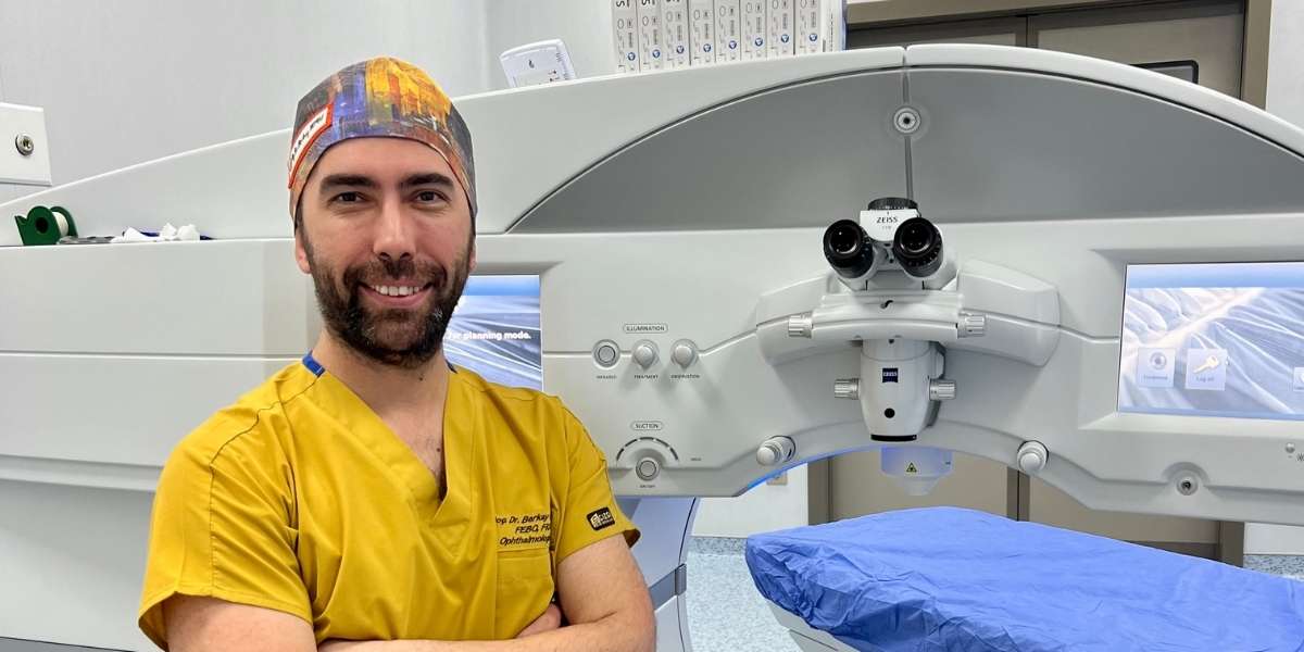
Turkey is one of the best country for ICL (Phakic Intraocular Lens) at a afforable price. If you are looking for a place for ICL (Phakic Intraocular Lens) abroad with best clinics and doctors, you are definitly on the right spot. Check Dr. Berkay Akmaz's ICL (Phakic Intraocular Lens) prices, reviews and packages.
Ophthalmology Specialist – Assoc. Prof. Dr. Berkay Akmaz was born in İzmir in 1985. Due to his father's profession, he spent his primary school years in different cities. He began his education at Gülhane Military Medical Academy in 2003 and graduated with the title of Medical Doctor from Ege University Faculty of Medicine in 2010. In 2010, Assoc. Prof. Dr. Berkay Akmaz ranked 25th in the Medical Specialization Exam (TUS) in Turkey and started his specialization in the Department of Ophthalmology and Surgery at Dr. Lütfi Kırdar Kartal Education and Research Hospital. He completed his specialization training in 2014.






| Definition | It is the procedure of implanting an artificial lens into the eye to correct high-degree refractive errors such as myopia, hyperopia, and astigmatism. The natural eye lens is not removed. |
| Applications | Treatment of myopia, hyperopia, and astigmatism; especially preferred for patients with high refractive errors, those unsuitable for laser treatment, or patients with thin corneas. |
| Mechanism of Action | A phakic intraocular lens is placed in the space in front of the natural lens. This lens increases the eye’s refractive power, providing clear vision and working in conjunction with the natural lens. |
| Advantages | 1. Provides successful results in high-degree refractive errors 2. Easier to reverse compared to laser procedures 3. Does not interfere with corneal tissue, leaves no permanent scars |
| Disadvantages | 1. Can be expensive 2. Includes risks such as increased intraocular pressure or development of cataracts 3. Requires regular intraocular pressure monitoring |
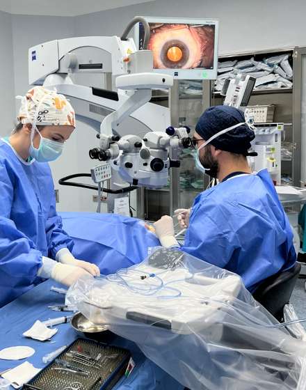
| Surgery Process | It is a procedure that takes approximately 20-30 minutes under local anesthesia. An intraocular lens is inserted through a small incision, and the incision typically heals on its own. |
| Recovery Process | Generally quick; there may be blurriness in the first few days. Most patients regain clear vision within a few days, with full recovery achieved within 1-2 weeks. |
| Side Effects | Mild pain, light sensitivity, temporary blurriness; rarely increased intraocular pressure, development of cataracts, or serious complications like retinal detachment. |
| Alternatives | 1. LASIK (Laser-Assisted In Situ Keratomileusis) 2. PRK (Photorefractive Keratectomy) 3. SMILE Laser |
| Eligibility Criteria | Suitable for individuals aged 21 and above, with stable refractive errors, specific corneal structures unsuitable for laser treatment, and patients with high myopia/hyperopia. |
| Care and Follow-up | Post-surgery use of eye drops, monitoring of intraocular pressure, and regular doctor check-ups are necessary. Attention should be paid to eye hygiene and protection from sunlight. |
What Is ICL?

Phakic intraocular lenses (ICL) are surgical implants used to correct refractive errors without removing the eye’s natural lens. Verisyse, which received the first FDA approval, was approved in September 2004 and began offering solutions to problems like myopia. Following this development, Visian ICL was also approved in December 2005 and began to be widely used.
These lenses, placed behind the iris but in front of the natural lens, offer a different option compared to laser vision correction procedures. Finally, with the approval of the EVO ICL model in March 2022, the development of phakic IOLs has advanced even further. This type of lens stands out by reducing the risk of corneal ectasia compared to LASIK.
What Are the Types of Phakic Intraocular Lens (IOL) Treatment?

Phakic intraocular lens treatment is a method used especially in patients with high levels of myopia, hyperopia, or astigmatism who are not suitable candidates for laser eye surgery. In this procedure, the natural lens remains in place, and an additional artificial lens is implanted into the eye. Depending on their position and characteristics, various types of phakic lenses are available:
- Posterior Chamber Phakic Lenses (ICL): Placed between the natural lens and the iris; the most common and reliable application.
- Anterior Chamber Phakic Lenses: Placed in front of the iris; preferred for eyes with a deeper anterior chamber.
- Toric Phakic Lenses: Used for patients who have astigmatism along with myopia or hyperopia to correct refractive errors.
- Spherical Phakic Lenses: Standard lenses designed to correct only myopia or hyperopia.
- Customized Phakic Lenses: Specially produced based on anatomical measurements of the eye to ensure maximum compatibility.
How Does ICL Work?
ICL is a type of intraocular lens placed inside the eye to correct refractive errors. This lens is positioned in the posterior chamber of the eye without removing the eye’s natural lens and eliminates vision problems. Posterior chamber ICL models, like the Visian ICL, are placed behind the iris and allow light to reach the retina. Thanks to this placement, the need for glasses or contact lenses is eliminated.
The materials used in the production of ICLs are generally biocompatible collagen copolymer or substances like polymethyl methacrylate (PMMA). These materials allow the lens to remain compatible with the eye and function for a long time without causing irritation. The new generation EVO Visian ICL features a central port that helps maintain intraocular pressure. This feature reduces the need for preoperative iridotomy by improving aqueous humor flow.
The vision correction function of the ICL works by changing the path of light refraction within the eye. It corrects refractive errors like myopia and astigmatism by compensating for irregularities in the eye. Unlike surface-based methods like LASIK that reshape the cornea, the ICL does not interfere with the corneal structure. Therefore, it has a high potential to provide contrast sensitivity and sharp vision.
When Is ICL Used?
ICL is designed as an alternative to laser-based surgeries to correct certain refractive errors and can be used according to various indications. High-degree myopia, which goes beyond the limits of laser surgeries, comes first among these indications.
The Implantable Collamer Lens (ICL) is preferred in myopes ranging from -3.0 to -20.0 diopters, with or without astigmatism up to 4.0 diopters. These types of lenses are especially suitable in cases where success is difficult to achieve with LASIK or PRK. Additionally, it is a good option for patients with thin corneas who are not suitable for laser surgery; since it does not change the corneal structure, it provides a safe solution by preserving corneal integrity.
ICL provides effective results in patients with stable refractive errors. Individuals with eye refractive errors that have not shown more than a 0.5 diopter change in the last 6 to 12 months can choose this method to achieve long-term effectiveness. The procedure is suitable for individuals aged 21 and over, where eye development has been completed. Still, in some cases, it may be considered for younger patients.
Other important indications are listed below:
- Adequate Anterior Chamber Depth: For ICL application, a depth of at least 3.0 mm is generally required. This depth is important for the correct placement of the lens and to prevent complications. Some new model lenses can also be placed at shallower depths.
- Adequate Endothelial Cell Density: The density of cells in the front part of the eye is important to reduce the risk of postoperative complications. Generally, a minimum of 1800 cells/mm² ECD is required.
- Contraindications to Laser Surgery: For individuals unsuitable for laser-based surgical methods, the ICL is suggested as an ideal alternative since it does not affect corneal tissue.
- Preference for a Reversible Procedure: For patients who do not want permanent changes, the ICL is a safe option as it can be removed and replaced.
- No History of Eye Diseases: The ICL should be preferred in patients without progressive eye diseases. Conditions like keratoconus, glaucoma, cataract, or uveitis can negatively affect ICL results.
Who Is Not Suitable for ICL?
ICL implantation may not be suitable for every patient. Due to certain contraindications, it is recommended not to use this lens in some individuals. Firstly, individuals under 21 years old and those with variable refractive errors are not among suitable candidates. Additionally, hormonal changes during pregnancy and breastfeeding can affect vision stability, limiting the application of these lenses. People whose refractions have fluctuated in the last year are also not considered suitable for the ICL.
Certain eye disorders also restrict the use of the ICL. Specifically:
- In individuals with glaucoma, this implantation is not recommended as intraocular pressure may be affected.
- In patients with Fuchs endothelial dystrophy or low endothelial cell count, the cornea may be damaged.
- In individuals with visually significant cataracts, if cataracts significantly affect visual ability, ICL application is not performed.
There are also certain anterior chamber depth requirements in terms of anatomical suitability. In myopic eyes, if the depth between the cornea and lens is below 2.8 mm, and in hyperopic eyes if this depth is below 3.0 mm, ICL implantation is not recommended. When this sufficient depth is not provided, risks such as vaulting of the ICL or increased intraocular pressure may arise.
Finally, if a person has severe amblyopia or permanent vision loss in one eye, ICL implantation is not preferred because the expected vision improvement may be limited.
How Is ICL Applied?
In the preoperative evaluation, the anterior chamber depth, horizontal white-to-white distance, and refractive error are measured in detail to determine the correct lens size. The anterior chamber depth should be at least 3.0 mm, thus preventing possible complications like pupillary block or cataract.
Additionally, a complete eye examination is completed to decide whether the patient is a suitable candidate for the ICL. If necessary, a laser iridotomy procedure can be applied before surgery to prevent an increase in intraocular pressure after surgery.
During the surgical procedure, a small incision is made in the cornea, and the anterior chamber is filled with a viscoelastic substance. This process allows for the protection of the eye’s internal structures and facilitates the lens placement process.
The ICL is carefully passed through the incision and is expected to unfold at this stage. The lens is positioned behind the iris, in front of the natural lens. To fix the lens, the surgeon places the footplates of the ICL under the iris. In this way, the lens is prevented from shifting and is kept in the correct position.
Another important goal for surgeons is to ensure the necessary vault between the ICL and the natural lens. The ideal vault should be approximately between 250 and 750 micrometers. This distance is critically important to prevent complications like cataract and glaucoma.
What Are the Side Effects of ICL?
Some potential side effects and complications of ICL surgery can occur both in the short and long term. The most common of these complications include cataract development, endothelial cell loss, increased intraocular pressure, and retinal detachment. Cataract formation poses a risk with age due to the ICL’s close position to the natural lens. Anterior subcapsular cataract development is common, especially in elderly individuals, and cataract surgery may be necessary. Additionally, endothelial cell loss can threaten corneal health and requires long-term monitoring.
Other significant risk factors of ICL surgery include increased intraocular pressure. In patients at risk of developing glaucoma due to pigment dispersion or obstruction of fluid flow, controlling intraocular pressure is of critical importance. In some of these patients, medication or additional surgical interventions may be necessary. Also, retinal detachment, though rare, can occur in highly myopic patients due to structural changes in the eye. Although this situation has a lower risk than cataract, it should not be forgotten that it is still a complication that needs attention.
Visual disturbances that may occur after ICL can affect the quality of life for some patients. Especially glare, halos, and night vision problems may result from errors in lens alignment or power. Additionally, pigment dispersion and iris atrophy are complications that increase the risk of pigmentary glaucoma. Pigment dispersion can lead to pigment release due to the lens irritating the iris. Sometimes iris atrophy can also be observed.
Finally, postoperative infections can also be seen in some patients. These inflammations are usually manageable with medication, but some severe cases can cause permanent vision problems. Furthermore, displacement or rotation of the lens is another condition that negatively affects visual acuity. The importance of correct surgical techniques and patient anatomy is significant in preventing this complication.
How Successful Is ICL?
The success rates of ICL lenses are quite high, offering effective results especially for myopia and astigmatism. While successful results are obtained in terms of visual acuity and long-term stability, 92–98% of patients gain significantly better vision after ICL implantation. In this process:
- Approximately 92% of eyes remain within ±1.00 diopters of the targeted prescription, while 73% achieve refractive accuracy within ±0.50 diopters.
- Most patients report uncorrected visual acuity of 20/20 or better.
The safety of these lenses is also impressive. Relevant studies show that the procedure is very safe with a low complication rate. Endothelial cell loss is about 3–4% within three years, which is considered minimal. Although conditions like cataract development or increased intraocular pressure rarely occur, rare cases like changes in intraocular pressure may require regular follow-up.
The long-term stability of toric ICLs offers an important advantage, especially in astigmatism correction. At the end of three years, 92% of toric lenses remain stable within 5 degrees, staying in the determined position. This presents a very positive picture in terms of rotational stability, strengthening long-term success.
How to Prepare for ICL?
Preparation for ICL surgery consists of several stages that need to be carefully followed to ensure the patient obtains better results during and after the surgery. Firstly, a detailed eye examination is conducted before the surgery. In this examination, the thickness and shape of the cornea and eye health are examined. At the same time, the patient’s prescription is evaluated to determine whether the person is a suitable candidate for the ICL.
Additionally, it is recommended to perform a laser iridotomy one to two weeks before the ICL surgery. This procedure allows the intraocular fluid to circulate freely by using a laser to create a small hole at the edge of the iris. In this way, it is aimed to prevent pressure buildup in the eye after surgery. The procedure is short-term and is usually completed in a clinical setting.
Some medications are also prescribed to patients before surgery. These medications include antibiotic and anti-inflammatory eye drops, aiming to prevent infection and reduce inflammation. Additionally, patients may need to stop using contact lenses according to the eye surgeon’s instructions.
Finally, some special instructions are given for the surgery:
- It is advised not to eat or drink fluids after midnight on the night before the surgery.
- Arrangements for transportation after the surgery are requested because patients cannot drive.
- Patients using blood-thinning medications may be asked to temporarily stop these medications.

How Should Post-ICL Care Be?
Postoperative care after ICL (Phakic Intraocular Lens) implantation is extremely important to ensure healthy healing and prevent complications. There are fundamental points that patients need to pay attention to during this process.
Regular monitoring of intraocular pressure after surgery plays a critical role. Intraocular pressure (IOP) is monitored within the first hour after surgery and lowered with medications like acetazolamide if high. Additionally, checks are performed on the first day, after one week, and after one month to ensure intraocular pressure remains stable. These controls make it possible to prevent problems like pupillary block or angle closure.
It is important for patients to use topical corticosteroids and antibiotics to reduce inflammation and prevent infection. To keep intraocular pressure under control, the use of brimonidine may be recommended for about a week. Regular use of medications contributes to minimizing postoperative complications.
Rest and eye protection also have great importance in postoperative care. Especially after the surgery, patients are advised to avoid strenuous activities like heavy lifting and exercise, and to wear protective glasses while sleeping to prevent trauma to the eye.
In long-term care, regular eye examinations are required. Potential problems like cataract, retinal detachment, or endothelial cell loss can be monitored through follow-up visits conducted especially after 1 week, 1 month, and later periods when necessary. By evaluating the space between the ICL and the natural lens, called the vault, the risk of cataract formation or pigment dispersion syndrome is prevented.
How Does Resistance to ICL Develop?
Mechanisms of resistance to ICL mostly involve postoperative complications and difficulties in the eye’s adaptation process to this lens. These difficulties usually arise from lens sizing and placement errors. Additionally, various biological responses that vary according to the lens’s position within the eye also affect this process.
- Endothelial Cell Loss: Endothelial cell loss, frequently seen in anterior chamber lenses, results from friction between the cornea and lens. If this condition is not managed, decompensation in corneal tissue may occur over time. This risk is lower with posterior chamber lenses. However, if such lenses are placed to contact the cornea, the risk of endothelial cell loss continues.
- Cataract Formation: Since posterior chamber lenses are placed in front of the natural lens, they carry the risk of cataract development. If sufficient space is not left between these lenses and the natural lens, anterior subcapsular cataract may occur. If proper placement is not achieved over time, mechanical friction or changes in the lens’s metabolic structure can increase the risk of cataract.
- Pigment Dispersion and Glaucoma: Incorrectly positioned or vaulted lenses can cause iris pigment dispersion. This condition increases intraocular pressure, triggering the development of secondary glaucoma. The lens pressing on the iris and preventing the outflow of aqueous humor leads to pigment release, which can block drainage pathways.
- Lens Dislocation and Misalignment: In some cases, the lens may dislocate or be improperly positioned. It usually occurs due to incorrect sizing or insufficient fixation during surgery. Lens dislocation can cause vision problems, pupil ovalization, and visual disturbances like glare. In this case, the risk of synechia development between the iris and lens increases in lenses fixed in the intraocular angle.
- Inflammation and Increased Intraocular Pressure: Postoperative inflammation can elevate intraocular pressure with persistent iritis and pupillary block. If this pressure is not treated in time, it can cause permanent vision loss. Improved lens technologies reduce the risk of increased intraocular pressure by eliminating the need for iridotomy.
Recent Posts

Dermatochalasis (Eyelid Sagging)
Anasayfa » Eyelid » Dermatochalasis (Eyelid Sagging)Dermatochalasis (eyelid sagging) is a condition caused by excess [...]
Read MoreMar

Congenital Cataract
Anasayfa » Cataract » Congenital CataractCongenital cataract is an eye disease that occurs when the [...]
Read MoreMar

Causes of Blurred Vision in One Eye
Anasayfa » Eye Health » Causes of Blurred Vision in One EyeThe causes of blurred [...]
Read MoreDec

Comments from Patients with Pseudotumor Cerebri
Anasayfa » Uncategorized » Comments from Patients with Pseudotumor CerebriComments from patients with pseudotumor cerebri [...]
Read MoreDec

Reviews of People Who Have Undergone Corneal Surgery
Anasayfa » Eye Health » Reviews of People Who Have Undergone Corneal SurgeryReviews from people [...]
Read MoreDec

Keratoconus Patient Reviews
Anasayfa » Eye Health » Keratoconus Patient ReviewsKeratoconus patient reviews provide important insights into the [...]
Read MoreDec

How Long Does Cataract Surgery Take?
Anasayfa » Cataract » How Long Does Cataract Surgery Take?The answer to the question of [...]
Read MoreDec

How Do Under-Eye Dark Circles Go Away?
Anasayfa » Uncategorized » How Do Under-Eye Dark Circles Go Away?The question of how under-eye [...]
Read MoreDec
Gallery